 |
| About Bioline | All Journals | Testimonials | Membership | News |
|
||||||
|
||||||
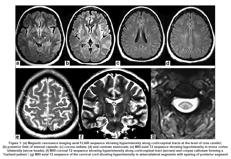
Neurology India, Vol. 57, No. 3, May-June, 2009, pp. 354-355 Letter To Editor 'Garland sign' in amyotrophic lateral sclerosis Atma Ram Bansal, Gopal Krishna Dash, Ashalatha Radhakrishnan, Chandrasekharan Kesavadas1 , Muraleedharan Nair Departments of Neurology, and 1 Imaging Sciences & Interventional Radiology, Sree Chitra Tirunal Institute for Medical Sciences and Technology, Trivandrum, Kerala, India.
Correspondence Address: Department of Imaging Sciences & Interventional Radiology, Sree Chitra Tirunal Institute for Medical Sciences and Technology, Trivandrum, Kerala Date of Acceptance: 31-Mar-2009 Code Number: ni09105 PMID: 19587488 DOI: 10.4103/0028-3886.53273 Sir, A 37-year-old lady presented with pure motor, asymmetrical onset, progressive quadriparesis with wasting of hands and feet of one year duration, with dysphagia, dysarthria, and emotional incontinence of four months duration. On examination, she had upper motor neuron (UMN) and lower motor neuron (LMN) signs in bulbar muscles, upper and lower limbs without involvement of extraocular movements. Rest of the neurological examination was normal. She satisfied the El Escorial criteria for definite amyotrophic lateral sclerosis (ALS). Her electrodiagnostic studies showed evidence of preganlionic neurogenic lesion involving bulbar, cervical, thoracic, and lumbosacral spinal segments. Her work up for secondary causes of anterior horn cell disease was negative. She was evaluated with magnetic resonance imaging (MRI) of the brain and spinal cord. MRI brain showed all the characteristic features of ALS including T2 hyperintensity extending along corticospinal tract from centrum semiovale to crus cerebri bilaterally. T2 hypointensity was noted in precentral gyrus. MRI cervical spine showed T2 hyperintensity involving the anterolateral column of the spinal cord. The coronal T2 image was forming a characteristic 'garland pattern' of hyperintensity extending along corticospinal tracts as well as involving corpus callosum [Figure - 1]. This peculiar imaging appearance has hitherto not been reported in a classical case of ALS. T2-weighted image showing high signal intensity involving the corticospinal tract extending into the anterolateral column of the spinal cord has been described. [1] Involvement of the corpus callosum has also been reported. [2] In addition, T2-weighted MRI typically demonstrates low signal intensity in the motor cortex; this finding has been attributed to T2 shortening due to iron deposition. [1] A lesion distribution simulating a garland has been described with Alexander disease, metastatic colorectal adenocarcinoma, and acute postinfectious glomerulonephritis. [3],[4],[5] References
Copyright 2009 - Neurology India The following images related to this document are available:Photo images[ni09105f1.jpg] |
| |||||||||
