 |
| About Bioline | All Journals | Testimonials | Membership | News |
|
||||||
|
||||||
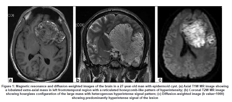
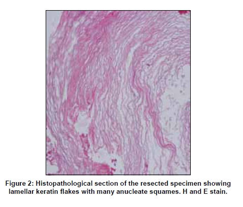
Neurology India, Vol. 57, No. 3, May-June, 2009, pp. 359-360 Neuroimage Intracranial epidermoid cyst: Magnetic resonance imaging features Rima Kumari 1 , Bhuvnesh Guglani 2 , Nitij Gupta 2 , Sujata Chaturvedi 3 Department of Neuroradiology, 1 Focus 3T MR Imaging and Research Center,
Correspondence Address: Dr. Bhuvnesh Guglani, 91, Geeta Apartment, Geeta colony, Delhi - 110 031. Date of Acceptance: 18-Mar-2009 Code Number: ni09108 PMID: 19587491 DOI: 10.4103/0028-3886.53266 A 27-year-old young male presented with a five- year history of headache and seizures with recent onset tingling and numbness in the right upper limb. Magnetic resonance imaging (MRI) revealed a large nonenhancing well-marginated extra-axial mass in the left frontotemporal region. On T1-weighted image the lesion showed mixed intensity reticulated appearance with interspersed hyperintense areas and it was hyperintense on T2-weighted (T2W) and diffusion-weighted images (DWI) [Figure - 1]. At surgery, a cyst with viscous, pearly white contents was removed. Microscopic examination of the cyst revealed keratinizing squamous epithelium with keratinous debris arranged in laminated layers with many anucleate squames [Figure - 2]. Epidermoids are rare benign, congenital, developmental tumors, usually located in the cerebellopontine angle and parasellar regions. The epidermoid cysts are thin walled and lined by stratified squamous epithelium. The typical MR image appearance of epidermoid tumors is hypo- to slight hyperintense on T1W images, iso- to hyperintense on T2W images, and hyperintense on DWI. [1],[2] Uncommonly on T1W images the epidermoids are hyperintense and on T2W images are of low signal intensity. Rarely on computed tomography (CT) epidermoids are hyperdense or calcified. [1],[2],[3] No definite relation between signal intensity on T1W images and the concentration of cholesterol and triglycerides within epidermoids has been found. However, high concentration of protein is thought to be responsible for an increase in signal intensity on T1W images. [2],[3] In this patient, the unusual MR imaging characteristics on T1W images may be explained by the high protein concentration with lamellar keratin flakes causing the honeycomb-like reticulated appearance. MRI with DW imaging is helpful to distinguish epidermoids from other cystic tumors, improving the specificity of diagnosis. [4] References
Copyright 2009 - Neurology India The following images related to this document are available:Photo images[ni09108f2.jpg] [ni09108f1.jpg] |
| |||||||||
