 |
| About Bioline | All Journals | Testimonials | Membership | News |
|
||||||
|
||||||
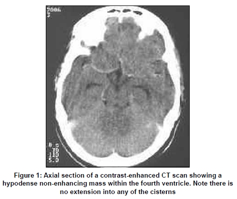
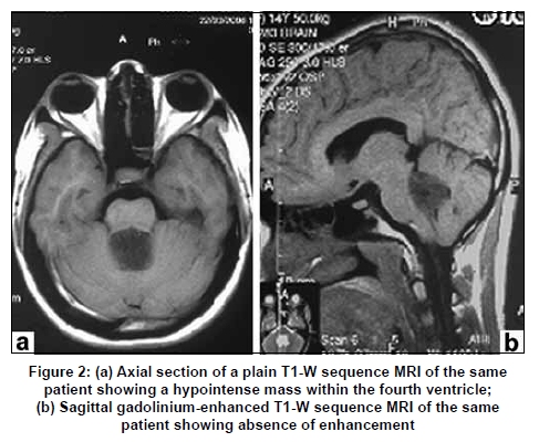
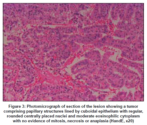
Neurology India, Vol. 57, No. 4, July-August, 2009, pp. 486-488 Case Report Choroid plexus papilloma presenting as a non-contrast-enhancing fourth ventricular mass in a child Ravindran Pratheesh, Ranjith K. Moorthy, Reecha Singh1, Vedantam Rajshekhar Departments of Neurological Sciences and 1Pathology, Christian Medical College, Vellore, India Correspondence Address: Dr. Vedantam Rajshekhar, Department of Neurological Sciences, Christian Medical College, Vellore - 632 004, India.rajshekhar@cmcvellore.ac.in Date of Acceptance: 26-Apr-2009 Code Number: ni09135 PMID: 19770555 DOI: 10.4103/0028-3886.55601 Abstract Choroid plexus papilloma (CPP) is a rare benign tumor of the central nervous system with a propensity for location within the lateral ventricle in children. We report a case of a 14-year-old girl who presented with transient facial paresis and ataxia. Her imaging showed a non-enhancing intra fourth ventricular mass, the histology of which was reported as CPP. The atypical clinical and radiological features in this case are discussed. Choroid plexus papillomas should be considered in the differential diagnosis of non-enhancing fourth ventricular masses.Keywords: Choroid plexus papilloma, computed tomography, fourth ventricle, magnetic resonance imaging Introduction Choroid plexus papillomas (CPP) are benign intraventricular tumors derived from the choroid plexus epithelium. These brilliantly contrast-enhancing tumors occur more commonly in children and in this age group the location is predominantly in the lateral ventricle. [1] This report highlights the unusual clinical and radiological findings in a patient who was diagnosed to have a CPP. Case Report A 14-year-old girl presented three months after an episode of transient left-sided facial paresis and imbalance while walking that had lasted for ten days. She was asymptomatic at the time of admission. She did not have any neurological deficits. Fundus examination was normal. Computed tomography (CT) showed a non-contrast-enhancing hypodense intra fourth ventricular mass. There were no calcifications [Figure - 1]. Magnetic resonance imaging (MRI) of the brain showed a hypointense fourth ventricular mass on short TR sequences that did not show enhancement after administration of gadolinium [Figure - 2]. There was no hydrocephalus. She underwent radical excision of the mass and she made an uneventful recovery. During surgery the mass was found to have a small attachment to the floor of the fourth ventricle. The histopathology showed a tumor composed of papillary structures lined by cuboidal epithelium with regular, rounded centrally placed nuclei with no evidence of mitosis, necrosis or anaplasia. This was reported as CPP [Figure - 3]. Contrast CT did not show any residual lesion at 16 months follow-up and she was asymptomatic [Figure - 4]. Discussion Clinical features of choroid plexus papillomas Choroid plexus papillomas account for 0.4-0.6% of all brain tumors and in children they account for 2-4% of brain tumors. [1] The reported annual incidence is 0.3 per 1,000,000 population. [2] The most common site of origin in the pediatric age group is within the atria of the lateral ventricles, whereas fourth ventricular tumors are more common in adults. [1] The commonest presentation is with raised intracranial pressure as a consequence of block of cerebrospinal fluid (CSF) pathways and/or overproduction of CSF. However, these tumors can have varied presentations including cerebellar dysfunction and cranial nerve involvement like hearing loss if it extends into the subarachnoid space. [3] Our patient had a transient left facial paresis that improved without any medical or surgical intervention, and she was asymptomatic at presentation to us. Radiological features On CT imaging, CPPs have been reported to be iso- to hyperdense and intensely contrast-enhancing with cystic areas and spotty calcifications. [4],[5] The MRI characteristics of these tumors have been described in literature as iso to hypointense in short TR sequences, variably hyperintense in long TR sequences and commonly associated with flow voids. [4],[5],[6],[7] The most important characteristic is the intense contrast enhancement which is homogeneous in most cases. Inhomogeneous enhancement has also been reported in a few cases.[6] A literature search revealed only one case of non-enhancing CPP, reported by Vasquez et al.[7] Imaging in our patient showed a hypodense fourth ventricular lesion with no areas of calcification and no enhancement after contrast administration. Pathology Macroscopically, these tumors are well-circumscribed cauliflower-like masses and may be adherent to the ventricular wall. At surgery, these tumors usually are free from the floor of the fourth ventricle except in rare instances. [8] There have been reports of CSF seeding in these benign tumors. [8] In our patient, the tumor was found to be adherent to the floor of the fourth ventricle and this was another atypical feature. Gross total excision is the treatment of choice and the reported five-year survival is 100%. [9] The presence of mitoses, less than 50% of the tumor cells heavily positive for S100, absence of TTR(transthyretin)-positive cells, brain invasion by cell nests, absence of marked stromal edema, and presence of necrotic areas have shown to correlate with a poor prognosis. [10] Conclusion Choroid plexus papilloma is a benign tumor seen in children and has a good prognosis. Although known to brilliantly enhance with contrast, a non-enhancing variant is present and should be included in the differential diagnoses for non-enhancing fourth ventricular masses in children. References
Copyright 2009 - Neurology India The following images related to this document are available:Photo images[ni09135f4.jpg] [ni09135f2.jpg] [ni09135f1.jpg] [ni09135f3.jpg] |
| |||||||||
