 |
| About Bioline | All Journals | Testimonials | Membership | News |
|
||||||
|
||||||
Neurology India, Vol. 57, No. 6, November-December, 2009, pp. 706-714 Review Article Transplantation and stem cell research in neurosciences: Where does India stand? Prakash N. Tandon Emeritus Professor, Neurosurgery, All India Institute of Medical Sciences, New Delhi and President, National Brain Research Centre, Manesar, Haryana, India Date of Acceptance: 15-Dec-2009 Code Number: ni09204 PMID: 20139497 DOI: 10.4103/0028-3886.59464 Abstract The nearly absent ability of the neurons to regenerate or multiply has prompted neuroscientists to search for the mean to replace damaged or dead cells. The failed attempts using adult tissue, initiated nearly a century ago, ultimately brought rays of hope when developing fetal neurons were used for transplantation in 1970s. The initial excitement was tempered by limited success and ethical issues. But these efforts unequivocally established the feasibility of successful neural transplantation provided appropriate tissue was available. The ability to derive embryonic stem cells with their totipotent potential by Thomson in 1998 rekindled the interest in their use for replacement therapy for damaged brain tissue. The present review surveys the current status of this promising field of stem cell research especially in respect to their therapeutic potentials for purposes of neural transplantation. A brief account is provided of the ongoing Indian efforts in this direction.Keywords: Adult neural stem cells, embryonic stem cells, neural transplantation Historical Background While attempts at neural transplantation for repair of damaged/diseased brain started a century ago [1] the real enthusiasm was stimulated after the pioneering work of Das and Altman (1972), [2] Bjorklund and colleagues (1971, 1979a,b, 1980), [3],[4],[5],[6] Olson (1970) [7] and Lund and Hauschka (1976). [8] These studies heralded an explosion of investigations which established that use of fetal neural tissue provides a reliable method of achieving a successful graft in an adult host. Investigators all over the world initiated experiments dealing with various aspects of neuronal grafting as a strategy to replace damaged areas of the brain in the late 1970s. It has been unequivocally demonstrated that such grafts "take", grow, develop at least limited two-way connections with the host brain, produce appropriate neurotransmitter, and to a variable extent restore functional deficits resulting from disease or damage to the host brain. [9] Further, these grafts have been found to induce "trophic" effects on the host nervous system. These results in experimental animals were so tantalizing that for once the neurosurgeons jumped straight from the "rat-to-man" without even waiting for the results of the studies in higher primates. The first neural transplants in humans were performed in Sweden by Backlund and his colleagues in 1982 and 1983. [10] In 1984-85, a multidisciplinary group was established at the All India Institute of Medical Sciences, New Delhi to study the neurobiological and behavioral consequences of the neural transplants in rats and in rhesus monkeys. Successful transplantation could be achieved in 80-85% of adult rats. Recognizing the limitations of transferring information gathered from rat to man, it was decided to study the fate of fetal neural transplants in sub-human primates. Rhesus monkeys were utilized for this purpose. Following a number of trials, ultimately successful transplants were observed in the caudate nucleus in two monkeys. [11] These studies revealed that the fetal neural transplant in rhesus monkey is successful in 20-30% cases only. At three to four months after transplantation most of the grafts had resorbed completely leaving behind a necrotic cavity heavily infiltrated by lymphocytes and macrophages. However, in the rodent model the transplanted neurons matured, differentiated and developed phenotypic characteristics, comparable to the normal adult nigral neurons. Electron microscopy revealed characteristic sub-cellular organelles and synapses. Golgi stain clearly demonstrated the growth and branching of neuronal processes up to 1-3 mm. Some of these neurons migrated into the surrounding brain. Neuronal processes could be demonstrated to cross the graft-host interface in either direction. Immunohistochemistry confirmed that these neurons and their processes were positive for tyrosine hydroxylase (TH) implying their capacity to produce the appropriate neuro-transmitter: Dopamine. This remained so for the first three to four months after the implant. Most investigators did not study the fate of these grafts beyond this period. The unique observations of our study were those related to morphological details in long term surviving grafts. At varying intervals, we observed changes compatible with premature aging leading to neuronal loss. These changes worsened with time so that at the end of 18 months and two years majority of the surviving neurons were showing extensive degenerative and ageing changes. [12] While it is impossible to demonstrate the development of intricate circuitry between the graft and the host, a number of observations would indicate that such circuitry, at best can only be very partial developing contact with neurons in the vicinity. In spite of the tremendous enthusiasm generated by the reports of the beneficial effect of adrenal medullary transplants in the head of the caudate nucleus of patients of Parkinson's disease by Backlund et al. (1985) [10] and Madrazo et al. (1987, 1991). [11],[13] It was generally accepted that the neurobiological basis of the observed clinical effects was not clear. Peterson et al. (1989) [14] could not find any surviving cells at autopsy of a Parkinson's disease patient treated with adrenal to brain transplant. The clinical trials so far conducted have provided enough evidence, at least for patients of Parkinson's disease, there can be mild to moderate improvement in their motor function, though it may be temporary. This necessitate search for alternate sources for transplantable cells, cultured, cryo-preserved or genetically modified. There are already leads in this direction. [15] Long before embryonic stem cells were demonstrated to give rise to all types of brain cells, an extensive experience had already accumulated in respect to use of fetal neural tissue, obviously containing neural progenitor cells or even stem cells for repair or replacement of damaged brain. [4],[5],[6],[9],[15] Similarly, stable clones of neural stem cells (NSCs) isolated from human fetal telencephalon were shown to replace neurons and respond to development clues when transplanted in new born mouse brain. [16] There has been rapid success in devising in vitro protocols for differentiating human ES cells to neuro-epithelial cells. Progress has already been made to guide these neural precursors further to more specialized neural cells such as spinal motor neurons or dopamine-producing neurons (or various types of glia). Neural Stem Cells In February 2001, only 15 months after the publication of two seminal papers by Thompson and Snyder and McKay in Science on isolation of human embryonic stem cells (ESC), the author published a general article-"Neural Stem cell research: A revolution in the making" [17] A decade after the Science papers, the prediction made in 2001 has been validated beyond imagination by the voluminous literature that has already accumulated, the number of specialized centers, departments, laboratories established globally, the scientific conferences held and the socio-political, ethical implications debated all over the world and even a large number of clinical trials initiated. At the same time the conclusion arrived at in the 2001 paper, [17] "it could be safely stated NSC (neural stem cell) research needs to be pursued with vigor for clinical use. While there is a lot of hope one should not be carried away by the hype and prematurely raise the expectations among those most in need of it" has continued to be echoed even today by a large number of distinguished researchers from all over the world. This has prompted a prestigious journal like Philosophical Transactions of the Royal Society to devote a complete issue in January 2008 on the subject of "Stem Cells and Brain Repair". I could do no better than to quote Magnus and colleagues in this volume,"Stem Cells, although difficult to define, hold great promise as tools for understanding development and as therapeutic agents. However, as with any new field, uncritical enthusiasm can outstrip reality". [18] Having followed the subject keenly during the past decade, having observed the interest it has generated among colleagues, both basic scientists and clinicians and concerned about the inadequate knowledge, often verging on gross misconceptions amongst some of them, has prompted this review of the existing knowledge on the subject primarily restricted to aspects of interest for neuroscientists. Grafting cells for therapeutic purposes has been ongoing for a long time in the case of bone marrow or skin transplantation. The recent advances in stem cell biology have opened up unprecedented possibilities to cure many hitherto untreatable diseases. [19] The first experimental demonstration that stem cells exist goes back to the early 1960s when the hematopoietic system was shown to harbor single cells responsible for the renewal of the circulating blood. [20] The pioneering work of Weissman and his colleagues on biology of hematopoietic stem and progenitor cells, [21],[22] the purification and characterization of mouse hematopoietic cells, [23] clonal analysis of hematopoietic stem cell differentiation in vivo[24] as also the isolation of a candidate human hematopoietic stem cell population [25] helped a great deal in understanding the basic biology of hematopoietic stem cells which has important lessons for stem cell biology in general. [26],[27] Embryonic Stem Cells The discovery of the embryonic stem cell constitutes one of the greatest achievements of modern biotechnologies. It was observed that, at the blastocyst stage, each embryonic cell is essentially as totipotent as the egg itself. Already in 1981 Evans and Kaufman [28] achieved establishment in culture of pluripotential cells from mouse embryo. However, it was only in 1998 when Thompson et al. published that they had been able to derive embryonic stem cell lines (ES) from human embryo that the technology attracted global attention. [17] In the same year it was shown that germ cells isolated from gonads of older human embryos can also give rise to permanent lines of embryonic stem cells. The stem cells that result from germ cell proliferation are designated EG cells in order to distinguish them from the classical ES cells. [29] Definitions Stem cells Cells able to reproduce themselves throughout the life span of the animal and able to give rise to differentiated cells. They have the ability to divide for indefinite periods in culture and give rise to specialized cells. Embryonal stem cells Cell derived from embryo-pre or post implantation-prior to their differentiation into specific cell types. Totipotent cells Cells which have the potential to differentiate into derivatives of all three embryonic germ layers i.e. ectoderm, mesoderm and endoderm. In addition they can also specialize into extraembryonic membranes and tissues. Pluripotent cells Cells which can give rise to different types of cells representing derivatives of two different germ layers e.g. skin (ectoderm) and muscle (mesoderm). Neural stem cell Cells which can generate neural tissue, either one or both neuron and glia, (astrocytes, oligodendrocytes). The term is also used for stem cells derived from embryonic or adult nervous system which normally differentiates into nervous tissue. These cells remain undifferentiated for long periods of time while retaining potentials to differentiate into nervous tissue. Progenitor cells Cells with a more restricted potential than a stem cell, and are generally destined to give rise to a specific cell type. A vast amount of new information has accumulated on human stem cell biology. The generation of human embryonic, fetal and adult stem cell lines has been standardized. It is proposed that the cells obtained from these different sources could contribute different but, perhaps, equally important properties of therapeutic relevance. [30] Stem cells from different sources have unique attributes that will differentially affect their suitability for use in therapeutic strategies. Sources of stem cells Human embryonic stem cells Derived from the blastocyst inner cell mass of excess embryos generated by in vitro fertilization can provide an unlimited source of cells for transplantation and can be directed into neural precursor which can generate neurons, oligos and glia both in culture and in-vivo. [31-33] Park et al. 2005 and Perrier et al. 2004 [34],[35] demonstrated in-vitro and in-vivo differentiation of human embryonic stem cells into dopamine neurons. It may be mentioned that over the years, culture conditions that rely on the use of various cytokines and growth factors, have made it possible to induce the differentiation of a high proportion of ES cells into selected cell types such as neurons, pancreatic islet cells, cardiomyocytes etc. [19] Fetal neural stem cells Harvested from the post-mortem human fetal brain maintain a normal karyotype for a significant number of passages in culture and can produce a large number of neurons and astrocytes. [36] These posses a relatively high proliferative capacity and yet do not generate tumor following transplant. These are really mostly progenitor cells and not true ESCs. Seth at National Brain Research Centre, Manesar has established a fetal brain derived cell culture system to obtain CNS stem/progenitor cells. These could be selectively differentiated to astrocytes and neurons by providing appropriate growth factors and defined media conditions. Adult neural stem cells Contrary to earlier belief neural stem cells persist throughout life, not just in the two now well-known sites of adult neurogenesis e.g. the sub-ventricular zone and hippocampus but also at the other sites. [37],[38],[39] It has been claimed that adult neural stem cells can be harvested from brain tissue, post-mortem or through biopsy and expanded in culture both in rodents and human. Lie et al. (2002) described the existence of progenitor cells with neurogenic potential in the adult substantia nigra. [40] Young and Black (2004) have provided a detailed review of adult stem cells. However, their proliferative capacity is somewhat limited. [41] Non-neural adult stem cells A number of recent studies have challenged the traditional view that stem cells present in somatic tissues are restricted to producing that tissue's cell types. The first such observation was in respect to bone-marrow derived stem cells (BHMSC), which could develop into liver cells [42],[43] muscle, [44],[45],[46] bone [47] and neural tissue. [48],[49],[50],[51] Woodbury et al. (2000) [52] claimed that adult rat and human bone marrow stromal cell differentiate into neurons. Zhao et al. (2002) demonstrated neural differentiation and functional recovery after transplanting human bone marrow stem cells into the ischemic brain of rats. [53] Sanchez et al. (2001) observed expression of neural markers in human umbilical cord blood. [54] Recently there was great excitement about the possibility of generating neural progenitor cells from such diverse sources as mesenchymal cells derived from skin, bone-marrow, or adipose tissue. [55],[56],[57],[58] It may be mentioned that reproducible technique to convert them into authentic CNS cells has not yet been accomplished. The skin derived precursors (SKPs) display multi-lineage differentiation potential, producing both neural and mesodermal progeny in vitro. This had led to much sensationalism as evident from such titles of reports as, "Blood to Bone" or "Bone to Brain" etc. However, there are many questions on the validity of a number of these studies, in a large part owing to loose criteria, which have been used to describe 'neural differentiation'. [59] However, differentiation of SKPs under neurogenic conditions resulted in the production of cells that fulfil most, but not yet all, criteria for neuronal differentiation. [60] Neuronal marker studies revealed that SKP-derived neurons are probably peripheral in nature. One type of stem cell that definitely differentiates into mature terminally differentiated and functional neural cells in vitro are NSC. [61],[62],[63] Criteria used to define differentiation of NSCs to mature neural cells should be used as the gold standard to gauge whether somatic stem cells of non-neuronal origin, such as HSCs or MSCs, can give rise to neural progeny. A series of criteria have been described. These include expression of neuronal or glia-specific markers at the RNA or protein level and that the differentiated cells should display function characteristics consistent with neurons and glia such as voltage-gated sodium channels, depolarizing response to neurotransmitters and release of neurotransmitters on depolarization. However, more recent studies have demonstrated that at least NSC-generated neural progeny have functional characteristics consistent with neurons. [64],[65] Pluchino et al. (2003) using syngenic culture derived adult NSCs in an animal model of multiple sclerosis demonstrated promotion of multifocal remyelination. [66] It needs to be kept in mind that the functional benefit following transplantation of such cells may not be due to integration of donor neural cells but due to trophic effects elaborated by the donor cells that promote endogenous neural cells to repair the deficit. [67],[68],[69] Induced pluripotent stem cells In November 2007, two groups headed by James Thompson at the University of Wisconsin-Madison and Shinya Yamanaka and his post doctoral fellow Kazutoshu Takahasi at Kyoto University, Japan, described methods to reprogram adult human cells from skin to a pluripotent state using genetic engineering techniques. These cells, called induced pluripotent stem cells (iPS) were found to be similar to human embryonic stem (ES) cells in morphology, proliferation, surface antigens, gene expression, epigenetic status of pluripotent cell-specific genes and telomerase activity. Further, these cells could differentiate into cell types of the three germ layers. [70] This technique could help generate patient and disease specific cells, which cannot be generated from ES cells. Excellent reviews on the subject are available. [71],[72] Reprogramming protocols that exclude cancer-associated gene c-myc have been developed. [73] However, at the moment iPS cells remain a research tool and not a potential therapeutic agent. [74] David Cyranoski (2008) has pointed out that the greatest challenge still exists: The generation of high-purity, clinically relevant cell populations. [75] It is not only going to be very time consuming but also extremely costly. Zhang (2005) [76] has provided detailed accounts and comparison of methods for neuro-epithelial differentiation from human ES cells. Ideally the method should be simple, efficient, chemically defined, scalable and reproducible, and the end product should be enriched or purified homogeneous neural progenitor population. Neuroepithelial cells produced from human ES cells using different methods may appear similar in morphology and expression of certain neural precursor markers. However, they in fact differ significantly from each other depending upon the culture conditions. Carefully designed strategies will be needed in order to direct ES cells to the vast array of neuronal subtypes that are harbored in the primate forebrain. The ability to differentiate into versatile neuronal subtypes in a neurogenic environment places human ES cell-derived neuroepithelial cells as a useful source of cells for neural replacement therapy in multiple neurological disorders. Recognizing the therapeutic potentials of stem cells a number of public and private organizations like International Stem Cell initiative, UK Stem Cell Bank, ES Cell International in Singapore, Cellartis in Sweden, Millipore in USA have developed ES cell lines to be made available to researchers. [74] Despite significant progress in an efficient differentiation of neuroepithelial cells (70-90%), and a few neural subtypes, such as dopamine neurons (approx 30%), spinal motor neurons (approx 20%) and oligodendrocytes more than 90%, protocols for generating many neuronal subtypes needs to be established. Demonstration of functional integration of human (ES derived) neural cells in the brain is still awaited (our experience with fetal neural transplant-lessons learnt are probably valid for ES derived cells also). [77] While functional improvement occurs it is obvious that intricate integration in host circuitry was not unequivocally observed. The functional improvement could be partially attributed to protective role of the graft on endogenous neurons. Under most circumstances, neural transplant acts as a local chemical replacement rather than due to integration in the host circuitry. As a consequence the release of neurotransmitters is uncontrolled. This may be the reason for dyskinesia observed in patients who received foetal neural transplants as reported by Olanow et al. [78] Nine myths about stem cells In a review, Magnus et al. (2008) have listed nine common myths that affect our approach to evaluating stem cells for therapy. [18] These myths include some of the well entrenched beliefs like:
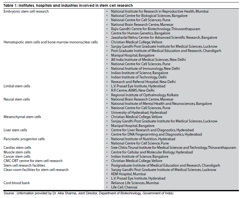
Other uses of stem cells Human ES cell-generated neural cells provide a tool for screening pharmaceuticals that may have therapeutic values in neurological disorders as also for toxicity screening and possibly drug discovery. The availability of human ES cells with natural diseases [90] (Verlinsky et al. 2005), via somatic nuclear transfer or through genetic alterations in laboratories, will provide not only disease-specific neural cells for drug screening but also a tool to unveil some fundamental pathological processes underlying individual neurological disorders. Expansion of HSC The number of HSC that one can isolate from mobilized blood or from umbilical cord, or from bone marrow limits the full application of HSC transplantation in man. Attempts to expand HSC with the known cytokines, stem cells factor, steel factor (SLF), thrombopoietic (TPO), interleukins 1, 3, 6, 11, plus or minus the myeloerythroid cytokines and erythropoietin have never resulted in a significant expansion of HSC. Weissman (2007) [93] reported, "We and others have attempted to reproduce the demonstration of production of critical neurons from marrow or hematopoietic stem cells precursors and have failed". Nevertheless, these experiments have led to extensive clinical trials in humans [94],[95],[96],[97] primarily for myocardial regeneration and more recently for patients with stroke. Unfortunately the claims made in this regard have not been universally accepted. [98],[99] Using strict criteria, the author concluded, "We do not believe there is sufficient evidence for any of the transplant claims of trans-differentiation. In fact, most if not all, reports of donor markers in unexpected tissues are the result of cell fusions and the rarity of cell fusions makes it questionable that such events are regeneration rather than reflect the functions of post-injury phagocyte cells. In addition to hematopoietic stem cells, the following stem cells have been prospectively isolated to homogeneity: Peripheral nervous system stem cells, central nervous system stem cells. [100],[101] In the case of human CNS stem cells, extensive experiments of transplanting them into the lateral ventricle, into the brain or into the spinal cord of SCID mice have shown that they contributed in a robust way to engraftment of the neurogenerative cells. These robust regenerations stand in contrast to microglial contributions and the rare Purkinje cell fusion derived from bone marrow and hematopoietic stem cells. Stem Cell Research in India Soon after the publication of the paper in Current Science [17] as Chairman of the Medical Biotechnology Task Force of DBT, a meeting was called to take stock of the interest in the field among biomedical scientists in the country and to prompt them to initiate studies in this emerging field by providing research grants. This had progressively emerged as a major national effort primarily steered by DBT. The key components of the strategy are-creation of centers of excellence (CoE); virtual network of centers; generation of adequate human embryonic stem cell (hESC) lines; human resources development through training; short and long-term overseas fellowships; study the biology of all types of adult stem cells and in parallel evaluate its safety and efficacy in animal models. Over 30 institutions, hospitals and industry are involved in SCR in the country. The government has invested about 8.0 million US$ for SCR in last two years. Draft guidelines for SCR in the country have been formulated and the same are currently being placed for public debate. The major ongoing programs include among others: Establishment of hESC lines, use of limbal stem cells to repair damaged cornea, isolation, purification and characterization of hematopoietic, mesenchymal and liver stem cells; differentiation of stem cells into neural, cardiac, β cell lineages, etc. Studies have been supported to explore the potential applications of adult stem cells in stroke, cardiac, pancreatic, spinal cord injury, use of lectins for hematopoietic stem cell preservation etc. Reliance Life Sciences, Mumbai has characterized several stem cell lines including two neuronal cell lines, Dopamine producing neurons and neurons for patients of stroke. One cell line has been deposited in the National Center for Cell Science (NCCS), Pune. Interaction between clinicians and basic scientists already exists in several centers in India such as the Christian Medical College, Vellore; L.V. Prasad Eye Institute (LVPEI), Hyderabad; National Institute of Mental Health and Neurosciences (NIMHANS), Bangalore, All India Institute of Medical Sciences (AIIMS), New Delhi. At CMC, Vellore a technology has been established for collection, isolation and purification of HSC for haploidentical hematopoietic stem cell transplantation. The first, haploidentical hematopoietic transplantation was carried out at CMC, Vellore in April 2003. The phase I multi-centric clinical trial and a pilot study using bone marrow mononuclear cells have been initiated in the country for myocardial infarction and stroke, respectively. A 'CMC-DBT Center for Stem Cell Research' has been created at CMC, Vellore. In India, some companies have started establishing the repositories of cord blood banking. Reliance Life Sciences, Mumbai, has a repository of 3,000 cord blood samples. Life Cell is a Chennai-based company and has a license agreement and knowledge-sharing tie up with Cyro-Cell International, USA. They have a repository of 1,000 cord blood samples and are offering to preserve stem cells for 30 years. The Department of Biotechnology, Ministry of Science and Technology and Indian Council of Medical Research have formulated draft guidelines for SCR in India jointly. National Brain Research Centre, Manesar has actively pursued basic science investigations on both commercial ES cells and neural precursor cells derived from human abortuses [Table - 1]. Neural progenitor cells The pre-requisite of a purified, continuous and sufficient population of well characterized human neural precursor cells (hNPCs) with ability to differentiate into glial cells and neurons remains the biggest hurdle. Several investigators claim success in isolation of purified population of hNPCs, but the procedure is typically difficult with ill-defined protocols. A detailed description of protocols for isolation, expansion and differentiation of hNPCs as well as characterization of glial and neuronal cells differentiated from hNPCs is largely unavailable. Seth and his team at National Brain Research Centre, Manesar has designed a standardized three week protocol that describes an ethically approved stepwise process for isolation, maintenance, expansion, differentiation and characterization of undifferentiated as well as differentiated hNPCs from human fetal brain samples collected from aborted material. Their work has yielded a unique resource in the country of human neural stem cells that can be used for various basic and transplantation studies. Use of ex-vivo expanded stem cells has been identified as new drug as per FDA, USA, i.e. investigational new drug (IND). This would require information about the source, number, purity, appropriate stage, optimum condition and criteria for harvesting stem cells; also standardization of doses in terms of concentration and number of stem cells for each application and minimal manipulation of cells for clinical use. Good animal models are required to address the issues of safety and efficacy before attempting clinical applications of stem cells. The basic requirements for clinical trial are: Adequate infrastructure, i.e. good manufacturing practices (GMP), clinical grade reagents, trained manpower, proper documentation, standard operating procedures (SOPs), quality control etc. Acknowledgment The author acknowledges the help provided by Dr. (Mrs.) Alka Sharma of the Department of Biotechnology, Government of India, Dr. Pankaj Seth and Dr. Shyamala Mani of National Brain Research Centre, Manesar in preparation of this review. References
Copyright 2009 - Neurology India The following images related to this document are available:Photo images[ni09204t1.jpg] |
| |||||||||
