 |
| About Bioline | All Journals | Testimonials | Membership | News |
|
||||||
|
||||||
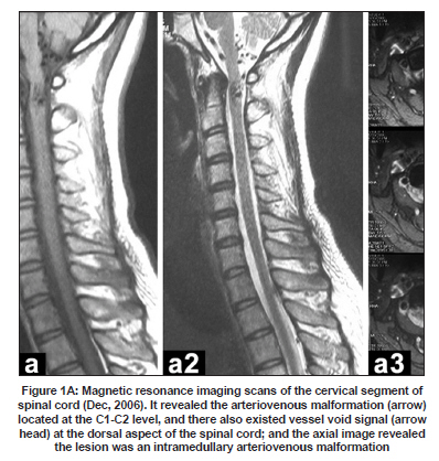
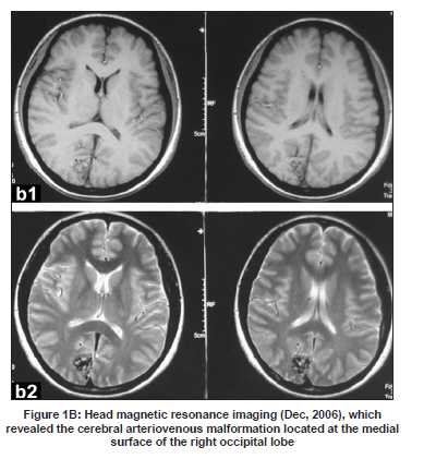
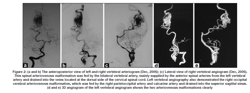
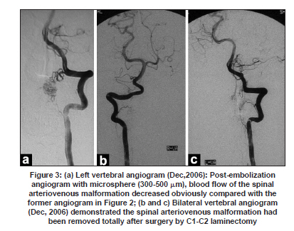
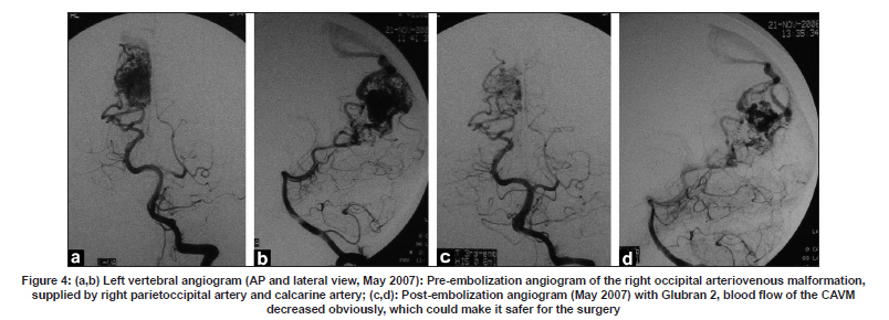
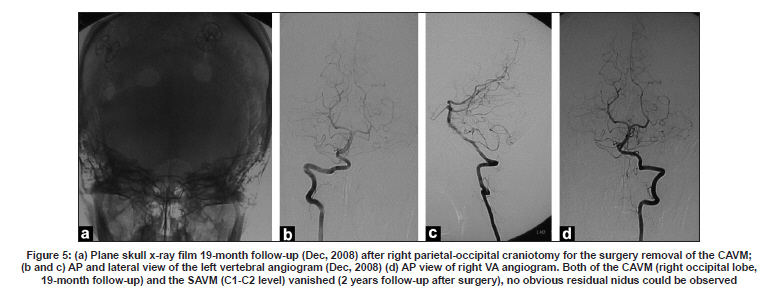
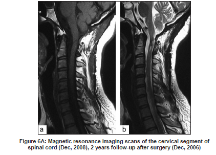
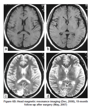
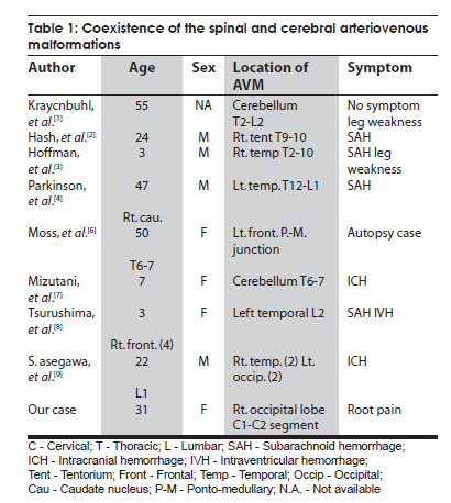
Neurology India, Vol. 57, No. 6, November-December, 2009, pp. 785-788 Case Report Coexistence of a single cerebral arteriovenous malformation and spinal arteriovenous malformation Yabing Wang, Hongqi Zhang, Feng Ling Department of Neurosurgery, Xuanwu Hospital, 45#, ChangChun Ave, Beijing - 100 053, PR. China Date of Acceptance: 23-Oct-2009 Code Number: ni09219 PMID: 20139512 DOI: 10.4103/0028-3886.59479 Abstract The coexistence of a cerebral and a spinal arteriovenous malformation (AVM) together is extremely rare. We present a 31-year-old woman, who suffered from severe root pains in the left upper extremity. Magnetic resonance imaging (MRI) revealed the abnormal vessels in the left occipital lobe and upper cervical segment of spinal cord. Cerebral angiography and spinal angiogram revealed two AVMs: One was in the right occipital lobe and the other was located in the C1-C2 segments of cervical cord. She had no other vascular lesions, and nor did her other family members. As the primary problem in her was left upper extremity root pains, which we considered was related to the spinal AVM, the first therapeutic treatment was focused on spinal AVM. The cerebral AVM of the right occipital lobe was surgically resected after part embolization.Keywords: Multiple; cerebral arteriovenous malformation; spinal arteriovenous malformations Introduction Coexistence of cerebral arteriovenous malformation (AVM) and spinal AVM is extremely rare. Since the first description in 1969, to the best of our knowledge, no more than 10 cases with single cerebral AVM combined with a single spinal AVM have been documented. We viewed all the 697 cases spinal vascular malformation since 1990 in our center; only one case was confirmed with single cerebral AVM and a spinal AVM, thus giving an incidence of 0.14% of concurrence of single cerebral AVM and a spinal AVM. Case Report A 31-year-old woman presented with severe root pain of the left upper extremity since 2001. On the first admission in December 2006, magnetic resonance imaging (MRI) revealed abnormal vessels in the spinal cord at C2 level and an abnormal lesion, a flow void signal in the right occipital lobe [Figure - 1 a and b]. Spinal and cerebral angiogram revealed a spinal arteriovenous malformation intramedullary at C2 segment and a cerebral AVM of the right occipital lobe [Figure - 2]a-d. The bilateral vertebral arteries, mostly the anterior spinal arteries from the left vertebral artery, fed the spinal AVM. The draining veins were into the veins located at the dorsal aspect of the cervical spinal cord. Left vertebral angiogram also demonstrated the right occipital cerebral AVM, mainly fed by the right posterior cerebral artery and drained into the superior sagittal sinus. The symptom of this patient was root pain of the left upper extremity, which we considered related to the spinal AVM located at the C2 segment. The spinal AVM was embolized using microsphere (300-500 µm) under local anesthesia. Postembolization angiography demonstrated a significant reduction in blood flow from the left vertebral artery to the AVM [Figure - 3]a. Several days later she had C1-C2 laminectomy and resection of the spinal AVM. Most of the AVM was intramedullary. The postoperative course was uncomplicated with no neurological deficits, and the root pain of the upper extremty gradually improved with carbamazepine over one week. The left vertebral angiogram revealed no obvious residual nidus [Figure - 3]b and c. The patient recovered well after the operation of the spinal AVM, but as we felt she still faced the risk of bleeding due to the cerebral AVM of the right occipital lobe, in consultation with the patient and her relatives she was readmitted for the treatment of the cerebral AVM in May 2007. Part embolization with Glubran 2 for the cerebral AVM before the surgery was performed [May 2007, [Figure - 4]a-d], and the cerebral AVM was totally excised by the right parietal-occipital craniotomy. Postoperatively she had good recovery without any neurological deficits. She had follow-up vertebral angiogram [Figure - 5] and MRI [including the cervical segment and head, [Figure - 6a and b]] in December 2008. Discussion The occurrence of multiple AVMs in the central nervous system is rare. [2],[3],[4] Furthermore, single cerebral AVM combined with a spinal AVM is extremely rare and the exact incidence is unknown. Up to now, not more than 10 such cases, single cerebral AVM combined with a single spinal AVM, have been reported since 1969. [1] Clinical details of the nine cases reported in the literature and our case are summarized in [Table - 1]. [1],[2],[3],[4],[6],[7],[8],[9] As compared to the other reported cases, the same artery the vertebral artery fed the two AVMs in our patient. In the other reported cases, the AVMs were fed by different artery system. Willinsky [5] reported a 3.9% incidence of multiple cerebral AVMs by reviewing the angiograms of 203 patients. In our center, the incidence of concurrence of single cerebral AVM and a spinal AVM is about 0.14% (1/697). The natural history of multiple AVMs remains unclear. At present there is no consensus regarding the appropriate treatment in patients with multiple AVMs and embolization has been suggested as a useful method combined with the surgical treatment in patients with multiple AVMs. The therapeutic strategy for multiple AVMs remains difficult. Based on our experience, we suggest a staged approach. References
Copyright 2009 - Neurology India The following images related to this document are available:Photo images[ni09219t1.jpg] [ni09219f6b.jpg] [ni09219f6a.jpg] [ni09219f4.jpg] [ni09219f1a.jpg] [ni09219f2.jpg] [ni09219f3.jpg] [ni09219f5.jpg] [ni09219f1b.jpg] |
| |||||||||
