 |
| About Bioline | All Journals | Testimonials | Membership | News |
|
||||||
|
||||||
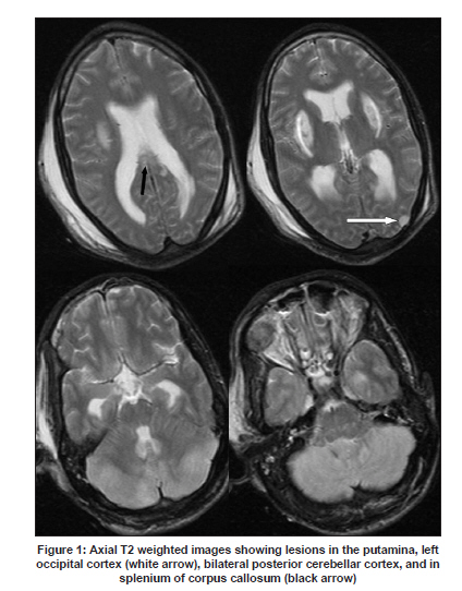
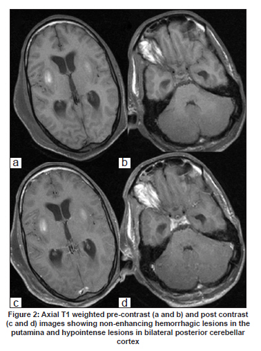
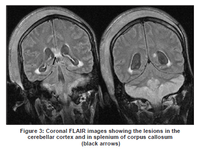
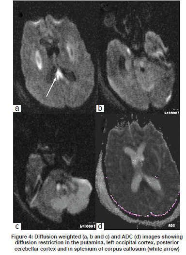
Neurology India, Vol. 57, No. 6, November-December, 2009, pp. 835-836 Neuroimage Methanol poisoning Sameer Vyas, Neeraj Kaur, Navneet Sharma1 , Paramjeet Singh, Niranjan Khandelwal Departments of Radiodiagnosis and 1 Internal Medicine, Postgraduate Institute of Medical Education and Research, Chandigarh, India Date of Acceptance: 30-Nov-2009 Code Number: ni09243 PMID: 20139536 DOI: 10.4103/0028-3886.59503 A 22-year-old male presented with acute onset vomiting, diarrhea, abdominal pain and altered sensorium. He had a history of acute alcohol intake (locally made with solvent) a day prior to the presentation. Biochemical investigations revealed severe metabolic acidosis. Diagnosis of methanol poisoning was made on the basis of history and biochemical abnormality. Magnetic resonance imaging (MRI) done on fifth day revealed hemorrhagic putaminal necrosis, left occipital, corpus callosum and cerebellar cortical lesions [Figure - 1],[Figure - 2],[Figure - 3],[Figure - 4]. Acute methanol intoxication can occur as accidental or suicidal ingestion. Patients present acutely with acute neurological, visual and gastrointestinal symptoms. [1] Neuroimaging helps in establishing the clinical diagnosis of the methanol poisoning. MRI findings in methanol poisoning are characteristic and include hemorrhagic putaminal necrosis (most characteristic), subcortical and deep white matter lesions, cerebral and cerebellar cortical lesions, and midbrain lesions. [1],[2],[3],[4] Basal ganglia involvement is likely due to direct effect of metabolites of methanol as well as selective vulnerability of the basal ganglia to acidosis, as compared to rest of brain. Selective basal ganglia and white matter lesions are not specific to methanol intoxication and can be seen in hepatolenticular degeneration, carbon monoxide poisoning, hypoxic-ischemic insult and Leigh's disease. [4] Optic nerve lesions are considered to be due to myelinoclastic effect of formic acid and due to axonal loss. [3] Hemorrhage in methanol poisoning is seen in up to 14% of patients and diffusion restriction may be seen in the involved areas. [4] Index case showed almost entire spectrum of the MRI finding seen in the methanol poisoning. References
Copyright 2009 - Neurology India The following images related to this document are available:Photo images[ni09243f4.jpg] [ni09243f1.jpg] [ni09243f2.jpg] [ni09243f3.jpg] |
| |||||||||
