 |
| About Bioline | All Journals | Testimonials | Membership | News |
|
||||||
|
||||||
Neurology India, Vol. 58, No. 1, January-February, 2010, pp. 53-57 Original Article Parkinson's disease: Functional changes in frontal and parietal cortex using 18 F- fluoro-deoxy glucose positron emission tomography/computed tomography Zhongyu Hou1,2 , Shuhui Hong3 , Bo Sun1 , Xiangtao Lin1 , Qingwei Liu2 , Shuzhan Yao2 , Shuwei Liu1 1 Research Center for Sectional and Imaging Anatomy, Shandong University School of Medicine, Correspondence Address: Dr. Shuwei Liu, Research Center for Sectional and Imaging Anatomy, Shandong University School of Medicine, 44 Wenhua Xi Road, Jinan - 250 012, China, liusw@sdu.edu.cn Date of Acceptance: 12-Jan-2010
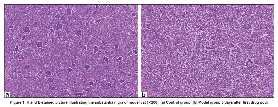
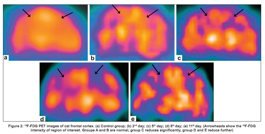
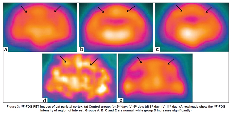
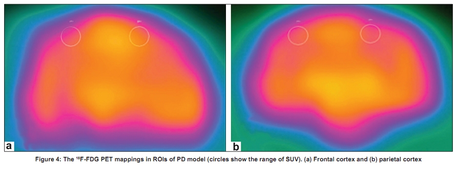
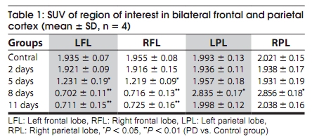
Code Number: ni10011 DOI: 10.4103/0028-3886.60397 Abstract Background : In Parkinson's disease (PD) there is increasing evidence to suggest motor function changes of the cerebral cortex occur in addition to the pathological changes in the extrapyramidal system. Keywords: Cat, frontal and parietal cortex, positron emission tomography/computed tomography, Parkinson′s disease, 18 F-Fluorodeoxyglucose Introduction Earlier studies in Parkinsons′s disease (PD) mainly involved nigrostriatal system. [1],[2],[3],[4],[5] However in the recent studies, there is increasing evidence showing motor functional changes of the cerebral cotex. [6],[7] Functionally frontal cortex is closely related to the basal ganglia. Thus investigations in this regard might show the pathophysiological changes and might also suggest possible explanation for the clinical manifestation of PD. Previous studies have detected metabolic and functional disturbance in the frontal cortex in patients with PD. However, patients recruited in these studies were on L-dopa for a long time, which may make the conclusions less persuasive. In this study, we developed a PD cat models by impairing bilateral dopamine neuron in substantia nigra selectively using 1-methyl-4-phenyl-1, 2, 3, 6-tetrahydropyridine (MPTP), and then examined the changes of glucose metabolism in the frontal and parietal cortex by positron emission tomography/computed tomography (PET/CT) to understand the complicated pathogenesis of PD. Materials and Methods All the cats used in this experiment were supplied by the Experimental Animal Center of Shandong University. The study was in accordance with Chinese Economic Community (86-609, EEC) guidelines for care of laboratory animals, and approved by the Local ethics committee at Shandong University School of Medicine. A total of 24 healthy male cats (3.0 ± 0.3 kg) were used for this study. Four animal model groups (dipl-PD) were developed by injection 5 mg/kg/day MPTP (Sigma, USA) intraperitoneally for 2, 5, 8 and 11 days, respectively. The third animal model group included six cats and the other three groups each included four cats. Control animal model group included siz cats, these cats received same dose of sodium chloride intraperitoneally. Two cats from the third animal model group and two cats from the controls were selected for pathological studies. The remainings cats in both the animal model groups and control group were examined by PET/CT. The behavior of the cats was observed independently by two experts and recorded every day. In the PET/CT research center of the Affiliated Provincial Hospital of Shandong University, these cats were anesthetized by halothane (1.5%) through a respiratory mask. The concentration of expired carbon dioxide, heart rhythm, and body temperature were continuously monitored during the PET experiments. All the PET/ CT examinations were performed by GE Discovery LS PET/CT equipped with MINI tracer (GE, USA). Four hours after food, the cats were anesthetized,and then injected with 18 F-FDG through femoral vein. After 30 minutes, the cats were fixed with the head in the prone position and scanned. Spiral CT was used with the following parameters: 140 kv, 90 mA, 0.8 s/circle, 22.5 mm/s Table speed, 512 x 512 matrix PET scanning was used with the following parameters: 128 x 128 matrix, 35 layers each bed, one slice iteration between two adjacent beds and 14.6 cm sweep length each bed. Order subset maximum expected value method (OS- EM) was applied for image reconstruction. The PET images were confluenced with CT images to obtain transverse, sagittal and coronal PET images, CT images and PET/ CT images. On the coronal images, we observed the radiation-gathering degree of bilateral frontal and parietal cortex of the cats in order to determine region of interest (ROI). Then, the standard absorption value (SUV) in ROI was detected. SUV = activity in the injured region (µmCi/cm 3 )/activity of injected medicine (mCi)/ weight (kg). The semiquantitative analysis of SUV could evaluate the changes of glucose metabolism in these areas. Statistical analysis used All the data were expressed as mean ± SD. Statistical significance was evaluated by ANOVA and P < 0.05 was considered as significant. Results Cats in the animal model groups (MPTP animals) behaved normal during the first day and from the second day onwards there were behavioral changes. They included: Increased rotation of eyes, decreased blinking, bradykinesia and loss of appetite. There was gradual worsening of these symptoms and on the fifth day of the injection, the cats became severly dull with instability of gait and obviously decreased activity of limbs. By day 8 to 11 of injection,the cats could hardly move or eat. Light microscope examination of the substantia nigra in the control group was normal. The neurons in the substantia nigra in the animal model group were degenerated or atrophic, with Nissl′s body dissolving. Cell count showed neuron drop out by 80% in the animal models when compared to the control animals. These changes were in parallel with the behavior changes, which supported that the MPTP PD models were successfully developed [Figure - 1]. On reconstructed coronal 18 F-FDG PET images of cats′ brains, we found the 18 F-FDG intensity of bilateral frontal and parietal cortex of PD models after two days was identical with the control group (P > 0.05). On the fifth day, the 18 F-FDG intensity reduced in the bilateral frontal cortex ( P < 0.05), while in the bilateral parietal lobes it was still normal. On the eighth day, it reduced further in the bilateral frontal cortex ( P < 0.01) but increased significantly in the bilateral parietal cortex ( P < 0.05). On the eleventh day, the 18 F-FDG intensity retained the level of the eighth day in the frontal cortex, while it returned to normal in the bilateral parietal cortex. There was no significant difference in the right and left frontal and parietal cortex among all the groups ( P > 0.05) [Figure - 2] and [Figure - 3]. To compare the glucose metabolism of the frontal cortex with that of the parietal cortex objectively and exactly, semi-quantitative analysis of the SUV in this region was detected [Figure - 4]. The SUV of ROI in the bilateral frontal and parietal cortex was coincident with the 18 F-FDG intensity [Table - 1]. Discussion In recent years there has been a paradigm sift in the basic science research in PD. To define the pathogenesis of the disease more precisely there has been a shift from nigrostriatal system to accompanying motor functional changes of the cerebral cortex [1],[2],[3],[4],[5],[6],[7] Cerebral cortical functions are extremely complex and data from the experimental studies may not be extrapolated to the complex behavior in human. The physiologic and biochemical changes in human can be quantitatively and dynamically studied at a molecular level to some extent by PET/CT studies. [8] The advantages with this technology include: (1) glucose serves as energy source for the cerebral activity and glucose metabolism can reflect the relative activity of cerebral neurons. (2) 18 FDG-PET has a quite high resolution for regional cerebral glucose metabolism. (3) 18 FDG-PET reaches a metastable state and has no constructed defect until 30-40 min after absorption. [9] In recent years, functional imaging like PET and functional magnetic resonance imaging (fMRI) have been employed to explore the changes of the cerebral cortex in PD patients. Using 18 F-FDG PET a decrease in the glucose metabolism has been shown in the frontal cortex in PD patients. [6] Similarly oxygen metabolic defect has been detected in the frontal cortex of PD patients using 15 O-PET. [7] the drop of the regional cerebral blood flow (CBF) in the supplementary movement area and frontal cortex has been demonstrated by PET studies. [10] Sabatini et al. using fMRI studies in six akinetic PD patients have demonstrated that the loss of dopamine in basal ganglia could change the movement path of the cerebral cortex in many ways, [11] the localized "low excitation" appeared in the rostrum of the supplementary movement area while "overexcitation" appeared in other motor area (e.g. M1 area). Activity in these multiple motor areas may be the reflection of the coping mechanisms for the lost motor function. Similar were the observations in some other studies also. [12],[13],[14],[15] All these observations suggest that the loss of dopaminergic neurons in the substantia nigra is associated with functional changes in the cerebral cortex. Nevertheless, there were some shortcomings in the above studies: Firstly, most of the studies were focused on PD patients who were on L-dopa for a long time and presented with akinesia. The data from these studies may not reflect the sequential changes in the frontal cortex associated with progression of the disease. Secondly, time course diversity of function changes in the cerebral cortex had not been investigated. Previous investigations have demonstrated that more than 70% dopaminergic neurons in substantia nigra are lost before obvious clinical symptoms appeared in PD patients, suggesting that the loss of dopamine neurons in substantia nigra is an early event in the development of PD. In this study, the PD cats did not show PD manifestations on the second day after first injection of MPTP. The dopamine neurons in substantia nigra were partly lost when the glucose metabolism in bilateral frontal and parietal cortex still remained normal. On the fifth day, of MPTP treatment obvious PD symptoms were observed and a remarkable reduction of glucose metabolism in the bilateral frontal cortex was noted, while the glucose metabolism was still normal.in the bilateral parietal lobes.These observations suggest that abnormal glucose metabolism in the frontal cortez occurrs simultaneously with the appearance PD symptoms, but not before the pathological changes in the substantia nigra. On the eighth day, glucose metabolism continued declining in the bilateral frontal cortex with the aggravation of PD symptoms, whereas in the bilateral parietal cortex, a significant increase of glycometabolism was observed. On the eleventh day, the glucose metabolism retained the level of the eighth day in the frontal cortex, while in the bilateral parietal cortex it returned to normal. There was no significant difference in the right and left frontal and parietal cortex among all the groups. Because the glucose metabolic map could reflect relative activity of cerebral neurons, our findings suggest that there is functional disturbance of frontal cortex in PD, which aggravates gradually the with time and reaches a stable state at certain point in the course of the disease. This functional disturbance might play an important role in PD development. On the other hand, the increasing glucose metabolism in the parietal cortex indicates that there is hyperfunction in this area, which perhaps is a compensation for the functional disturbance in the frontal cortex, but it only lasts for a short time. Acknowledgments Source of Support: This study was supported by Specialized Research Fund for the Doctoral Program of Higher Education of Chinese Ministry of Education; Open Foundation of State key Laboratory of Magnetic Resonance and Atomic and Molecular Physics of the Chinese Academy of Sciences. References
Copyright 2010 - Neurology India The following images related to this document are available:Photo images[ni10011f2.jpg] [ni10011t1.jpg] [ni10011f4.jpg] [ni10011f3.jpg] [ni10011f1.jpg] |
| |||||||||
