 |
| About Bioline | All Journals | Testimonials | Membership | News |
|
||||||
|
||||||
Neurology India, Vol. 58, No. 1, January-February, 2010, pp. 146-147 Letter To Editor Lumbosacral epidural lipomatosis due to prolonged steroid intake causing cauda equina syndrome Nabil S. Mahmood, Shiran Shetty1 , Sajan Andrews Departments of Radiodiagnosis and Imaging, 1 Medicine, Father Muller Medical College, Mangalore, Karnataka, India. Correspondence Address: Nabil S. Mahmood, Department of Radiodiagnosis and Imaging, Father Muller Medical College, Mangalore, Karnataka, India, nabilsherifmahmood@rediffmail.com Date of Acceptance: 30-Nov-2009
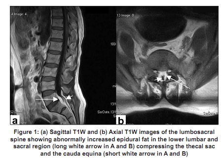
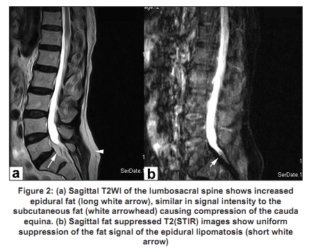
Code Number: ni10038 DOI: 10.4103/0028-3886.60416 Sir, Epidural lipomatosis is a well-recognized complication of prolonged steroid intake. The condition can occur in obese individuals and sometimes may even be idiopathic. [1] Nearly all the cases described so far involved the thoracic and lumbar region. [2] Isolated sacral epidural lipomatosis is extremely rare and only one idiopathic case has been reported. We describe a second such case in a male who was on prolonged steroid therapy for the last four years for interstitial lung disease. An 84-year male presented with increasing low backache over the last few months which was followed by radiating pain along the lateral aspect of the left lower limb, one month in duration. The patient also had complaints of bladder and bowel dysfunction. His most worrying symptoms that made him seek medical attention were weakness of both lower limbs, predominantly the left, and inability to walk without help. Examination revealed motor power of Grade 3 in the left ankle and great toe dorsiflexors and motor power of Grade 4 in the corresponding muscles on the right side. There was sensory blunting in the perineal region and along the lateral aspect of the left leg and sole of the left foot. The anal as well as bilateral ankle reflexes were absent. Magnetic resonance imaging (MRI) study of the lumbosacral spine revealed an abnormal amount of epidural fat in the lower lumbar and sacral canal compressing the thecal sac and the cauda equina [Figure - 1] and [Figure - 2] suggestive of epidural lipomatosis. The conus medullaris was normal and there was no disc pathology. The patient was managed conservatively with steroid tapering. After one month he reported symptomatic improvement of low backache as well as radiating pain. Examination revealed Grade 4+ power in the ankle and toe dorsiflexors in both lower limbs. Minimal sensory blunting persisted in the perineal region. A follow-up MRI was suggested, however, the patient himself was not keen for a repeat MRI and opted for a clinical follow-up. To conclude, lumbosacral epidural lipomatosis is extremely rare, but nevertheless has to be considered as one of the causes of lumbosacral plexopathy, especially with predisposing factors such as obesity or prolonged steroid intake and should be evaluated further with MRI. References
Copyright 2010 - Neurology India The following images related to this document are available:Photo images[ni10038f1.jpg] [ni10038f2.jpg] |
| |||||||||
