 |
| About Bioline | All Journals | Testimonials | Membership | News |
|
||||||
|
||||||
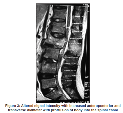
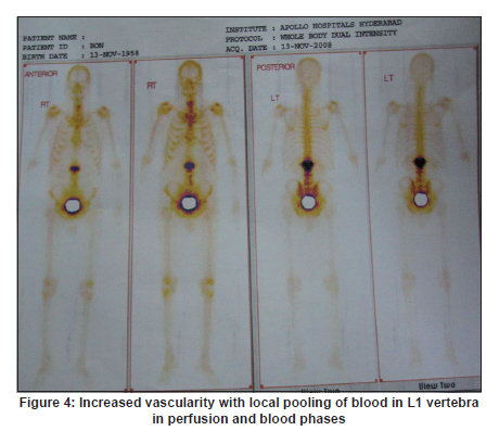
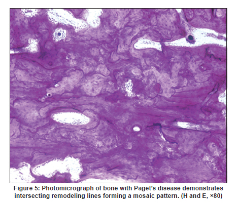
Neurology India, Vol. 58, No. 3, May-June, 2010, pp. 499-500 Neuroimage Does Paget's disease affect a single vertebra? Pavan K Avadhanam, Praveen Ankathi, Purohit A Kumar Department of Neurosurgery, Nizam's Institute of Medical Sciences, Punjagutta, Hyderabad, India Date of Acceptance: 09-Oct-2009 Code Number: ni10132 PMID: 20644296 Paget's disease is a metabolic disorder characterized by abnormal osseous remodelling and is an uncommon cause of back pain We are reporting a rare case of mono-osteotic presentation of Paget's disease in a vertebral body, in which back pain is the main symptom. A 50 year old male, presented with one year history of lower back pain. The pain was insidious in onset and gradually progressed with pain radiating along the medial aspect of both the thighs and knees. Pain used to aggravate with standing, walking and relieved with rest and medication. Pain was associated with paraesthesias radiating along the medial aspect of both thighs and knees. He had no other systemic complaints. Physical examination revealed gibbus deformity at the lumbar region and the paraspinal muscles were taut and tender. The movements of the lumbar spine were restricted. Neurological examination revealed bilateral hip flexor weakness (grade 4/5). There was a 25% decrease in sensations in both the L3 dermatomes. Erythrocyte sedimentation rate first hour was 70 mm and serum alkaline phosphatase was 165 IU (normal 80-120 IU). Radiographs of the lumbar spine showed decrease in the height of first lumbar vertebra and increase in the anteroposterior and transverse diameters. There was an increased density of the vertebral body [Figure - 1]. Computed tomography (CT) scan of the spine showed expansive sclerosis, which was more in the periphery than in the centre [Figure - 2]. Magnetic resonance imaging (MRI) revealed altered signal intensity of entire L1 vertebra including transverse processes, laminae and spinous process with hypointensity on T1 and hyperintensity on T2 weighted and on STIR sequences. There was an increase in anteroposterior and lateral dimensions with decreased height of the body and protrusion of the body into the spinal canal. Reduced disc space was seen between D12-L1 and L1-L2 [Figure - 3]. Bone scintigraphy showed increased vascularity and focal pooling of the tracer in the upper lumbar region in perfusion and blood phase. In delayed phase, there was a physiological distribution of the tracer in axial and appendicular skeleton with focal intense tracer concentration in L1 vertebra and transverse process, which were suggestive of metastasis or Paget's disease [Figure - 4]. Fine needle aspiration failed to reveal any specific pathology. Patient underwent L1 laminectomy, decompression and D12-L2 posterolateral fusion. Intraoperatively, there was an altered texture of the bone with increased softness and vascularity. Postoperatively, patient was relieved of back pain and radiculopathy, but paraesthesias still persisted. During the follow-up examination at 6 months, the patient was completely relieved of back pain, radiculopathy and paraesthesias. Histopathological examination revealed irregularly thickened interconnecting woven bone trabeculae with loose fibrovascular tissue. The trabeculae showed mosaic pattern of arrangement with prominent cement lines, which confirmed Paget's disease [Figure - 5]. The diagnosis of Paget's disease of spine in India is likely to be missed due to the rarity of the disease [1],[2],[3],[4] and the clinicoradiological mimic of this condition is metastases. This case illustrates that Paget's disease should always be kept in mind as one of the diagnostic possibility when evaluating back pain, more show radiological studies are suggestive. [5] References
Copyright 2010 - Neurology India The following images related to this document are available:Photo images[ni10132f5.jpg] [ni10132f2.jpg] [ni10132f3.jpg] [ni10132f1.jpg] [ni10132f4.jpg] |
| |||||||||
