 |
| About Bioline | All Journals | Testimonials | Membership | News |
|
||||||
|
||||||
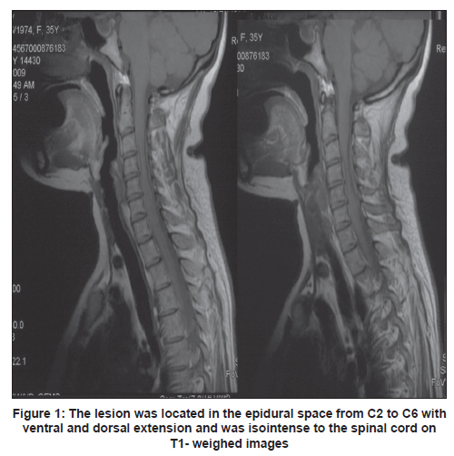
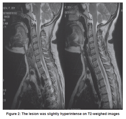
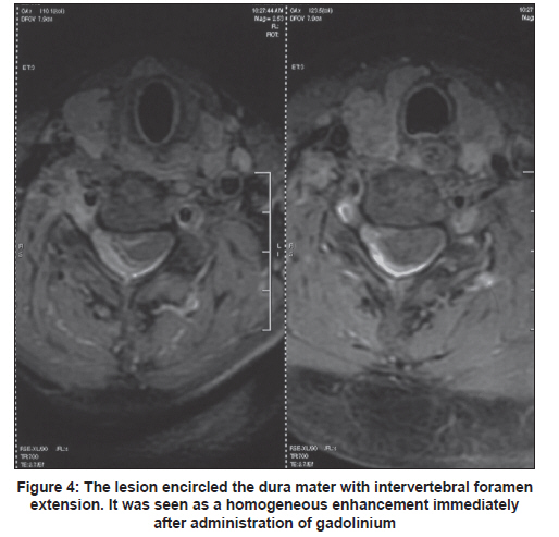
Neurology India, Vol. 59, No. 1, January-February, 2011, pp. 132-134 Letter to Editor Cervical en-plaque epidural meningioma Weiying Zhong, Haifeng Chen, Siqing Huang, Chao You Department of Neurosurgery, West China Hospital, Sichuan University, NO 37, GuoXue Xiang, Chengdou, Sichuan Province, China A 35-year-old woman was admitted for neck pain of 2 years' duration. Neurological examination revealed motor weakness of 3/5 on the right side and 4/5 on the left side and sensory loss below C7 segment. Bilateral Hoffmann and Babinski signs were positive. Cervical magnetic resonance imaging (MRI) revealed an epidural mass on the right side with ventral and dorsal extension from C2 to C6. The lesion was isointense on T1-weighted and slightly hyperintense on T2- weighted images with homogeneous enhancement [Figure - 1],[Figure - 2],[Figure - 3],[Figure - 4]. Spinal angiography revealed no abnormality of cervical spinal vessels. Subtotal resection was performed. At operation, the lesion was located in the epidural space adherent firmly to the dura mater with intervertebral foramen extension. Histology confirmed the diagnosis of epidural meningioma. Meningiomas are the second most common intraspinal tumors and usually arise from the intradural-extramedullary space and are located in the thoracic region of the spine. Solitary epidural spinal meningiomas account for 2.7% to 10% of all spinal meningiomas, [1],[2] more common in children and men and more aggressive than intradural meningiomas. [3] En-plaque epidural meningiomas are sheet-like or collar-like meningiomas encircling and infiltrating the dura mater, and only a few cases have been reported. [2],[3],[4] Epidural spinal meningiomas are usually isointense to the spinal cord on T1-weighted and isointense or slightly hyperintense on T2-weighted images with homogeneous enhancement. [1],[4] They can arise in any part of the dural sac with dura mater encirclement and several vertebral levels extension, especially en-plaque ones. The lesions can erode vertebra with intervertebral foramen widening and vertebral body arc changes but rarely with vertebra destruction. The dural attachment stalk is an essential component for the diagnosis. [2],[3],[4] Other epidural lesions should be excluded. Surgical resection is the main treatment for spinal meningioma. The recurrence rate mainly depends on the degree of resection, but complete resection is more difficult to achieve in epidural en-plaque meningiomas because of the location and extent of the lesion, and the attachment to dura mater. Recurrence rates are high, and the prognosis of en-plaque spinal meningiomas remains poor. [2],[3],[4] References
Copyright 2011 - Neurology India The following images related to this document are available:Photo images[ni11038f3.jpg] [ni11038f2.jpg] [ni11038f1.jpg] [ni11038f4.jpg] |
| |||||||||
