 |
| About Bioline | All Journals | Testimonials | Membership | News |
|
||||||
|
||||||
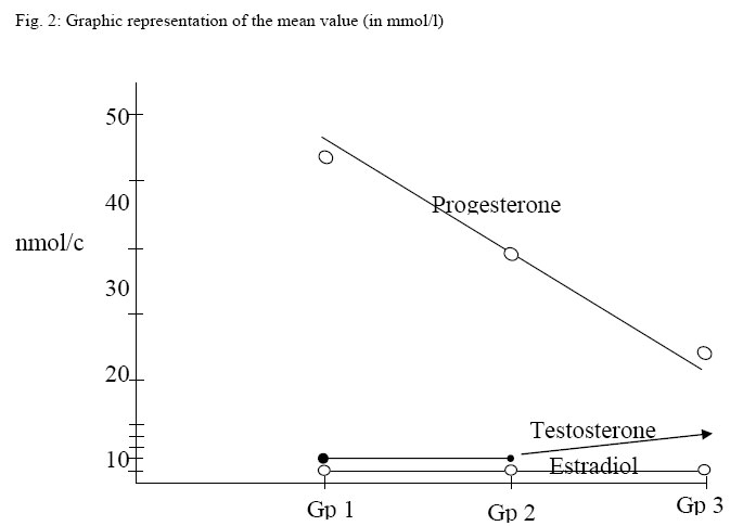
Nigerian Journal of Physiological Sciences, Vol. 20, No. 1-2, 2005, pp. 90-94 EFFECT OF OVARIECTOMY ON THE LEVELS OF PLASMA SEX HORMONES IN ALBINO RATS * E. A. ALAGWU., ** R. O. NNELI Departments of Physiology, College of Medicine and Health Sciences, *Imo State University, Owerri and **Abia State University, Uturu, Nigeria. Received:21/10/2005 Code Number: np05017 Summary: The study was carried out to evaluate the levels of sex hormones (testosterone, progesterone & estradiol), six weeks post normal ovariectomy as against estimating the levels immediately or less 48 hours after operation. 28 adult female albino rats of Wistar strain were used. They were fed twice a day with Guinea pellets and tap water was offered ad libitum. The animals were grouped into three. Group one was sham operated and served as the control, group two was unilaterally ovariectomised (right removed), group three was bilaterally ovariectomised. The animals were sacrificed at the end of the study by decapitation and the blood collected and assayed for sex hormones. The levels of the sex hormones were measured using radioimmunoassay technique. The results showed significant increase in testosterone level in group 3 with decrease in group 2 which was not significant when compared to group 1. On the progesterone levels, there were significant decrease in group 3 and not 2 when compared to group 1. Estradiol level showed significant decrease in only group 3. The decrease in group 2 was not significant when compared with the control. This work therefore does not propose a longer period of hormonal estimation beyond 48 hours following ovariectomy as the results obtained 6 weeks post ovariectomy did not differ or alter significantly beyond 48 hours when compared with the work of Bast and Greenwald 1977, Bulcher 1977, De Jong et al 1978. It is therefore concluded that sex hormone values obtained 6 weeks after normal ovariectomy did not differ significantly as was observed by various workers and confirmed by the present study. Key Words: ovariectomy, sex hormone, Estradiol. Introduction
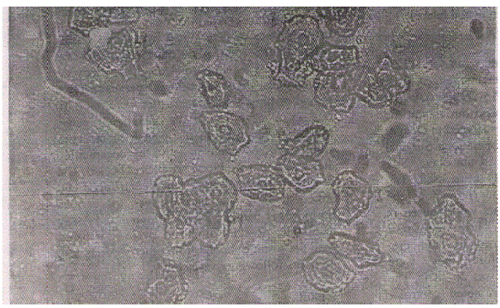
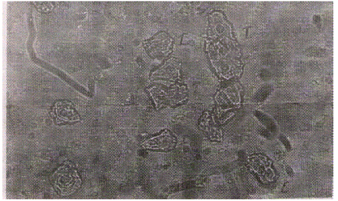
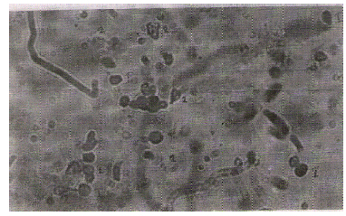
Ovariectomy is a term used for ovarian removal. It could be unilateral (partial) or bilateral (complete) when one ovary or both ovaries are removed respectively. The aims of various workers in the field differed. Bast and Greenwald (1977) observed that small follicles proliferate to large ones after unilateral ovariectomy (ULO). Howland and Skimer (1973) observed that FSH levels are increased only during a short period of 24 hours after ULO and not during later days. Also Welshen and Dullaart (1974) in the rat, Best and Greenwald (1977) in the hamster showed that FSH levels are increased from 4 hours after ULO. On the estrogen level after ULO, Baranczuk and Greenwald (1973) found increase and unchanged plasma estradial levels in hamsters at 7 and 31 hours respectively after ULO, and estradial levels in the rat after ULO at diestrus appeared not to decrease in comparism to those in control rats, while progesterone is lower between 4 and 20 hours after ULO. De Jong et al (1978) observed minor changes in circulating estradial level after bilateral ovariectomy (BLO) on the days of proestrus and metestrus. Battey and Romeo (1951) evolved ovariectomy as a remedy of otherwise incurable ovarian diseases. Apart from the aims expressed above by the above workers other aims include (1) to study the ovarian functions as they relate to the reproductive ages. (2) Determine the ovulation rate in the remaining intact ovary. (3) Determine the mechanism of ovarian hypertrophy (4) To determine whether litter sizes are affected by removing one ovary. (5) Evaluate the effects of performing the ovariectomy at particular stage of the cycle and measuring serial hormonal levels immediately after ovariectomy and a high period up to 31 hours. (6) Measurement of gonadotrophins and plasma steroid hormones. (7) Inducing menopause and monitoring the extra-ovarian sex hormonal levels and gonadotrophin levels. The aims generally varied. The present study was aimed at evaluating the levels of sex hormones (testosterone progesterone, and estrogen) and to determine whether it is necessary to prolong the study time beyond 48 hours as practiced by many workers as the period of study (48 hours or less) might be affected by stresses due to surgery and anaesthesia including environmental factors. Materials and Methods: Twenty-eight adult albino rats of Wistar strain were used. Their weights ranged from 115-120gms, aged between 5 weeks to 6 weeks. Guinea pellets feeds were used throughout the experiment. The rats were distributed in 3 cages of 10, 10, 8 before the operation. Surgical instruments were sterilized in autoclave. Silk and chronic suture materials were used. Tap water was provided ad libitum throughout the period. After surgery, the rats were housed in group of 4 and 5 in a cage. Three weeks post operation; they were returned to their pre-operational groupings before they were scarified. Room temperature was 26-28OC. The mean weights at sacrifice were noted (control – 155 ± 2.7, unilateral = 162.6 ± 1.8 and bilateral = 172.5 ± 2.6g). Method A single longitudinal skin incision was made on the dorso-lateral area at the level of the lower poles of the kidney after shaving the furs with scissors and cleansing with 70% alcohol. The skin was retracted laterally toward one side and the ovary exposed through a thin muscle mass just below the dorsal muscle mass. Each incision was of minimum length to allow the extrusion of ovary. Ligation of the upper horn of the uterus, including the arteries with chromic catgut was carried out. Ovary was excised and wounds were closed. High degree of aseptic procedure was maintained throughout the operation. Operations were carried out in estrus phases as determined by vaginal smears using Shorr’s stain. Light ether anaethesia was used to put the animal to sleep. After recovery, the animals were put 4-5 rats per cage for a period of three weeks thereafter were put 8-10 rats in a cage as in pre-operational stage. Markers were used to distinguish them. They were sacrificed while in estrus by decapitation at 6 weeks and the blood samples collected. The sera were collected after being centrifuged and stored at 10OC and later assayed for various hormones using Radio Immuno Assay technique. Results obtained were compared with the result obtained from other workers less 48 hours following ovariectomy. ResultsVaginal Cytology: The vaginal smears from the groups were studied (fig, 1a, b & c). First under low power (x10) to access the cell yield and finally under high power (x40) with microscope. The smears of the group 1 and group 2 were similar in pictorial outlook with the background showing many cornified, non-nucleated epithelial cells without leucocytes indicating estrus phase. Group 3 failed to show estrus picture rather showed many leucocytes with nucleated epithelial cells indicating diestrus phase. (Figure 2) Discussion Various workers – Baranczuk, Welschen and Dullaart (1974), Bast and Greenwald 1977, De Jong et al 1978 and others estimated the levels of steroid hormones before 48 hours of surgery but our study was extended to six weeks with hope that this period was long enough to allow the effects of stress induced surgery and anaesthesia including other environmental factors that could interfere with the result to wane. The results were then compared with their own. Barancz and Greenwald (1973) found an increase and unchanged plasma estradiol levels in hamster at 7 and 31 hours respectively after ULO in the rat. Estradiol levels at diestrus was not significantly decreased when compared with the control. Progesterone level was found to be lower between 4 to 20 hours after ULO. Mandl (1951) observed that ovariectomy, irrespective of the time of hormonal estimation caused significant increase in the levels of testosterone and decrease in progesterone and estradiol levels. Also Noble (1939) showed that in complete ovariectomised rats, the high testosterone observed maintained the female rats in diestrus phase. The result of the present study showed that testosterone level was significantly elevated in group 3 as demonstrated by Noble (1939). Vaginal cytology also showed that the smears taken from this group showed persistent diestrus phase as was also observed by Noble (1939). Progesterone and estradiol levels were significantly decreased in group 3. The decrease in group 2 was not significant when compared with control group. This agreed with the work of Mandl (1951) who found significant decrease in progresterone and estradiol. This work does not therefore propose a longer period of sex hormone estimation following ovariactomy as results obtained six weeks post ovariectomy did not differ significantly from the ones obtained before 48 hours by Mandl (1951) Noble (1939) and Greenwald et al (1977). It is concluded therefore that surgical and anesthetic stresses have no effect on ovariectomy irrespective of the time of sex hormonal estimation as sex hormone values obtained before 48 hours and 6 weeks post normal ovariectomy did not differ significantly. References
© Physiological Society of Nigeria 2005 |