 |
| About Bioline | All Journals | Testimonials | Membership | News |
|
||||||
|
||||||
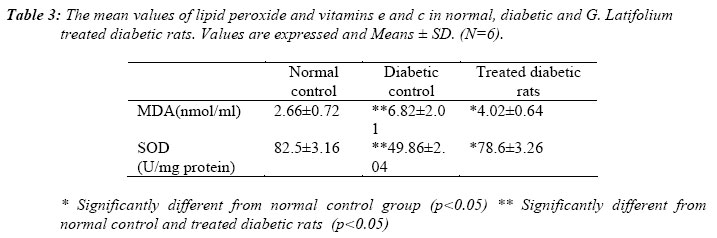
Nigerian Journal of Physiological Sciences, Vol. 21, No. 1-2, 2006, pp. 61-65 ANTI-LIPID PEROXIDATIVE ACTIVITY OF GONGRONEMA LATIFOLIUM IN STREPTOZOTOCIN-INDUCED DIABETIC RATS. H. U. NWANJO, M. C. OKAFOR and G. O. OZE College of Medicine & Health Sciences,Imo State University, Owerri, Imo State, Nigeria .E-mail: harrissonnwanjo@yahoo.com Received: 24/7/06 Accepted: 15/11/06 Code Number: np06012 Summary: This study was designed to investigate the anti-lipid peroxidative effects of aqueous extracts from Gongronema latifolium (Utazi) leaves in non-diabetic and streptozotocin-induced diabetic rats. We evaluated plasma lipid peroxidation product (malondialdehyde, MDA) and antioxidant enzyme, superoxide dismutase (SOD) in all the animals. Result demonstrated that the untreated diabetic rats were subjected to oxidative stress as indicated by significantly (P<0.05) low level of their antioxidant enzyme, superoxide dismutase (49. 86 ± 2.04 U/mg protein) and high plasma lipid peroxidation index (6.82 ± 2.04 nmol/ml) when compared with non-diabetic rats value of 82.5 ± 3.16 U/mg protein and 2.66 ± 0.72 nmol/ml respectively. The aqueous extract of G. latifolium leaves reversed this situation as shown by significantly (P<0.05) increase in the activity of superoxide dismutase (78.6 ± 3.26 U/mg protein) and decreased levels of plasma malondialdehyde (4.02 ± 0.64). In conclusion, the results suggest that aqueous extract of G. latifolium leaves possesses hypoglycemic as well as anti-lipid peroxidative properties.Key Words: Diabetes, lipid peroxidation, G. latifolium.
Introduction
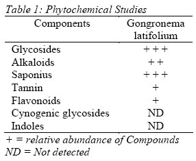
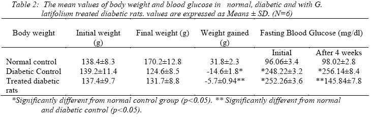
Diabetes mellitus is a multifactorial disease, which is characterized by hyperglycemia and glucosuria (Scoppola et al, 2001) among others. These traits are hypothesized to damage cell membranes, which results in elevated production of oxygen species (ROS). This generation of oxygen-free-radicals during cellular metabolism, and by certain environmental factors, including life style, appears to play a critical role in the pathogenesis of diabetes mellitus (Hartnett et al, 2000). Hyperglycemia, the main symptom of diabetes, not only increases the production of ROS but also affects antioxidant reactions catalyzed by ROS scavenging enzymes (Uchimura et al, 1999). Defects in ROS scavenging enzyme system have been reported in diabetes mellitus (Kesavulu et al, 2000). Many minor components of foods such as secondary plant metabolites have been shown to alter biological processes, which may reduce the risk of chronic disease in human. Gongronema latifolium, Benth, (Asclapiadaceae) is a perennial edible plant with soft and pliable stem. It is widely used in the West African sub-region for a number of medicinal and nutritional purposes (Dalziel, 1937). In Sierra Leone, a decoction or cold infusion of the pounded stem is used for colic and intestinal symptoms usually associated with worms (Deighton, 1957). In Ghana, the boiled fruits are used as laxative. In Eastern States of Nigeria, the plant locally known as “Utazi” is a popular spice. The leaves are used to prepare food for mothers that have recently put to bed, where it is believed to stimulate appetite, reduce post-partum contraction and enhance the return of the menstrual cycle. It is well known that G. latifolium not only possesses hypoglycemic activity, but also hypotensive, hepatoprotective and hypolipidemic effects (Ugochukwu and Badaby, 2003; Ugochukwu et al, 2003; Nwanjo, 2005; Nwanjo and Alumanah, 2005). In the present study, we investigated the protective effect of aqueous extracts from G. latifolium leaves on biomarkers of oxidative stress in streptozotocin-induced diabetic rats. Materials and Methods Plant Materials and ChemicalsFresh and apparently uninfected leaves of G. latifolium were collected from plants growing within Owerri, Imo State. The botanical identification of the plant leaf was done at the Department of Plant Biology and Biotechnology, Imo State University, Owerri, where voucher samples are kept for reference. All chemicals used in this study were of analytical grades purchased from British Drug House (Poole, U.K). Preparation of the Extract Fresh leaves of G. latifolium were dried in carbolite moisture extraction drying oven (Grant instruments, Cambridge, England) at 450C – 500C for 3 hours. Grinding was done using Thomas contact mill (pye Unicam, Cambridge, England). The ground materials were sieved through a 1mm sieve. One hundred gramme (100 g) of fine power was boiled in about 500 ml of water for 3 hour (wet extraction). The extract was cooled and filtered. The filtrate was evaporated by hot air oven (Grant instrument, Cambridge, England) treatment at 450C – 500C to a yield of 2g plant material, which was dissolved with appropriate volume of water. Phytochemical Studies Preliminary photochemical test as described by Harbone (1973) and Trease and Evans (1996) were carried out on the aqueous extract of G. latifolium. In general, tests for presence or absence of phytochemical compounds using the above methods involved the addition of an appropriate chemical agent to the aqueous extracts of the plant in a test tube. Summary of the methods are as below: Analysis for alkaloidsAcidified (H2SO4) ethanolic extract of the vegetable sample was cooled, filtered and spotted on TLC plates. The plates were developed in chloroform and dil. H2SO4 and spots located with Dragendroff’s reagent. Analysis of Cyanogenic Glycosides Acid (HCI) extract of the vegetable sample was heated and a piece of picrate paper was dropped into the test tube as an indicator. Analysis for FlavonoidsEthyl acetate extract of the vegetable was poured into two test tubes. Ammonia solution was added to one of the test tubes while 1% FeCl3 was added to the other. Colour changes were observed. Analysis for Glycoside and IndolesHot acid H2SO4 hydrosylate of the vegetable was cooled and then neutralized with KOH. Fehling’s solutions (A & B) were used to identify glycosides. Methanolic extract of the vegetable was spotted on TLC plates and developed in chloroform: ethyl acetate: formic acid solvent system. Colour was developed using Erhlich (modified) and Dragendroff’s reagents. Analysis for saponins:Hot water extract of the vegetable was filtered and cooled. An aliquot of the filtrate checked for frothing by shaking while the remaining aliquot was tested with olive oil for emulsification. Analysis for Tannins: Drop of FeCl3 were added to an aliquot of cooled hot water extract of the vegetable. To another aliquot some drops of lead sub acetate were added. Colour changes in the different portions were observed. The mixture was then shaken vigorously or gently as the case may be. The presence or absence of saponins, flavonoids, glycosides, tannins, alkaloids e.t.c. was observed. The results of Phytichemical analysis is presented in Table 1. Acute Toxicity TestsThe acute toxicity (LD50) test of the extract was carried out to define the range of the lethal dose and the safe range for the extract. Thirty six (36) Albino rats of both sexes weighing 150 – 200g were randomly divided into 6 groups of 6 rats each. Groups were treated with extract (100, 200, 500, 1000, 2000 mg/kg) by oral compulsion. Deaths within a period of 24 hours were recorded and the median lethal dose LD50 of the extract was determined according to the method of Miller and Tainter (1944). The LD50 of the aqueous extract was calculated to be 1050 ± 45 mg/kg body weight and doses up to 500 mg/kg bodyweight were observed to be safe (with no recorded deaths). All the doses used in this study were carefully chosen to exclude the lethal range. Animals Albino rats weighing 150-200g were used in this study. All the rats were kept at an average room temperature of 300C in the animal room of College of Medicine and Health Science, Imo State University, Owerri. They were allowed free access to water and feed (product of Pfizer Nigeria Ltd.) throughout the period of the experiment. Eighteen rats, included for the study, were divided into 3 groups, 2 were made diabetic by intraperitoneal injection of 65mg/kg body weight of streptozotocin (STZ) (Sigma, St. Louis, MO, USA) dissolved in citrate buffer (0.01M, pH 4.5). Diabetes was confirmed by the determination of fasting blood glucose concentration on the third day post administration of STZ showing fasting blood glucose levels above 250mg/dl. Body weight and fasting blood glucose levels of all the rats were determined before the start of the experiment. Rats were divided into the following groups. Group 1: Control, given only the citrate buffer (0.01M, pH 4.5). Group 2: Streptozotocin induced diabetes, made with a single dose of streptozotocin (65mg/kg body weight) by intraperitoneal route. Group 3: Diabetic rats treated with G. latifolium 200mg/kg/once a day, daily. Treatment was by oral compulsion. After 4 weeks of treatment the body weight and fasting blood glucose of the animals were again determined. Blood was collected on the third and fourth days through the rat tail artery for glucose estimation . Experimental and Analytical Procedure Twelve hours after the last treatment and after the last feed, the animals were weighted and anaesthetised with chloroform vapour, quickly brought out of the jar and sacrificed. 5ml of blood was collected by cardiac puncture and transferred into an EDTA tube. The anticoagulated blood was then centrifuged using Wisperfuge model 1384 centrifuge (Tamson, Holland) for 15min to facilitate separation. The plasma thus obtained was used for malondialdehyde (MDA) (product of lipid peroxidation) estimation. Plasma MDA was measured by a thiobarbituric acid assay procedure (Albro et al, 1986), which was calibrated using 1, 1, 3, 3 – tetraethoxypropane (Sigma Chemicals, St. Louis, MO, USA) as a standard. Results were expressed as nanomoles of MDA per millimetre of serum. The remaining packed RBCs were washed thrice with normal saline. Haemolysis was performed by pipetting out 1ml of washed red blood suspension in ice-cold distilled water. Erythrocyte ghosts were sedimented in a high-speed refrigerated centrifuge at 12000 rpm for 40min. The cell content was separated out carefully and used for superoxide dismutase estimation (McCord and Fridovich, 1969) Statistical AnalysisThe results were analysed using Duncan Multiple Range Test. All data were expressed as mean ± SD. Differences between groups were considered significant at p<0.05. Results Table 2 showed the results of the analysis, in which there was a significant increase (p<0.05) in fasting blood glucose and a comparative decrease (p<0.05) in body weight when compared with the normal control. There was a slight increase in body weight and a significant decrease in fasting blood glucose in diabetic rats treated with G. latifolium. In Table 3 there was a significant increase in lipid peroxide levels (p<0.05) in streptozotocin induced diabetic rats with respect to normal controls. It also shows a statistically significant decrease in lipid peroxide levels in diabetic rats treated with extract of G. latifolium leaves. The activities of SOD were significantly higher in diabetic rats treated with extract of G. latifolium leaves compared to diabetic control rats (p<0.05). There was no significant difference among the treated diabetic and normal groups but there was significant difference between diabetic and normal controls. Discussion Impaired glucose metabolism leads to oxidative stress (Ceriello et al, 1992), and protein glycation produce free radicals (Wolff et al, 1991). Therefore, the elevated plasma lipid peroxide levels in streptozotocin-induced diabetic rats recorded in this work, along with a significant decrease in antioxidant enzyme, superoxide dismutase activity could be at least in part result from inactivation of the enzyme by H2O2 or by glycation, which are known to occur during diabetes (Sozmen et al, 2001; Searle and Wilson, 1980). Earlier there have been many reports documenting elevated serum lipid peroxide levels and diminished antioxidant status in diabetic subjects (Sato et al, 1977, Oberley, 1988). As diabetes and its complications are associated with free radical mediated cellular injury (Asayama, 1993), aqueous extract of G. latifolium was administered to diabetic rats to assess their antioxidant potential. Our results showed that G. latifolium not only has hypoglycemic activity (Nwanjo, 2005). But it also significantly reduced the plasma lipid peroxide levels and increased antioxidant enzyme, superoxide dismutase activity in diabetic rats. SOD is for primary defense against reactive oxygen metabolites (Mahdi, 2002). Such metabolites have been implicated in the damage brought about by ionizing radiation, as well as in the effects of several cytostatic compounds (Marklund et al, 1982). The decreased activity of antioxidant molecules along with elevated lipid peroxide levels in diabetic rats could probably be associated with oxidative stress and/or decreased antioxidant defense potential (Mahdi et al, 1996). The reversal in their content following treatment may be due to decreased oxidative load. The extracts of G. latifolium may also act by either directly scavenging the reactive oxygen metabolites, due to the presence of various antioxidant compounds (Gupta et al, 2002), or by increasing the synthesis of antioxidant molecules. Further studies are required to evaluate the levels of metal ions such as copper, zinc, magnesium, manganese and selenium in diabetic rats following treatment with G. latifolium extracts, as altered metabolism of these metals have been reported to occur in diabetes mellitus (Walter et al, 1991). References
© Physiological Society of Nigeria 2006 The following images related to this document are available:Photo images[np06012t2.jpg] [np06012t1.jpg] [np06012t3.jpg] |
| |||||||||