 |
| About Bioline | All Journals | Testimonials | Membership | News |
|
||||||
|
||||||
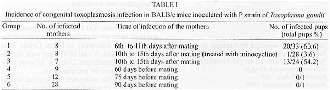
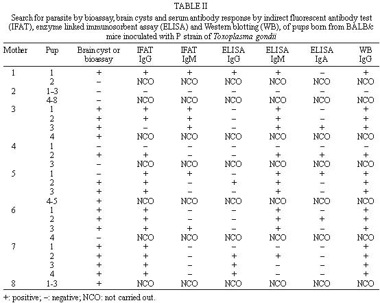
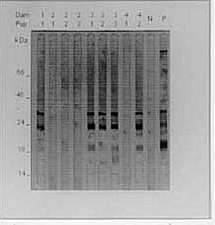
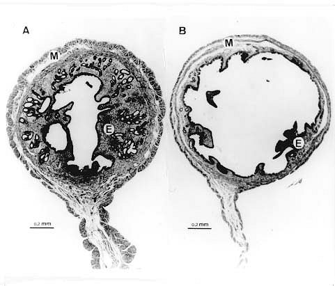
Mem Inst Oswaldo Cruz, Rio de Janeiro, Vol. 95(1): 121-126 Experimental Toxoplasmosis in Balb/c Mice. Prevention of Vertical Disease Transmission by Treatment and Reproductive Failure in Chronic Infection B Fux, AM Ferreira, GD Cassali, WL Tafuri, RWA Vitor+ Departamento de Parasitologia, Instituto de Ciências Biológicas, Universidade Federal de Minas Gerais, Caixa Postal 486, 31270-901 Belo Horizonte, MG, Brasil + Corresponding author. Fax: +55-31-499.2970. E-mail: vitorrwa@mono.icb.ufmg.brReceived 21 July 1999 Code Number:OC00020 In a study of congenital transmission during acute infection of Toxoplasma gondii, 23 pregnant Balb/c mice were inoculated orally with two cysts each of the P strain. Eight mice were inoculated 6-11 days after becoming pregnant (Group 1). Eight mice inoculated on the 10th-15th day of pregnancy (Group 2) were treated with 100 mg/kg/day of minocycline 48 h after inoculation. Seven mice inoculated on the 10th-15th day of pregnancy were not treated and served as a control (Group 3). Congenital transmission was evaluated through direct examination of the brains of the pups or by bioassay and serologic tests. Congenital transmission was observed in 20 (60.6%) of the 33 pups of Group 1, in one (3.6%) of the 28 pups of Group 2, and in 13 (54.2%) of the 24 pups of Group 3. Forty-nine Balb/c mice were examined in the study of congenital transmission of T. gondii during chronic infection. The females showed reproductive problems during this phase of infection. It was observed accentuated hypertrophy of the endometrium and myometrium. Only two of the females gave birth. Our results demonstrate that Balb/c mice with acute toxoplasmosis can be used as a model for studies of congenital T. gondii infection. Our observations indicate the potential of this model for testing new chemotherapeutic agents against congenital toxoplasmosis. Key words: Toxoplasma gondii - Balb/c mice - congenital transmission Toxoplasma gondii infection is a common congenital disease in humans and domestic animals. Congenital toxoplasmosis is one of the principal causes of abortion, foetal death, and stillbirths in sheep, goats and pigs (Dubey & Beattie 1988). Severe cases occur with greater frequency when the mother is infected during the first two trimesters of pregnancy. When acquired later, symptoms tend to be subclinical or even lacking in the foetus (Desmonts & Couvreur 1974). Numerous animal models, including primates, rabbits, guinea pigs, hamsters, mice and rats, have been used in recent years to study the pathology of the infection and the efficacy of vaccines and new drugs for the treatment of congenital transmission, chorioretinitis and toxoplasmic encephalitis. Of these, mice have been found to be the most susceptible to T. gondii and are particularly interesting model to study congenital infection. Roberts and Alexander (1992) demonstrated that vertical Toxoplasma transmission only occurs in Balb/c mice infected with T. gondii for the first time during pregnancy. It was observed that vaccination with a T. gondii antigen enveloped in lipid vesicles significantly reduced the incidence of abortion in mice (Roberts et al. 1994). Recently Alexander et al. (1998) observed aspects of cellular immunity using this model, and demonstrated that IL-4 is associated with pregnancy-induced immunosuppression. In this paper, we report on a study of congenital toxoplasmosis in Balb/c mice infected with a strain of T. gondii isolated in Brazil, which gave particular high ratio of congenital transmission during acute toxoplasmosis. The incidence of congenital transmission in Balb/c females treated with minocycline was evaluated. MATERIALS AND METHODS Mice - Female Balb/c mice were maintained and bred under conventional laboratory conditions at the Federal University of Minas Gerais, Belo Horizonte, Brazil. A total of 97 mice were used in the study: 72 females and 25 males. All were serologically negative for toxoplasmosis. Infection - The cysts used for the experimental infection were obtained from the brains of Swiss mice after 30 days of infection with the P strain of T. gondii. This strain was isolated from a dog in Brazil (Jamra & Vieira 1991). The mice were sacrificed by cervical dislocation; the brains were removed and homogenised in phosphate buffered saline (PBS) pH 7.2. Mice were infected orally with two cysts either eight weeks prior to mating or between the 6th and 15th day of pregnancy. Eight mice inoculated between the 10th and 15th days of pregnancy were treated with 100 mg/kg/day of minocycline until the birth of their offspring (Chang et al. 1991). Details of each experimental group are shown in Table I. Mating - Uninfected female mice, mice infected between 6th and 15th days after mating, and those with chronic infections of 60, 75 or 90 days, were maintained with a male for seven days. Immediately after giving birth, the experimentally infected mothers were substituted by uninfected foster mothers to avoid possible transmission through suckling. Detection of congenital infection - Detection of congenital infection was carried out directly through examination for cysts in the brain of the newborn mice, by bioassay of neonate tissues and by the following serologic tests: indirect fluorescent antibody test (Ifat), enzyme linked immunosorbent assay (Elisa) and Western blotting (WB). For the direct examination, the brains of the newborn offspring were removed and homogenised in PBS pH 7.2, and the suspension was placed on a slide, mounted under a cover slip, and viewed under a light microscope. The bioassay of neonate tissues was carried out on the newborn offspring, which died during the experimental period (the incidence of neonate mortality was high due to rejection after fostering). The brain, heart, lungs and liver were macerated in 1 ml of PBS pH 7.2 and inoculated into the peritoneal cavity of Swiss mice. These mice were evaluated after 30 days by parasitological and serologic tests. Ifat was carried out, using the procedure outlined by Camargo (1964), on the appropriate slides containing formalin fixed Toxoplasma tachyzoites. Plasma was diluted in PBS pH 7.2 for the IgM class antibody (1:4 to 1:256), and for IgG class antibodies (1:16 to 1:16000). Elisa was carried out using a modification of the procedure described by Voller et al. (1976). Antigen was obtained by sonication of tachyzoites in a Branson sonicator at 20 watts for 30 sec at 4°C. Plasma was diluted 1:64 in PBS-tween-20 at 0.05%, in duplicate and incubated at 37ºC for 45 min. The plaque was then washed and 100 µl of peroxidase conjugated IgG, IgM or IgA mouse anti-immunoglobulin was added to each well. The reaction was visualised with orto-phenylenodiamine and stopped with 4N-H2SO4. Absorbance was read at 490 nm on a Microplaque Reader Biorad 3550. The values were considered positive when they were above the mean absorbance obtained for four non-reactive plasma, plus three standard deviations (cut off). WB was performed by a modification of the procedure described by Towbin et al. (1979). Purified tachyzoites were dissolved in sample buffer and eletrophoresed on 12.5% polyacrylamide gels. The standards used for molecular weights (66, 45, 24, 18 and 14 kDa) were from Sigma (MW-SDS-70L). After separation by sodium dodecyl sulphate-polyacrylamide gel electrophoresis, proteins were transferred to nitro-cellulose membranes at 20 h at 30V and 40 mA. The detection of proteins was carried out by an immunoenzymatic method in 3 mm strips. Proteins were shown up by developing strips using 4-chloro-1naphthol. Positive (plasma of mice inoculated orally with two cysts 60 days after infection) and negative (plasma of normal mice) controls were used in all the tests. Histopathology - A histological examination was carried out on the reproductive systems of nine female Balb/c mice infected for 60 days (Group 4). Five normal females of the same age were sacrificed at the same time. The uterus, ovaries and brain were removed immediately and fixed in 10% formalin/PBS pH 7.2. Following processing, the samples were set in paraffin and sectioned in slices of 4 µm width. The material was then stained with haematoxilin-eosin and examined under a light microscope. Mice of Groups 5 and 6 were not subjected to histological analysis. RESULTS Congenital transmission of T.gondii during the acute phase - The incidence of congenital toxoplasmosis in the experimental groups is summarised in Table I. Congenital toxoplasmosis in Group 1 was observed in seven of the eight females inoculated (87.5%) with 20 (60.6%) pups positive for brain cysts or by bioassays. Antibody response was evaluated in 19 30-day-old mice ( Table II). IgG antibodies detected by Ifat showed 95% concordance with the parasitological diagnosis. Fourteen of 19 pups (73.7%) showed titres of between 1:16 and 1:256 for the Ifat-IgG, 13 also with positive parasitological tests. The Ifat-IgM detected six (31.6 %) of the mice with a titre of 1:4, five of them with positive parasitological results. In the Elisa-IgG, only five pups (26.3%) showed an absorbance of ³ 0.094 (cut off-IgG). In the Elisa-IgM, 12 pups (63.2%) showed an absorbance of ³ 0.079 (cut off-IgM), and in the Elisa-IgA four pups (21.0%) showed an absorbance of ³ 0,068 (cut off-IgM). In the WB-IgG, 15 (78.9%) of the pups evaluated recognised T. gondii antigens. The principal bands had molecular weights of 32, 22 and 19 kDa. Representative results obtained from mothers 1, 2, 3 and 4 are shown in Fig.1. The mothers' plasma recognised the largest number of antigens, with a profile similar to that detected in the positive control mice. Fig. 1: Western blotting obtained from Balb/c mice born from mothers inoculated with Toxoplasma gondii 6-11 days of pregnancy. Pups from mothers 1, 2, 3 and 4 (Group 1). N: negative control; P: positive control. The apparent molecular weight (kilodaltons) of protein standards are given on the left side. In Group 2, eight females were inoculated between the 10th and 15th day of pregnancy and treated with 100 mg/kg/day of minocycline. Congenital transmission was observed in only one (3.6%) of the 28 pups (Table I). All of the pups evaluated by Ifat and Elisa gave negative results, except for one, which was positive for Ifat-IgG, IgM and Elisa-IgM, but negative for Elisa-IgG and IgA. Eight females which had not been treated with minocycline were evaluated simultaneously (Group 3). Congenital toxoplasmosis was recorded for six (85.7%) of the seven females inoculated, with 13 (54.2%) of the 24 pups positive. The antibody response was evaluated by Ifat and Elisa in 20 pups 30 days after they were born (Table II). Similar to Group 1, 55% of the offspring gave titres ³ 1:16 in Ifat-IgG and 50% showed titres ³ 1:4 in Ifat-IgM. All the 13 pups had brain cysts and showed a positive bioassay, with a high concordance with Ifat. All of the pups evaluated by the Elisa-IgG, showed absorbance < 0.094 (cut off). Five of them, evaluated 60 days after birth, showed absorbance values ³ 0.094. Only 16 pups were evaluated at birth with the Elisa-IgM, and 31.2% showed absorbance values ³ 0.079. Thirteen pups were evaluated with the Elisa-IgA, and only 1 (7.7%) showed absorbance ³ 0.068. Congenital transmission of T. gondii during the chronic phase of infection - Reproductive failure during, and probably related to, the chronic phase of T. gondii infection was recorded in Groups 4, 5 and 6 (Table I). Only two females gave birth. The pups of these two mothers showed no signs of infection when tested for cysts in the brain, and were negative by bioassay and Ifat. Histological examination of Group 4 females showed accentuated hypertrophy of the endometrium and myometrium (Fig. 2). A tendency for a reduction in folliculogenesis and the formation of corpora lutea in the ovaries when compared to the uninfected animals was also noted. No cysts were found in the uterus or ovaries of these animals. Fig. 2: histopathology of the uterus of Balb/c mice with Toxoplasma gondii in the chronic phase. A: uninfected Balb/c mice _ uterus without apparent alteration; B: chronically infected Balb/c mice _ uterus with accentuated hypertrophy of the endometrium (E) and myometrium (M) (hematoxilin-eosin). DISCUSSION Congenital toxoplasmosis was observed in Balb/c mice during the acute phase of infection with the P strain of T. gondii. The incidence of infected pups was 60.6% (Group 1). These results are in agreement with those of Roberts and Alexander (1992) who observed approximately a 50% rate of infection in Balb/c pups examined using mice infected orally with 20 cysts of the RRA (Beverley) strain of T. gondii. All of the eight mice from Group 2 infected during pregnancy but treated with minocycline [using the scheme proposed by Chang et al. (1991)], gave birth. This treatment was, therefore, sufficient to prevent the vertical transmission of T. gondii. Only one pup (3.6%) was born infected, while in Group 3 (untreated control) 13 (54.2%) of the pups were congenitally infected. These results indicate the potential of this model for testing new chemotherapeutic agents against congenital toxoplasmosis. Pups of Group 1 were tested for T. gondii antibodies at 30 days old. Ifat-IgG showed a concordance of 95% with the presence of brain cysts, while the Elisa showed a concordance of 53%. The Elisa-IgM and IgA also showed a low concordance with the presence of brain cysts. A concordance of 100% between the Ifat-IgG and IgM and the presence of brain cysts was recorded in Group 3. In this group, Elisa-IgG gave false-negative results for the plasma collected from all of the 30-day-old pups. At 60 days old, 50% of the congenitally infected pups were Elisa-IgG positive, suggesting a delay in the initiation of the specific antibody response to T. gondii. Roberts and Alexander (1992) used an Elisa-IgG to diagnose infection in eight-week-old Balb/c pups, and obtained a 100% correlation with the bioassay. The results obtained from WB in Group 1 may, in part, help in our understanding of the low sensitivity of the Elisa in our model, as well as the discrepancies with the results of Roberts and Alexander (1992). The WB showed a high concordance with the examination for brain cysts. However, few antigens were recognised, the majority of them between 15 e 35 kDa. These antigens, probably some of the principal components of the surface of the tachyzoites (SAG1 and SAG2), are highly immunogenic and responsible for the intense antibody response in the acute phase of toxoplasmosis (Wong & Remington 1993). Positive results in the Ifat for the 30-day-old pups are probably due to the recognition of these tachyzoite surface-dominant antigens. It is probable that the low sensitivity of the Elisa test in diagnosing the infection in 30-day-old pups is related to deficiencies in the extraction of membrane components by sonication. In our study of vertical transmission during the chronic phase of toxoplasmosis [defined by Suzuki et al. (1993) as the period 30 days after infection], only two females gave birth. After 60 days of infection, the females showed signs of uterine and ovarian atrophy, probably resulting in the reproductive failures we observed. However, T. gondii cysts were not found in the uterine tissue of Balb/c mice in the chronic phase. This fact may be related to the size of the inoculum, the strain used, or be due to the sections examined. It is possible that examination of the sections used in immunoenzyme assays would have detected the parasites. Roberts and Alexander (1992) did not record reproductive problems with this model. They showed that Balb/c pups born during the chronic phase of toxoplasmosis were protected from congenital infection, contrary to the situation with other mouse lineages (Remington et al. 1960). Stahl and Turek (1988), however, also recorded reproductive failures and uterine atrophy in Nya: NYLAR mice inoculated with the CORNELL strain of T. gondii. It was probably also occurring in our model, and may be related to the virulence or the tropism of the T. gondii strain used in this study. In conclusion, Balb/c mice present a satisfactory model for research on congenital toxoplasmosis, suitable for testing new chemotherapeutic agents to prevent the vertical transmission of T. gondii during acute phase. During chronic infection Balb/c mouse is a good model to study the toxoplasmosis pathology, but not congenital transmission using P strain. ACKNOWLEDGMENTS To Ricardo T Gazzinelli for critical reading of the manuscript, Anthony B Rylands for english review and Rosálida EN Lopes for technical assistance. This study was supported by Fundação de Amparo à Pesquisa do Estado de Minas Gerais (Fapemig). REFERENCES
Copyright 2000 Fundacao Oswaldo Cruz - Fiocruz The following images related to this document are available:Photo images[oc00020a.jpg] [oc00020d.jpg] [oc00020c.jpg] [oc00020b.jpg] |
| |||||||||
