 |
| About Bioline | All Journals | Testimonials | Membership | News |
|
||||||
|
||||||
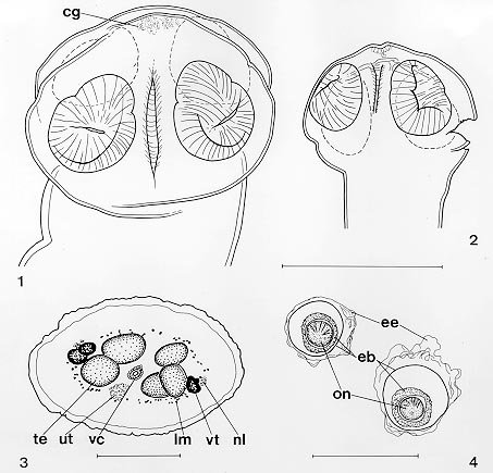
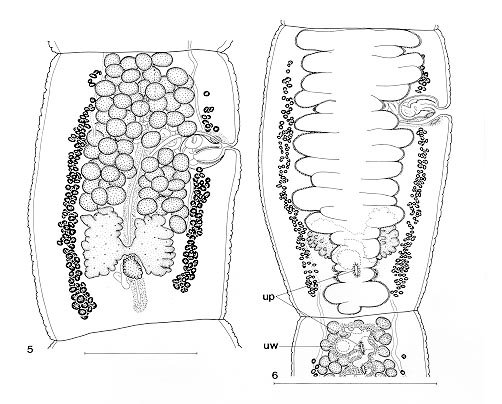
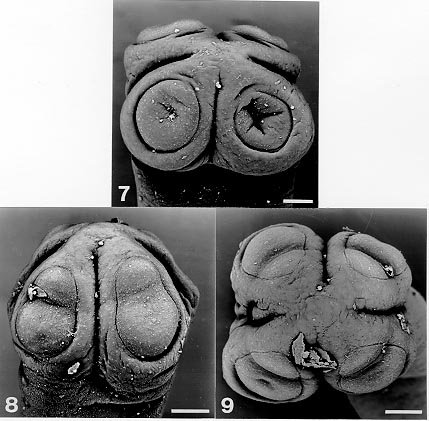
Mem Inst Oswaldo Cruz, Rio de Janeiro, Vol. 95(2): 161-165, Mar./Apr. 2000 Redescription of Tejidotaenia appendiculata (Baylis, 1947) (Cestoda: Proteocephalidea), a Parasite of Tupinambis teguixin (Sauria: Teiidae) from South America Amilcar Arandas Rego/+, Alain de Chambrier* Departamento de Helmintologia, Instituto Oswaldo Cruz, Av. Brasil 4365, 21045-900 Rio de Janeiro, RJ, Brasil *Natural History Museum, P. O. Box 6434, 1211, Geneva 6, Switzerland + Corresponding author and Research fellow of CNPq (Cat. I-A). Fax: +55-21-872.2917.E-mails: arego@gene.dbbm.fiocruz.br and arego@openlink.com.br Received 19 April 1999 Code Number:OC00028 The species Tejidotaenia appendiculata (Baylis, 1947), a parasite found in teju, Tupinambis teguixin is redescribed and a new diagnosis is provided. The species is characterized by the anterior position of the ovary and the peculiar shape of suckers. It is the first record of this species in Brazil. Key words: Tejidotaenia - cestode - Proteocephalidea - Lacertilia - Neotropical region - Brazil Proteocephalidean cestodes have been recorded from lizards in Australia, Africa and India, most of them belonging to Acanthotaeniinae (Rego 1994). In America, only one acanthotaeniine species, Acanthotaenia overstreeti, was described by Brooks and Schmidt (1978) from a lizard in Puerto Rico. In South America the only proteocephalidean species reported from lizards are members belonging to the genera Proteocephalus Weinland, 1858 and Ophiotaenia La Rue, 1911. Tejidotaenia appendiculata (Baylis, 1947) is a species described from the lizard, Tupinambis nigropunctatus [= T. teguixin, according to Avila-Pires (1995)] from Surinam (Dutch Guiana). This species has an unusual position of the ovary and a peculiar type of suckers, with a constriction resembling biloculate suckers. The senior author, on revising the collection of proteocephalids from Helminthological Collection of the Instituto Oswaldo Cruz, Fiocruz, found some specimens collected from Tupinambis teguixin. The specimens were identified as Baylis' species due to the unequivocal characters of the suckers. This is the second record of T. appendiculata and the first reference in Brazilian varanids. The species is redescribed here because Baylis (1947) did not give figures of the proglottides, or reproductive organs, and some details of sucker morphology were unresolved. In this paper a new generic diagnosis is provided. MATERIALS AND METHODS The specimens were collected and fixed by the Solution of Railliet and Henry (alcohol, acetic acid and formaldehyde - Travassos 1951); some specimens were compressed. Samples were stained with Delafield haematoxylin or hydrochloric carmine solution, dehydrated in an ethanol series, cleared in beechwood creosote or Eugenol, and mounted in Canada balsam. Pieces of strobila were embedded in parafin wax, sectioned at 5-8 µm, and stained with Delafield haematoxylin and eosin. Specimens for scanning electron microscopy (SEM) were dehydrated in graded ethanol series, then dehydrated in graded amyl-acetate series, critical point dried in CO2 (lower index), sputtered with gold and examined in a Zeiss DSM 940S. Drawings were made with a camera lucida. Eggs were drawn after tranfer to distilled water. Scanning electron micrographs were made at the Museum of Natural History, Geneva. All measurements are in mm. RESULTS Tejidotaenia appendiculata (Baylis, 1947) Freze, 1965 Syn.: Proteocephalus appendiculatus Baylis, 1947 Ophiotaenia appendiculata (Baylis, 1947) Yamaguti, 1959 Host : Tupinambis teguixin (Linnaeus, 1758) [(Syn.: Tupinambis nigropunctatus Spix, 1825 (cf. Avila-Pires 1995)] Material studied: Helminthological Collection of Instituto Oswaldo Cruz, vials, No. 30.956 (immature worms) from Serra do Navio, Amapa and 17,548 (mature and gravid) from Linhares, Espírito Santo, Brazil. Material mounted in Canada balsam Nos. 34.002 a-e, 34.003, 34.004 and 34.005 a-b, and the Natural History Museum, Geneva Nos. INVE 25588-25589, 25659-25664, 25780. Site of infection: intestine. Description (based on ten specimens): small worms. Strobila acraspedote, 5-6, bearing 10-12 proglottides. Unsegmented zone 0.27-0.33 long. Immature proglottides wider than long. Mature proglottides longer than wide, 0.54-0.60 x 0.42-0.45. Gravid proglottides anapolytic, elongated, 0.75-1.00 x 0.42-0.45. Scolex tetralobulate, 0.42-0.51 x 0.33-0.60; wider than strobila, and with an apex bearing conspicuous glandular cells (Figs 1, 2). Suckers pleomorphic, frequently seen with a distinct constriction separating an anterior and a larger posterior part with a sucker cavity (Figs 8-9); when relaxed, suckers appearing as flat discs and showing no evidence of distinct loculi (Fig. 7). Suckers measuring, 0.16-0.24 x 0.15-0.17 (mean = 0.20 x 0.16). Internal longitudinal musculature weakly developed, forming small bundles of muscular fibers. Osmoregulatory canals not easily observable (Fig. 6). Vitelline follicles and gonads medullary (Fig. 3). Sixty to eighty oval testes in one or two layers, in single compact field, more compact anteriorly (Fig. 6). Cirrus pouch globose, 0.07-0.10 x 0.05- 0.08 (m = 0.08 x 0.06). Cirrus pouch/proglottis width = 19-29%, x = 26%, n = 13. Cirrus not spinose. Genital pores irregularly alternating, opening anteriorly in 1/3 of proglottis length. Ovary bilobate, not basal, at a distance anterior in mature and gravid segments, 0.19-0.22 wide, occupying 45-51% of proglottis width (Figs 5, 6). Vagina opening anterior or posterior to cirrus pouch, irregularly alternating, with terminal vaginal sphincter clearly visible (Figs 5, 6). Vitelline follicles oval, arranged in two lateral fields, not extending to entire length of proglottis. Uterine primordium like a medullar cylindrical mass of chromophil cells. Uterine branches occupying up to 82% of proglottis width, with 16-20 lateral branches on each side, occupying entire proglottis length, part of uterus axis and diverticula posterior to ovary. Uterus, after laying eggs, displaying a thick wall composed of elongated cells situated on its outer surface (Fig. 6) Eggs not contained in capsules (Fig. 4), laid through several pore-like structures. Eggs with three membranes: hyaline external membrane collapsed, up to 0.050 in diameter; embryophores with two layers, external 0.030-0.032 and internal 0.016-0.018 in diameter; oncospheres 0.010-0.012 in diameter with hooks 0.005-0.008 long (Fig. 4). REMARKS The position of the ovary in T. appendiculata is substantially more anterior than the basal position which is commonly found in the Proteocephalidea. In addition, a part of the uterus axis is situated posterior to the ovary. All these features are newly recognized. The shape of the suckers constitutes another interesting feature of T. appendiculata in that the suckers are pleomorphic. Apparently, there is only a single suctorial cavity (Fig. 7); however, sometimes it is possible to observe a small hollow on the surface of the small "anterior sucker". Most commonly, each sucker appears to be pyriform with a constriction dividing them into a small anterior prolongation and a larger posterior part (Figs 8-9). Frontal sections of the scolex did not indicate any kind of particular musculature that could explain this amazing shape. We assume that the shape of the sucker could depend on its attachment to the intestine wall during fixation with chemical. Consequently, the structure seen in Figs 8-9 may be resulting from the contact with the intestinal villosity. The same kind of situation has been observed with Amphoteromorphus piraeeba Woodland, 1934 (see de Chambrier & Vaucher 1997). However, this hypothesis needs to be verified on recently collected material. Yamaguti (1959) placed this species in the genus Ophiotaenia, but Freze (1965) proposed a new genus, Tejidotaenia, based mostly on the presence of biloculate suckers. Our examination demonstrated the existence of other important characters to characterize this genus: the position of ovary is not basal, as in most species of Proteocephalidea; the vitellaria do not reach the anterior and posterior parts of the proglottis, and the axis of the uterus is similar to Proteocephalus sophiae de Chambrier & Rego, 1994, i. e, it is prolonged posterior to the ovary. The uterus, after the laying eggs, displays a thick wall composed of elongated cells situated on its outer surface. The presence of such cells was already observed in Crepidobothrium spp., parasites of neotropical snakes, by de Chambrier (1989). We propose a new diagnosis for the genus in order to include these characteristics. Genus Tejidotaenia Freze, 1965 Diagnosis: Proteocephalidea. Worms of small size. Strobila acraspedote, with few proglottides. Mature and gravid proglottides longer than wide. Suckers pyriform, pleomorphic, uniloculate, sometimes divided into two parts, but not forming true loculi. Testes medullary in one or two fields, more compact anteriorly. Genital pores alternating irregularly. Cirrus pouch small, globose. Ovary medullary, bilobed in dorso-ventral view, not basal, situated at level of posterior third of proglottis. Vitellaria medullary, lateral, not reaching to anterior and posterior borders of proglottis. Vagina anterior or posterior. Uterus medullary. Part of uterus axis and diverticula posterior to ovary. Parasites of South American Teiidae. Type and only species: Tejidotaenia appendiculata (Baylis, 1947). REFERENCES
Copyright 2000 Fundacao Oswaldo Cruz - Fiocruz The following images related to this document are available:Photo images[oc00028c.jpg] [oc00028b.jpg] [oc00028a.jpg] |
| |||||||||
