 |
| About Bioline | All Journals | Testimonials | Membership | News |
|
||||||
|
||||||
Mem Inst Oswaldo Cruz, Rio de Janeiro, Vol. 96(7) 2001, pp. 951-954 SHORT COMMUNICATION Report of Didymocystis wedli Ariola, 1902 (Digenea; Didymozoidae) from Thunnus albacares in Brazil A Kohn+, AL Santos, MFD Baptista-Farias Laboratório de Helmintos Parasitos de
Peixes, Departamento de Helmintologia, Instituto Oswaldo Cruz-Fiocruz, Av. Brasil
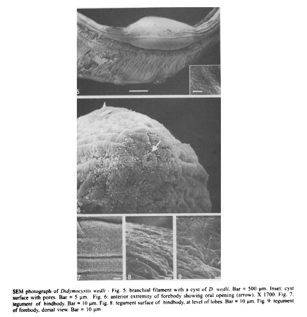
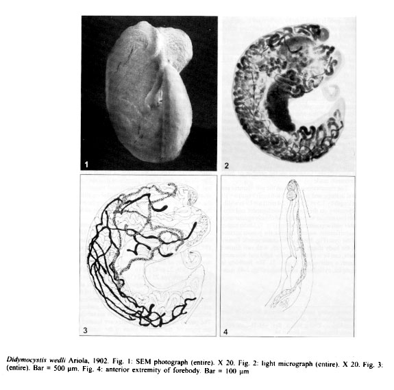
4365, 21045-900 Rio de Janeiro, RJ, Brasil Received 5 December 2000 Code Number: oc01185 Didymocystis wedli a parasite from the gills of Thunnus albacares from the coast of the State of Rio de Janeiro, is described by use of light and scanning electron microscopy. This is the first report of this species in Brazil and South America. New data are presented on the surface topography as demonstrated by scanning electron microscopy. Key words: Didymocystis wedli - Digenea - Thunnus albacares - Brazil During a survey of helminth parasites of tunas from the coast of the State of Rio de Janeiro, Didymocystis wedli was found parasitizing gills of 26 Thunnus albacares out of 72 examined (prevalence: 36%, intensity: 4-86), which represents the first report of this species in Brazil and in South America. The fishes were obtained from fishermen and brought to the laboratory to be examined; gills were removed and examined in a saline medium under stereoscopic microscopy and cysts were dissected to release the worms.The methodology used was described in Kohn et al. (1997). Measurements are in micrometer unless otherwise stated, with means in parentheses, followed by the number of measurements made (n). The membranous cysts on the gill filament are oval in shape and measure 3 to 6 mm (5 mm) n = 19 in length by 1.5 to 2.8 mm (2.4 mm) n = 19 in width, depending on the stage of development and contain two similar worms applied closely against each other with opposite extremities (Fig. 5). Under higher magnification we can observe the porous surface of the cyst (Fig. 5 inset). The characteristic feature of the body shape of D. wedli, with two distinct parts, can be well observed by light and scanning electron microscopy (Figs 1-3). The forebody is elongate, cylindrical, and slender (Figs 1-4), measures 1-2.2 mm (1.4 mm) n = 13 long by 0.1-0.3 mm (0.2 mm) n = 17 wide. The tegument of ventral and dorsal surfaces of forebody is wrinkled cobblestone-like, without spines or papillae (Fig. 9). At higher magnification the oral opening is seen in the retractil tip of the forebody (Fig. 6). Sensory papillae were not observed around the oral opening. The large hindbody has two symmetrical rounded lobes on the anterior end, forming a groove, from which emerges the elongated forebody; the posterior third of the hindbody is curved ventrally forming a somewhat pointed tail (Figs 1-3). The hindbody measure 3.4-10.2 mm (5.8 mm ) n = 24 in length by 0.9-3.1mm (1.8 mm) n = 24 largest widht. The dorsal surface is more prominent than the ventral, forming a cover with tegumental transversal striations. There are no spines or papillae on either the ventral or the dorsal tegumentary surface of the hindbbody, which presents the same wrinkled cobblestone-like appearance (Figs 7-8). The structures of the worms agree with previous descriptions (Ariola 1902, Kobayashi 1921, Okada 1926, Yamaguti 1934, Ishii 1935, Madhavi 1982). The small rounded to piriform oral sucker opens in a retractil depression situated in the anterior extremity of the forebody and measure 27 to 45 (37) n = 19 by 22 to 42 (31) n = 19; it is followed by a globular pharynx 25 to 62 (38) n = 19 by 17 to 50 (36) n = 19. The oesophagus is very slender and long, bifurcating at the level of the middle of the forebody, in two ceca that extend to the posterior end of the hindbody (Fig. 4). The ventral sucker is absent. The testes, ovary and vitellaria are tubular. The paired elongate tubular testes are located at the edges of the anterior lobes of the hindbody. The ovary consists of a small compact mass and two to three sinuous tubes which may have short branches, extending close to the testes. The vitellaria are formed by narrow and sinuous tubes with side branches, that extend into dorsal area of the hindbody (Figs 2-3). The Mehlis' gland and seminal receptacle are present. The Laurer's canal was not seen, the uterus is convoluted and fills most of the hindbody; it has a large reservoir filled with eggs and opens in the anterior extremity of the forebody. The eggs are bean-shaped, very small, contain miracidia and measure 15 to 17 (15) n = 34 by 10 to 12.5 (12.5) n = 34. Measurements of non compressed worms - Forebody: 0.9-1.6 mm (1. 2 mm) n = 13 x 0.1-0.3 mm (0.2) n = 20. Hindbody: 2.6-6.2 mm (4.2 mm) n = 27 x 0.6-1.9 mm (1.4 mm) n = 24. Oral sucker: 30-47 (37) n = 21 x 25-42 (32) n = 21. Pharynx: 30-57 (41) n = 20 x 25-45 (37) n = 20. Eggs: 15-17 (15) n = 34 x 10-12.5 (12.5) n = 34. D. wedli was reported in Italy from Euthynnus alleteratus, Thynnus vulgaris, Thunnus thynnus and Katsuwonus pelamys (Ariola 1902, Dollfus 1926, Okada 1926); in Japan from Scomber japonicus, K. pelamys, T. thynnus, T. orientalis, Seriola quinqueradiata (Kobayashi 1921, Yamaguti 1934, Ishii 1935); in India from Auxis thazard and T. tonggol (Madhavi 1982, Murugesh & Madhavi 1995) and from T. albacares and T. obesus in a non specific place in Pacific (Pozdnyakov 1990). In Brazil, only three species of Didymozoidae were refered: Unitubulotestis sardae (MacCallum & MacCallum, 1916) by Hsu (1968) and Nematobothrium scombri (Taschenberg 1879) by Rêgo and Santos (1983) from Rio de Janeiro and Brasicystis bennetti Thatcher 1979 from Amazonas. We found only 36% of T. albacares infected with D. wedli, whereas a 100% prevalence rate was found in the same host examined in Gulf of Mexico (Nikolaeva 1968 in Nikolaeva 1985).The morphology of our specimens agrees with previous descriptions (Ariola 1902, Kobayashi 1921, Okada 1926, Yamaguti 1934, Ishii 1935, Madhavi 1982, Pozdnyakov 1990, Murugesh & Madhavi 1995). The large variation in body size can be explained by the number of specimens measured. Murugesh and Madhavi (1995) reported that the variation in the number and distribution of the ovarian and vitelline branches depended on the maturity of the worms; in younger specimens the testes are seen as thick tubes but with maturity they become thin, spent and moniliform. This variation was also observed in our material. Using SEM we can obtain a tridimensional image of the parasite, showing the external appearence of the parasite. The pattern of the tegumental surface of body show some variations depending on the state of body contraction, as shown in Figs 7-8. This is the first study of the genus Didymocystis using SEM. Another didimozoid species studied by SEM was the adult and the egg with miracidia of Allodidymozoon operculare and the cyst and the egg of A. gasterale by Abdul-Salam and Sreelatha (1995). The same authors studied by SEM a didymozoid larva from Apogon uninotatus from Kuwait Bay (Abdul-Salam & Sreelatha 1993). As with A. operculare, papillae and spines were not observed in the tegument of D. wedli. ACKNOWLEDGEMENTS To Dr Monika Barth from Departamento de Virologia, Instituto Oswaldo Cruz, for the facilities offered for the use of the electron microscope. To Dr Gustavo Wilson Nunan and Dr Décio Ferreira de Moraes Jr. from Museu Nacional, Universidade Federal do Rio de Janeiro, for the identification of fish hosts. To Dr Antonia Maria de Andrade Rabello from "Quaker Brasil Ltda", Rio de Janeiro, for providing the hosts. To Dr Simone C Cohen and to the referee for English review. REFERENCES
Copyright 2001 Instituto Oswaldo Cruz - Fiocruz. The following images related to this document are available:Photo images[oc01185f1-4.jpg] [oc01185f5-9.jpg] |
| |||||||||
