
|
Memórias do Instituto Oswaldo Cruz
Fundação Oswaldo Cruz, Fiocruz
ISSN: 1678-8060 EISSN: 1678-8060
Vol. 97, Num. 1, 2002, pp. 123-126
|
Mem Inst Oswaldo Cruz, Rio de
Janeiro, Vol. 97(1) 2002, pp. 123-126
SHORT COMMUNICATION
An Autochthonous Case of Echinococcus
vogeli Rausch & Bernstein, 1972 Polycystic Echinococcosis in the State
of Rondônia, Brazil
Rosângela Rodrigues-Silva/+,
José Resende V Peixoto*, Regina Maria Figueiredo de Oliveira**, Roberto
Magalhães Pinto/++, Delir Corrêa Gomes/++
Laboratório de Helmintos Parasitos de Vertebrados,
Departamento de Helmintologia, Instituto Oswaldo Cruz-Fiocruz, Av. Brasil 4365,
21045-900 Rio de Janeiro, RJ, Brasil *Santa Casa de Misericórdia, UFRJ,
Rio de Janeiro, RJ, Brasil **Disciplina de Parasitologia, Departamento de Patologia
e Laboratórios, Faculdade de Ciências Médicas, Uerj, Rio de Janeiro,
RJ, Brasil
+Corresponding author. Fax: +55-21-2598.4363. E-mail: rsilva@gene.dbbm.fiocruz.br
++CNPq research fellows, Proc. nos 300374/80-1 and 303124/89-0, respectively.
Received 3 May 2001
Accepted 2 July 2001
Code Number: oc02023
The present case report refers to a patient
from the State of Rondônia, North region of Brazil, attended with clinical
suspicion of hepatic echinococcosis. Examination by imaging (ultrasonography
and computerized tomography) revealed a conglomerate of cystic lesions, with
mobile contents within the cyst. The serology (immunoblot) for Echinococcus
sp. was positive (21 and 31 kDa bands). This case is the first reported in
Rondônia, suggesting the need to investigate the polycystic echinococcosis
in individuals with hepatic cysts from areas of tropical forest and hunting
habits where wild life was present as wild dogs, cats and rodents, particularly
Agouti paca (paca) and Dasyprocta aguti (agouti).
Key words: polycystic echinococcosis - Echinococcus
vogeli - human report - Rondônia - Brazil
Polycystic echinococcosis is an emergent zoonosis
induced by Echinococcus vogeli Rausch & Bernstein, 1972 and E.
oligarthus (Diesing, 1863) Lühe, 1910 (D'Alessandro 1997, Pawlowski,
1997). In Brazil its intermediate hosts are either Agouti paca (Linnaeus,
1766) (= Cuniculus paca) commonly named paca or Dasyprocta aguti
(Linnaeus, 1758) (= Dasyprocta leporina), commonly named agouti, "cutia".
The finding of cysts of E. vogeli in pacas was reported from Sena Madureira,
State of Acre (D'Alessandro et al. 1981, Meneghelli et al. 1990), Serra do Navio,
State of Amapá (Rausch et al. 1984), while polycystic larvae in the liver
and spleen of agoutis from Jacutinga, State of São Paulo (Lutz 1907) and
Ilha de Marajó, State of Pará, have been reported (Soares et al. 1999).
The polycystic larval form mainly develops in the liver and can destroy the
hepatic parenchyma simulating a malignant neoplasia (D'Alessandro et al. 1979).
The bush dog Speothus venaticus (Lund,
1842) and the domestic dogs are the only animals reported as natural hosts of
E. vogeli (D'Alessandro et al. 1996).
As accidental hosts, humans can be infected by
the ingestion of eggs of the parasite contaminating either faeces of pet dogs
(definitive host) or hunt that previously have ingested paca viscera with polycystic
larval forms (Meneghelli et al. 1990, D'Alessandro 1997).
Human echinococcosis is insidious and difficult
to diagnose since the presence of the parasite can not be easily detected, if
deeply located. As an auxiliary tool for conclusive or presumptive diagnosis,
laboratory investigation is necessary with immunoserological assays (Craig 1994,
Romani 1995).
In this study, we report an authoctothonus human
case of polycystic echinococcosis due to E. vogeli in a patient from
the State of Rondônia, North region of Brazil.
The term echinococcosis was adopted in accordance
to WHO (1996) and Pawlowski (1997).
Case report - Woman, 48 years old, mulatto,
married, born in the State of Maranhão but having migrated out from this
area when she was under 5. She was first attended at the Hospital Militar de
Porto Velho and afterwards (August 1996) sent to the ambulatory of the Gastroenterology
Department in the Santa Casa de Misericórdia (SCM), State of Rio de Janeiro.
During her first visit to the SCM, the patient declared to live in the suburban
area of Porto Velho, State of Rondônia, and her main complaint was a pain
in right hypochondria, that had started two years before. The patient was neither
alcohol nor tobacco addict. She reported to be aware of echinococcosis, since
one of her sons presented a history of pulmonary echinococcosis. She was accustomed
to hunting wild animals and among them, the paca. In Porto Velho, in July 1995,
she was submitted to an abdominal ultrasonography, that revealed a cystic structure
in the right lobe. Computerized tomography (CT) confirmed a conglomerate of
cystic lesions in the liver, indicating a hepatic echinococcosis.
In the SCM, clinical examination of the patient
revealed good general conditions, eupnea, absence of jaundice and adenomatosis.
Respiratory and circulatory systems were not altered. Under palpation the abdomen
was flaccid, painful and with an evident mass formation in the right hypochondria,
at 4 cm from the right costal rim in the hemiclavicular line.
The following laboratory tests were performed:
complete blood count, blood biochemistry (glucose, urea, creatinine, sodium,
potassium), hepatic profile (direct and indirect total bilirubin, aminotransferases,
total proteins and fractions, alkaline phosphatase), urine and fecal examination,
thoracic X-ray. Results of the above referred procedures were in accordance
with normal patterns. Viral hepatitis serology was positive with anti-HVA IgG
and negative to viral hepatitis B with AgHbs, anti-Hbc, anti-Hbc IgM and anti-Hbs.
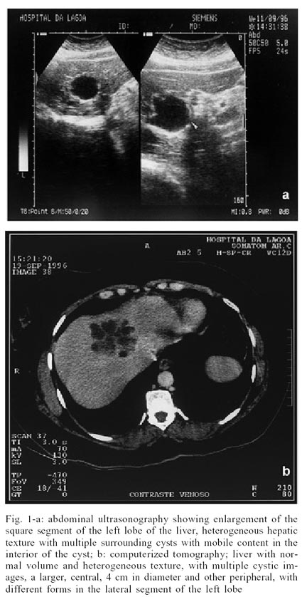
Two months later abdominal ultrasonography revealed
the enlargement of the square segment of the liver left lobe, inducing a high
lateral displacement of the median superior hepatic vein, heterogeneous hepatic
texture with multiple surrounding cysts and the presence of a mobile content
in the interior of the cyst (Fig. 1a).
In CT liver appeared within a normal volume, heterogeneous texture, with multiple
cysts with regular margins and with anechogenic structures inside. There was
a larger central image 4 cm in diameter and other radiate peripheral in the
lateral segment of the left lobe (Fig. 1b).
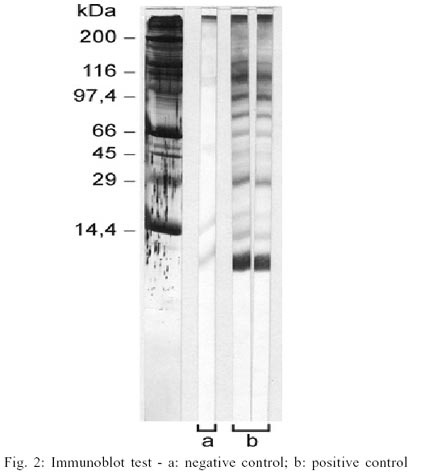
Serological tests for Echinococcus sp.
were performed by immunoblot (Western blot) assay with total antigen of lyophilized
sheep hydatic cyst fluid (ATLH-O) supplied by the Laboratório de Serologia,
Departamento de Parasitologia del Centro de Referência de Laboratórios
de Salud Publica, Instituto Nacional de Salud, Peru. Bands with molecular weights
of 21 and 31 kDa were observed (Fig. 2).
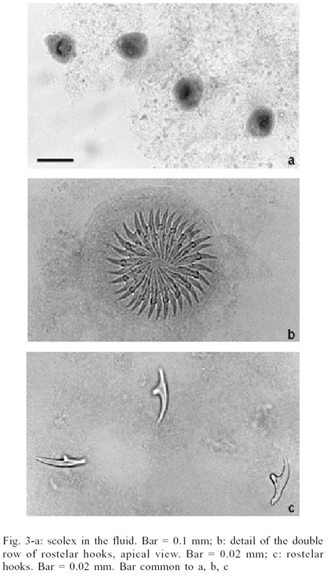
In 1996, the patient was submitted to an albendazole
treatment (400 mg/2x/daily) for three months with no improvement. Then a surgical
procedure was decided, with drainage of the entire cavity of the cyst, due to
its close connection to the median superior hepatic vein and vena cava. The
cyst fluid was collected and scolex were stained with Mayer's Carmalum, cleared
with beechwood creosote and preserved in Canada balsam and analyzed under a
brightfield microscope (Figs 3a,b).
The species was identified to E. vogeli taking into account the number
(34) and shape of the hooks, greater dimensions of the blade (0.0178 mm) in
relation to the guard (0.0158 mm) and total length (0.0372 mm) and the comparison
with other species of the genus Echinococcus, according to Rausch and
Bernstein (1972), Rausch et al. (1978) D'Alessandro et al. (1981) and Meneghelli
et al. (1990) (Fig. 3c). Specimens were
deposited in the Helminthological Collection of the Instituto Oswaldo Cruz (CHIOC)
no. 34336 (whole mount).
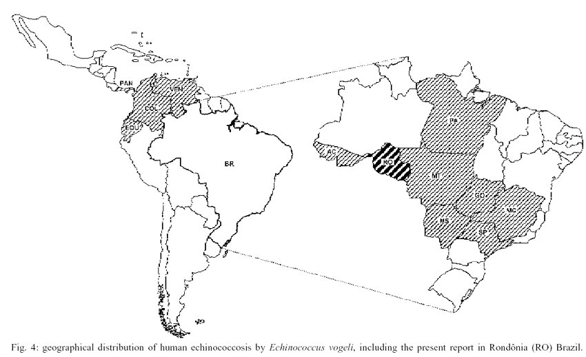
Human cases of polycystic echinococcosis due
to E. vogeli have been described from Panama (1 case), Colombia (13 cases),
Ecuador (6 cases) and Venezuela (2 cases) (D'Alessandro et al. 1979, D'Alessandro
1997). In Brazil related cases of this disease were referred to occur in the
vicinity of State of Amazonas (10 cases) (Meneghelli et al. 1986, 1992, Pacheco
et al. 1986, Timmerman et al. 1986, Meneghelli 1989, Ferreira et al. 1995) and
in the mid west (2 cases) and southeast (6 cases) regions (Meneghelli 1985,
Ferreira et al. 1987, 1995, Soares & Amaral 1998) indicating that the zoonosis
may be widely spread. Although another case was detected in the neighborhood
of Amazonas, it is to be supposed that other cases remain unreported in this
region, considering that other states near Amazonas and bordering countries
have been assignaled on what refers to the geographical distribution of echinococcosis
(Fig. 4).
The reported case assembles the necessary epidemiological
conditions to the transmission of polycystic echinococcosis, since the patient
was used to hunting wild animals, including pacas that may have E. vogeli
(Rausch et al. 1981). Moreover, she came from an area where the pacas and the
wild dogs occur as previosly referred by D'Alessandro (1997) and D'Alessandro
et al. (1981).
The immunoserological assays data are in accordance
to Romani (1995) and Ayadi et al. (1995) and detected the same molecular weights
bands, appearing in sera from patients infected with E. granulosus. Maddison
et al. (1989) identified a specific antigen of E. granulosus with a relative
mass of about 8 kDa. The reactivity of this antigen with sera from infected
patients showed 91% of sensibility and was 100% specific, in despite of the
cross-reactions observed in sera from patients harboring E. multilocularis
and E. vogeli. Cross-reactions were also observed by Ferreira and Zaha
(1990), Verástegui et al. (1992) with the same antigen with sera from patients
with schistosomiasis, filariasis and cysticercosis. Seropositivity cystic echinococcosis
was observed even when E. vogeli antigen fraction was employed (Gottstein
et al. 1995).
Nevertheless, molecular weights of 21 and 31
kDa seem to be specific to the genus Echinococcus and not to a particular
species. From the epidemiologic point of view, differential diagnosis may be
irrelevant because E. vogeli and E. mul-tilocularis are allopatric
in distribution (Gottstein et al. 1995). Thus, the present specific identification
was based on morphological data only.
The present findings may be included among those
whith the most common clinical aspects of the polycystic echinococcosis in Brazil:
abdominal localization affecting the liver with palpable aching mass, loss of
weight and the thoracic localization represented by the involvement of the vena
cava (D'Alessandro 1997). The parasitosis seems to be neither sex nor age related
and the period of manifestation varies from one month to 13 years. The average
age (44 years) of the patients is close to the present case (48 years old),
according to previous reports (D'Alessandro 1997). Nevertheless, the patient
did not present splenomegaly, jaundice, portal hypertension and alteration of
laboratorial tests, observed in other cases (D'Alessandro et al. 1996).
Most surprising is a lack of data on the cases
in the State of Amazonas, suggesting that this zoonosis is under-diagnosed (D'Alessandro
1997). There is an urgent need to investigate the ethiology of abdominal masses
detected in individuals from the Brazilian Mid-west and North regions, mainly
in those that are either fond of hunting wild rodents or that are in close contact
with wild or pet carnivores that are feed with viscera of pacas and agoutis.
In conclusion, our data confirm that reported cases of polycystic echinococcosis
are tip of an iceberg (D'Alessandro 1997).
ACKNOWLEDGEMENTS
To Genilton José Oliveira and Heloísa
Maria Nogueira Diniz, Education Department, Oswaldo Cruz Institute, for their
technical support with the photographs.
REFERENCES
- Ayadi A, Dutoit E, Sendid B, Camus D 1995.
Specific diagnostic antigens of Echinococcus granulosus detected by
western blot. Parasite 2: 119-123.
- Craig P 1994. Current research in echinococcosis.
Parasitol Today 10: 209-211.
- D'Alessandro A 1997. Polycystic echinococcosis
in tropical America: Echinococcus vogeli and E. oligarthrus. Acta
Trop 67: 43-65.
- D'Alessandro A, Moraes MAP, Raick AN 1996.
Polycystic hydatid disease in Brazil. Report of five new human cases and a
short review of other published observations. Rev Soc Bras Med Trop
29: 219-228.
- D'Alessandro A, Rausch RL, Cuello C, Aristizabal
N 1979. Echinococcus vogeli in man, with a review of policystic hydatid
disease in Colombia and neighboring countries. Am J Trop Med Hyg 28:
303-317.
- D'Alessandro A, Rausch RL, Morales GA, Collet
S, Angel D 1981. Echinococcus infections in Colombian animals. Am
J Trop Med Hyg 30: 1263-1276.
- Ferreira MS, Nishioka SA, Rocha A, D'Alessandro
A 1995. Echinococcus vogeli polycistic hydatid disease: report of two
Brazilian cases outside the Amazon region. Trans R Soc Trop Med
Hyg 89: 286-287.
- Ferreira MS, Rocha A, Gonçalves EG, Carvalho
AM, Nishioka SA, Andrade NB 1987. Relato de caso: um caso de hidatidose policística
autóctone de Minas Gerais, Brasil. Rev Soc Bras Med Trop 20:
181-186.
- Ferreira H, Zaha A 1990. Analysis of different
antigen sources in the diagnosis of human hydatid disease by immunoblot. In
R Ehrlich, A Nieto, L Yarzábal (eds), Basic Research Helminthiases,
LOGOS, Montevideo, p. 189-201.
- Gottstein B, D'Alessandro A, Rausch RL 1995.
Immunodiagnosis of polycystic hydatid disease/polycystic echinococcosis due
to Echinococcus vogeli. Am J Trop Med Hyg 53: 558-563.
- Lutz A 1907. Observação de uma cutia
infeccionada com Echinococcus. Rev Soc Ci São Paulo
2: 113-114.
- Maddison S, Slemenda S, Schantz P, Fried J,
Wilson M, Tsang V 1989. A specific diagnostic antigen of Echinococcus granulosus
with an apparent molecular weight of 8 kDa. Am J Trop Med Hyg 40: 377-383.
- Meneghelli UG 1985. Calcificações
hepáticas múltiplas decorrentes de doença hidática policística.
Rev Goiana Med 31: 53-60.
- Meneghelli UG 1989. Hidatidose na Amazônia
brasileira. Arch Int Hidatid 29: 79-80.
- Meneghelli UG, Barbó MLP, Magro JE, Belluci
AD, Velludo MASL 1986. Policystic hydatid disease (Echinococcus vogeli):
clinical and radiological manifestations and treatment with albendazol of
a patient from the Brazilian Amazon region. Arq Gastroenterol 23:
177-183.
- Meneghelli UG, Martinelli ALC, Velludo MASL
1990. Cistos de Echinococcus vogeli em fígado de paca (Cuniculus
paca) originária do Estado do Acre, Brasil. Rev Soc Bras Med
Trop 23: 153-155.
- Meneghelli UG, Martinelli ALC, Velludo MASL,
Belluci AD, Magro JE, Barbó MLP 1992. Polycistic hydatid disease (Echinococcus
vogeli) clinical, laboratory and morphological fidings in nine Brazilian
patients. J Hepathol 14: 203-210.
- Pacheco PRG, Komma MD, Pinto RNL, Souza LCS,
Pereira LIA 1986. Doença hidática policística. Relato de um
caso procedente do Pará. Rev Soc Bras Med Trop 19: 67.
- Pawlowski ZS 1997. Terminology related to
Echinococcus and echinococcosis. Acta Trop 67: 1-5.
- Rausch RL, Bernstein JJ 1972. Echinococcus
vogeli sp.n. (Cestoda: Taeniidae) from the bush dog, Speothus venaticus
(Lund). Z Tropenmed Parasit 23: 25-34.
- Rausch RL, D'Alessandro A, Ohbayashi M 1984.
The taxonomic status of Echinococcus cruzi Brumpt and Joyeux, 1924
(Cestoda: Taeniidae) from an agouti (Rodentia: Dasyproctidate) in Brazil.
J Parasitol 70: 295-302.
- Rausch RL, D'Alessandro A, Rausch VR 1981.
Characteristics of the larval Echinococcus vogeli Rausch & Bernstein,
1972 in the natural intermediate host, the paca, Cuniculus paca L.
(Rodentia: Dasyproctidae). Am J Trop Med Hyg 30: 1043-1052.
- Rausch RL, Rausch VR, D'Alessandro A 1978.
Discrimination of the larval stages of Echinococcus oligarthrus (Diesing,
1863) and E. vogeli Rausch and Bernstein, 1972 (Cestoda: Taeniidae).
Am J Trop Med Hyg 27: 1195-1202.
- Romani ELS 1995. Determinação
de Antígenos Relevantes da Forma Larvar do Echinococcus granulosus: Padronização
e Aplicação do "Immunoblot" no Diagnóstico da Hidatidose
Humana, MSC Thesis, Instituto Oswaldo Cruz, Rio de Janeiro, 87 pp.
- Soares MCP, Amaral ISA 1998. Polycistic echinococcosis
by E. vogeli in the Amazon region. J Hepathol 28: 908.
- Soares MCP, Cruz ERM, Cartágenes PRB,
Alves MM, Bensabath G 1999. Hidatidose policística e cisticercose em
fígado e baço de cutias (Dasyprocta aguti) da Ilha de Marajó,
Pará, Brasil. Rev Soc Bras Med Trop 32: 317.
- Timmerman A, Andrade DR, Hutzler RU, Marinho
IS, Uliana SRB 1986. Terapêutica da hidatidose hepática com albendazol:
relato de 1 caso. In Abstracts of the IX Congresso Brasileiro de Hepatologia,
São Paulo, p. 85.
- Verastegui M, Moro P, Guevara A, Rodriguez
T, Miranda E, Gilman, RH 1992. Enzyme-linked immuno-electrotransfer blot test
for diagnosis of human hydatid disease. J Clin Microbiol 30:
1557-1561.
- WHO-World Health Organization 1996. Guidelines
for treatment of cystic and alveolar echinococcosis in humans. WHO Bull
74: 231-242.
© 2002
Instituto Oswaldo Cruz - Fiocruz
The following images related to this document are available:
Photo images
[oc02023f4.jpg]
[oc02023f3.jpg]
[oc02023f2.jpg]
[oc02023f1.jpg]
|
