 |
| About Bioline | All Journals | Testimonials | Membership | News |
|
||||||
|
||||||
Mem Inst Oswaldo Cruz, Rio de Janeiro, Vol. 97(3) 2002, pp. 431-433 SHORT COMMUNICATION
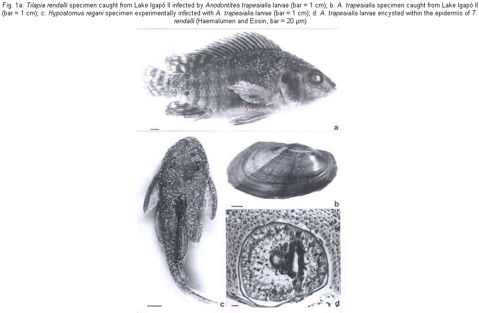
The Histopathology of the Infection of Tilapia rendalli and Hypostomus regani (Osteichthyes) by Lasidium Larvae of Anodontites trapesialis (Mollusca, Bivalvia) Ângela Teresa Silva-Souza, Jorge C Eiras*/+
Universidade Estadual de Londrina,
Londrina, PR, Brasil *Departamento de Zoologia e Antropologia, Faculdade de
Ciências, Universidade do Porto, 4099-002 Porto, Portugal Received 25 June 2001 Code Number: oc02083
It is described the histopathology of the infection of Tilapia rendalli (Osteichthyes, Perciformes, Cichlidae) and Hypostomus regani (Osteichthyes, Siluriformes, Loricariidae) by lasidium larvae of Anodontites trapesialis (Mollusca, Bivalvia, Mycetopodidae). The larvae were encysted within the epidermis of the host, being surrounded by a thin hyaline membrane, 3-6 µm thick, of parasite origin. A proliferative host cell reaction did not occur. The histopathology of the infection shows that the lesions induced by the parasites are minimal. However, the numerous small lesions produced by the release of the larvae may provide optimal conditions for the infection by opportunistic pathogens, namely fungus, which may eventually cause the death of the host.
Key words: fish - bivalve larvae - parasitism The life cycle of a number of freshwater bivalve molluscs involves the infection of fish by their veliger larvae. The development and metamorphosis of the larvae are completed only if they attach to a particular species of fish, therefore demonstrating host specificity (if the larvae do not reach a compatible host they are sloughed off before the metamorphosis is completed). Depending on the parasite species the larvae attach to the gills, or fins and integument of the host, and become encapsulated and develop the juvenile stage (Waller & Mitchell 1989). This infection extends till the metamorphosis is accomplished, and during that period the fish disperse the rather sedentary freshwater bivalves. The consequences of the parasitization can cause the deadth of the host (Meyers & Millemann 1977, Moles 1983), increase the sensitivity of fish to pollutants, such as crude oil (Moles 1980), and reduce the host growth even in the case of small intensity of infection (Moles 1983). The South American Mutelidae have a particular type of larvae, called lasidium by Ihering (1891-1892). A similar kind of larvae occurs also in Africa at least in the species Mutela bourguignali (Fryer 1959). There are some studies on the development of the lasidium larvae (Fryer 1959, 1961), and Bonetto and Ezcurra (1962) provided a detailed description of the development of Anodontites trapesialis forbesianus infecting different hosts. However, almost nothing is known about the infection of fish, and the host-parasite relationships are virtually unknown. In this note some data on the histopathology of the infection of Tilappia rendalli and Hy-postomus regani by A. trapesialis larvae are presented. The T. rendalli specimens were caught from lake Igapó II at Londrina, Paraná, Brazil. Seven out of 117 specimens (5.9%) were found infected in August 1999. One out of 52 (1.9%), and two out of 26 specimens (7.7%) were found infected respectively in September and October 1999. Sampling in other months did not show the presence of the parasite. The infected specimens had a great number of conspicuous white spots irrregularly scattered all over the body surface (Fig. 1a), occurring also in the lateral fins, and ocasionally in the other fins. The number of white spots varied from a few to hundreds in some specimens. The parasitized H. regani specimen was obtained by experimental infection. Some specimens of A. trapesialis (Fig. 1b), caught from lake Igapó II in March 2000 were introduced in an aquarium containing, for about sixt months, a specimen of H. regani. About one week latter the fish presented hundreds of white spots scattered all over the body, including the fins (Fig. 1c) and died. Examination of the white spots contents from both fish species allowed the identification of lasidium larvae of A. trapesialis. Observation of the gils of both host species showed they were not infected. Apparently, each species of larvae infects primarily on a specific site of the host: Lampsilis radiata siliquoidea infects the gills (Waller & Mitchell 1989), as well as Margaritifera margaritifera (Karna & Millemann 1978, Meyers et al. 1980) and Unio spp. (Bauer 1987), while Anodonta oregonensis develops in the fins of the host (Moles 1983). The gills of our specimens were not infected, result which agrees with data from Bonetto and Ezcurra (1962) concerning A. trapesialis forbesianus infecting eleven different host fish species where gill infection was observed occasionally only. Integument samples from infected fish were fixed in buffered formalin, routinely processed for histology, and stained with haemalumen and eosin, and Masson's trichrome. Microscopical observation showed that, in both host species, the larvae were encysted within the epidermis, not surpassing the epidermal basal layer (Fig. 1d). They were surrounded by a thin 3-6 µm thick hyaline membrane of parasite origin, and a proliferative host cell reaction to the parasites did not occur. The height of the epidermis above the larvae was reduced to about one fifth of the normal, and the cells of the epidermal basal layer disappeared under the parasite in the contact zone of the parasite and the dermis. A similar feature was described for the infection of the fins of Osmerus eperlanus by the larvae of A. anatina (Anders & Wiese 1993). The histopathology of the infection shows that the lesions induced by the parasites are minimal, especialy taking into acccount that the larvae are temporary parasites, being within the host epidermis only till completion of the metamorphosis. However, the great intensity of the infection, with hundreds of larvae, may be indirectly significant to the hosts survival. The numerous small lesions produced by the release of the larvae may provide optimal conditions for the infection by opportunistic pathogens, namely fungus, which may eventually cause the deadth of the hosts. Mortality of salmonids experimentally infected with glochidia of M. margaritifera was attributed partially to secondary fungus infection (Meyers & Millemann 1977). Mass mortalities of farmed fish at Brazil have been attributed to infection by bivalve larvae (P Cecarelli, pers. commun.).
ACNOWLEDEGEMENTS
To Dr Inés Ezcurra de Drago, from Instituto Nacional de Limnologia (Santa Fé, Argentina) for the identification of the larvae. REFERENCES
© 2002 Instituto Oswaldo Cruz - Fiocruz The following images related to this document are available:Photo images[oc02083f1.jpg] |
| |||||||||
