 |
| About Bioline | All Journals | Testimonials | Membership | News |
|
||||||
|
||||||
Mem Inst Oswaldo Cruz, Rio de Janeiro, Vol. 97(4) 2002, pp. 511-516 The Zymovars of Vibrio cholerae: Multilocus Enzyme Electrophoresis of Vibrio cholerae Fernanda S Freitas, Hooman Momen, Carlos Andre Salles This project received financial support from Faperj. +Corresponding author. Fax: +55-21-2598.3495. E-mails: csalles@gene.dbbm.fiocruz.br ; caqps@ig.com.br Received 18 July 2001 Code Number: oc02097 Laboratório de Sistemática
Bioquímica, Departamento de Bioquímica e Biologia Molecular, Instituto
Oswaldo Cruz-Fiocruz, Zymovars analysis also known as
multilocus enzyme electrophoresis is applied here to investigate the genetic
variation of Vibrio cholerae strains and characterise strains or group
of strains of medical and epidemiological interest. Key words: Vibrio cholerae - enzyme electrophoresis - genetic diversity The electrophoretic mobility of enzymes has been used to detect allelic variation of the respective genes in several microorganisms including Vibrio cholerae (Salles & Momen 1991, Chen et al. 1991, Wachsmuth et al. 1993, Beltran et al. 1999). In our previous papers we focused on the problem of finding a reliable genetic distinction between the biotypes classical and El Tor and to evaluate the degree of genetic variation prevalent among this species. Then we defined a zymovar as the set of strains having the same electromorphs among the loci investigated and zymovar analysis the procedures required to determine it. This approach is also known as Multi-Locus Enzyme Electrophoresis and zymovar as electrophoretic type, or ET. The study of V. cholerae using molecular tools introduced considerable change in our views affecting the bacteriology and the epidemiology of cholera. Here we present results obtained with 14 loci from a large sample of strains. MATERIALS AND METHODS Geographical origin of strains - Algeria, Australia, Burma, Bangladesh, Brazil, Bolivia, Colombia, Chile, Czech Republic, China, Sri Lanka, France, Ghana, Guatemala, Guiana, Hong Kong, India, Indonesia, Israel, Italy, Japan, Kuwait, Malaysia, Marroc, Mexico, Nepal, Nigeria, Pakistan, Peru, Philippines, Russia, Rwanda, Singapore, Tanzania, Thailand, Turkey, UK, USA, Venezuela, Zimbabwe. Sources of strains - B Davies, Peggy Pereira, P Desmarchelier, R Colwell, Glenn Morris Jr, GB Nair, AC Ghose, B Said, JV Lee, NICED (India), JM Fournier, L Amenuvor, and our own collection. Most of the general bacteriological, electrophoretical and computational procedures have been described previously (Salles & Momen 1991). The enzyme loci mentioned in the tables and text have been described previously (Salles & Momen 1991). They are: ACO (aconitase hydratase); ADH (alanine dehydrogenase); IDH (isocitrate dehydrogenase); ME (malic enzyme); NSE (carboxilesterase); PGD (6-phosphogluconate dehydrogenase); MDH (malate dehydrogenase); PGM (phosphoglucomutase); GPI (glucose phosphate isomerase); G6P (glucose-6-phosphatedehydrogenase) ; PD (proline dipeptidase); P1 (peptidase leucyl-leucyl-leucine); P2 (peptidase leucyl glycil glycine) with the addition of the locus LAP: leucyl leucyl aminopeptidase (Pasteur et al. 1990). Genetic diversity was estimated according to Selander et al. (1986). The genetic diversity per locus GDL is calculated from allele frequencies as
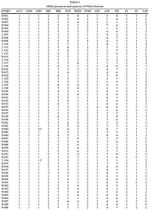
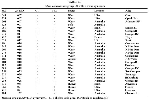
were Ai is the frequency of the ith allele in locus A and n the number of zymovars. The expression n/n-1 is a correction for small samples. Mean genetic diversity MGD is the arithmetic average of GDL over all loci. RESULTS Table I shows the electromorphs of 134 zymovars obtained from 155 strains of V. cholerae non O1 non O139 plus the zymovars of El Tor 7th pandemic (14A), ElTor S. American pandemic (14B) and classical (13). For the present purpose the O139 strains (Bengal) are equated with El Tor 7th Pandemic with the same zymovar 14A. The genetic diversity per locus was as following: ACO-0.092 ADH-0.064 IDH-0.133 ME-0.003 NSE-0.222 PGD-0.525 MDH-0.453 PGM-0.392 GPI-0.334 G6P-0.588 PD-0.690 P1-0.553 P2-0.314 LAP-0.461. The mean genetic diversity (MGD) was 0.339, sd = 0.2. Table III list selected O1 serogroup strains isolated from clinical and environmental sources with zymovars other than 13 or 14 showing the correspondence or lack of it between zymovar, serogroup, CT, TCP and sources. DISCUSSION The present estimate of MGD is 0.339. Our previous estimated MGD was 0.326 using nearly half the strains of the present study. Chen et al. (1991) found 0.311 and Beltran et al. (1999) from a large sample of American non-O1 isolates found a higher diversity of 0.436. A possible source of variation in MGD seems to be the choice of loci. Our choice was aimed at finding loci diagnostic of the species V. cholerae with low genetic diversity GD and this may have biased in part the MGD. This diversity is considerable and is of the same level of Escherichia coli but still less than some Gram negative bacteria. We have been unable to find significant correlation between zymovars and pathogenic/epidemic potential except , partially, in the case of the zymovars 13,14 A and 14 B (O1-O139 epidemic strains). It is however interesting to note that the allele NSE 4 present in zymovars 13 and 14 is not frequent (10/136) among non-O1 non-O139 and other O1 isolates. NSE 4 group include some strains of the most frequent serogroups of non-O1 V. cholerae isolated from human sources, O37 and O5 (Donovan 1984). This locus seems to be useful for a preliminary screening of new strains. Most genes investigated are housekeeping genes necessarily conserved for the survival of the organism. The know pathogenicity islands are phages or phage-like mobile elements responsible for the lateral transfer of non essential genes coding cholera toxin (CT) and toxin co-regulated pilli (TCP) (Karaolis et al. 1999, Faruque et al. 1999) and genes coding somatic O antigens as in the Bengal O139. This latter strain is genetically an El Tor 7th pandemic zymovar 14 A which received from a non-O1 vibrio, genes coding a modified LPS (Johnson et al. 1994, Bik et al. 1995). It is therefore unlikely that strong correlation may be found between zymovars and antigenic or virulence factors carried by these mobile elements. The strain responsible for the American pandemic is not the same strain causing the 7th pandemic. As show by Wachsmuth et al. (1993) and our results, it differ in locus LAP, with allele 3 while the Old World pandemic strains have allele 1. The 7th pandemic in the Old World is caused by zymovar 14A and the American pandemic by zymovar 14B. Both pandemics are usually described as the same 7th pandemic but the genetic distinction between the two agents may suggest an independent origin of the later. This poses an interesting epidemiological problem, the origin of the American pandemic, the agent and source of the first cases of cholera in Peru. LAP 3 is not common in our sample and may be equally rare among V. cholerae in nature. Locus LAP has epidemiological interest since cholera on the Atlantic coast may be due to the prevalent clone but may be the result of import from the Old World. A novelty brought by the American pandemic was the detection of epidemic V. cholerae O1 zymovar 14B sucrose negative. It was first isolated in French Guiana (JM Fournier, pers. commun.) and later spread to Brazilian Amazon region (De Paula et al. 1997). The production of acid from sucrose in cholera diagnostic medium TCBS is a characteristic of most V. cholerae used for the initial isolation and identification procedures. Although many sucrose negative strains have been described by Desmarchelier and Reichelt (1984), none were O1 and epidemic. This may be the first occurrence of epidemic O1 with such phenotypic variation. Table III list few strains of O1 serogroup with diverse genetic profiles. Strains from South India and Amazon Brazil have been studied in detail by Saha et al. (1996) and Coelho et al. (1995) respectively. The India strains with the same zymovar of the epidemic strain (14A) seems to be derived from the later with loss of the CT cluster but conserved TCP. The Amazon strains belong to a diverse genetic group (zymovar 54) and do not have CT and TCP. Therefore CT, TCP and the O1 antigen may not be linked to any particular zymovar. An interesting consequence of the fact that the O1 antigen does not define a clone and is found in many genetically diverse strains is that past epidemiological accounts of cholera outbreaks and epidemics (Pollitzer 1960, Wachsmuth et al. 1994) must be reviewed with caution when the isolation of O1 organisms from the environment is connected to clinical cases. Attempts to type strains of V. cholerae have been made involving detection of differences in the Vibrio's genome (RAPD, ribotyping etc). The RAPD applied to protozoa gave results in agreement with multilocus data (Tibayrenc et al. 1993) and may be a promising tool while the shortcomings with ribotyping (Lan & Reeves 1998) and the possibility of intense lateral gene transfer between organisms (Doolittle 1999) including V. cholerae (Cruz & Davies 2000) renders the interpretation of rybotyping and similar techniques rather problematic. A combination of multilocus enzyme electrophoresis with DNA sequencing of the loci studied proposed by Maiden et al. (1998) may show promising results. ACKNOWLEDGEMENTS To the scientists mentioned in Materials and Methods section for providing strains, their serogroup and clinical-epidemiological information. To Ana Carolina Vicente and Veronica Vieira (Genetic Department, IOC) for data on Cholera Toxin gene and Toxin Co-regulated Pili gene of several strains and helpful advice. REFERENCES
Copyright 2002 Instituto Oswaldo Cruz - Fiocruz The following images related to this document are available:Photo images[oc02097t3.jpg] [oc02097t1.3.jpg] [oc02097e1.jpg] [oc02097t2.jpg] [oc02097t1.2.jpg] [oc02097t1.1.jpg] |
| |||||||||
