 |
| About Bioline | All Journals | Testimonials | Membership | News |
|
||||||
|
||||||
Mem Inst Oswaldo Cruz, Rio de Janeiro, Vol. 97(4) 2002, pp. 599-600 SHORT COMMUNICATION Experimental Neuroschistosomiasis - Inadequacy of the Murine Model Luciana M Silva, Carla Neves de Oliveira,
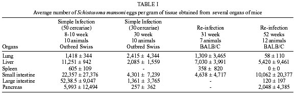
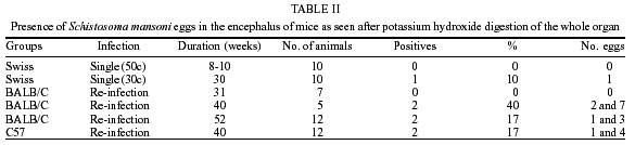
Zilton A Andrade+ Laboratório de Patologia Experimental, Centro de Pesquisas Gonçalo Moniz-Fiocruz, Rua Valdemar Falcão 121, 40295-001 Salvador, BA, Brasil Received 13 September 2001 Code Number: oc02114 Neuroschistosomiasis is rarely observed in human pathology, but it is of considerable importance. To investigate its pathogenesis, consequences and response to treatment, an experimental model would be desirable, but is not yet available, in spite of a few indications of a suitable mouse model in the literature. Severe, recent and late Schistosoma mansoni infections in outbred and inbred strains of mice revealed widespread distribution of parasite eggs in several organs, but only exceptionally did eggs reach the encephalus, thus revealing the inadequacy of the mouse as an experimental model for neuroschistosomiasis. Key words: neuroschistosomiasis - Schistosoma mansoni - murine model Neuroschistosomiasis can be a serious complication for patients infected with Schistosoma mansoni. The parasite eggs can reach the central nervous system either by embolization or by abnormal migration of adult worm pairs. Worms may pass through Batson's plexus, migrating from the portal to paravertebral veins, helped by retrograde blood flow generated by an eventual and sudden increase in abdominal pressure. It has been suggested that deposition of immune complexes, especially at the choroid plexus, can also result in central nervous system involvement in schistosomiasis (Pitella & Bambirra 1989). However, severity of clinical manifestations is certainly related to the presence of adult worms and their eggs, which correlate with the signs and symptoms of myelitis, radiculitis, tumor and meningeal irritation attributed to neuroschistosomiasis (Andrade 1986). In a few human cases, worms and eggs have been found in leptomeningeal veins in the brain and spinal cord, especially at the lumbo-sacral and thoracic regions, associated with phlebitis, arteritis, and chronic granulomatous inflammation (Gama & Sá 1945, Pondé et al. 1960, Ferreira et al. 1998). Due to difficulty in obtaining pathological material, diagnosis of neuroschistosomiasis in the large majority of cases relies on positive serology in the liquor and/or suggestive imaging (Ferrari et al. 1995, Ferrari 1999). Considering the important problems posed by neuroschistosomiasis, it is surprising that only a few attempts have been made to experimentally study it. The present investigation was conducted to find out whether the murine model would be suitable for such study. The literature has very little information concerning neuroschistosomiasis in mice. Aloe et al. (1996) described finding schistosome eggs in histological sections from the brain of mice and reported that mice with S. mansoni periovular granulomas in the brain showed decrease of nervous growth factor expression. Based on such findings, Fiore et al. (1996) reported that mice infected with S. mansoni exhibited behavioral disturbances, probably associated with modifications in the levels of nerve growth factor and cytokines induced by granulomas. These two reports did not indicate how frequently S. mansoni eggs reach the central nervous system of the mouse. In the present report, we attempted to do that, as follows: (1) outbred Swiss mice, 20-25 g, of both sexes, were infected with either 30 or 50 S. mansoni cercariae, Feira de Santana strain (Andrade & Sadigursky 1985) by the transcutaneous route. For early infection, animals were sacrificed at 8-10 weeks of cercarial exposure. For late infection, animals with 30-31 week old infection were sacrificed; (2) inbred BALB/C mice, 15-18 g, of both sexes, were infected with 30 cercariae. When the infection was proved patent by the presence of viable eggs in the stools, these animals were submitted to re-infection five times with 15 cercariae once a week. This procedure increases the severity of schistosomiasis, with higher probability to develop pipe-stem fibrosis in the liver (Araújo Santos et al. 2000). Mice were sacrificed at 31, 40 and 52 weeks after first cercarial exposure; (3) inbred C57 mice were infected with 20 cercariae, re-infected with 10 cercariae, and sacrificed at 40 weeks following the first cercarial exposure. For all animals sacrificed, except C-57 and 5 BALB/C mice, the liver, lung, small and large intestines, pancreas, kidney and spleen were removed and weighed. Fragments of these organs were digested in 60 ml of a 4% potassium hydroxyde solution for the counting of eggs, according to Cheever (1970) (Table I). For the encephalus, the entire organ was always digested and the samples for the microscopic seach of eggs were collected after sedimentation for several hours. Only a few schistosome eggs were found in the nervous tissue (Table II). The chance of finding eggs somewhat increased after prolonged and more severe infections. These results are difficult to reconcile with those of Aloe et al. (1996). They found between 20 and 50 schistosome eggs per mouse after examining 40 frozen brain sections. We did not succeed in finding eggs in histological sections of the brain, and because of that we resorted to the digestion method of the whole encephalus. Fragments of the liver were fixed in formalin and 5 µ-thick paraffin sections obtained were stained with hematoxylin and eosin, and sirius-red for collagen. The majority of the animals showed pipestem fibrosis, especially those of re-infection groups. In conclusion, our results indicate that mice with severe infection, with widespread distribution of schistosome eggs in several organs, failed to show significant involvement of the central nervous system. Therefore, the murine model that has been so successfully applied to investigate multiple aspects of human schistosomiasis did not appear to be suitable for experimental studies about neuroschistosomiasis. REFERENCES
Copyright 2002 Instituto Oswaldo Cruz - Fiocruz The following images related to this document are available:Photo images[oc02114t2.jpg] [oc02114t1.jpg] |
| |||||||||
