 |
| About Bioline | All Journals | Testimonials | Membership | News |
|
||||||
|
||||||
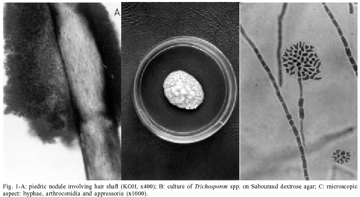
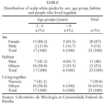
Mem Inst Oswaldo Cruz, Rio de Janeiro, Vol. 97(5) July 2002, pp. 747-750 Clinical and Mycological Study of Scalp White Piedra in the State of Paraíba, Brazil Zélia Braz Vieira da Silva Pontes/+, Adriano Lira Ramos, Edeltrudes de Oliveira Lima*, Maria de Fátima de Lacerda Guerra**, Neuza Maria Cavalcante Oliveira*, Jozemar Pereira dos Santos*** Laboratório de Micologia *Departamento de Ciências Farmacêuticas, Centro de Ciências da Saúde, Universidade Federal da Paraíba, Cidade Universitária, Castelo Branco III, 58038-910 João Pessoa, PB, Brasil **Secretaria de Saúde do Município, João Pessoa, PB, Brasil ***Departamento de Matemática e Estatística, Centro de Ciências e Tecnologia, Universidade Estadual da Paraíba, Campina Grande, PB, Brasil, +Corresponding author. Fax: + 55-83-216.7094. E-mail: pesqccs@ccs.ufpb.br Received 8 October 2001 Code Number: oc02142 White piedra is a superficial mycoses characterized by nodules on the hair shaft, caused by the basidiomycetous yeasts. In the present study, clinical and mycological findings of scalp white piedra caused by Trichosporon spp. are related. Twenty three cases of scalp white piedra were observed with a high incidence in women (87%) and preschool children from 2 to 6 (74%) years old. These groups presented a relationship of dependence with this infection. Despite the low socio-economic status, poor standards of hygiene, (48% of the patients) as well as the fact that 30.4% of the children shared the same nursery, these factors were not significant for the transmission of the mycosis. These were the first reports of scalp white piedra in João Pessoa city, Paraíba, Brazil. Key words: mycosis - scalp white piedra - Trichosporon spp. - Paraíba - Brazil Piedra is a fungal infection of the hair shaft. Two varieties of piedra are recognized: black piedra caused by Piedraia hortai, and white piedra. Classically white piedra was considered to be produced by an asexual yeast-like fungus, Trichosporon beigelii (Rippon 1974). A taxonomic revision using molecular data has shown that the genus Trichosporon Behrend consists of six human pathogenic species: T. asahii, T. mucoides, T. ovoides, T. asteroides, T. cutaneum and T. inkin and all of which belong to the class Basidiomycetes (Guého et al. 1992). These species are considered causative agents of mucosa-associated, systemic mycosis, and superficial infections, including white piedra (Guého et al. 1994). White piedra is characterized by the presence of irregular nodules along the hair shaft. The nodules are white or brown, of medium to soft consistency and their fungal elements which comprise artroconidia and/or blastoconidia can be easily detached from the hair shaft. White piedra is a cosmopolitan infection being found in the hair of the beard, moustache, genitals and axilla. Eyebrow and eyelash involvement can occur while on the scalp white piedra was described less frequently. It is described as being commonly associated with bacteria (Thérizol-Ferley et al. 1994). Direct microscopic examination of infected hair and culture enables a clear differential diagnosis to be made (Figueras & Guarro 2000). This study sought to investigate related cases of scalp white piedra among patients of both sexes and to determine whether factors such as low socio-economic status, poor standards of hygiene and people living together would have any influence upon the transmission of this mycosis. MATERIALS AND METHODS The study included 23 patients with clinical presentation of piedra examined between 1998 to 2000. All these patients were from João Pessoa, a warm and humid region in the Northeast of Brazil. The patients were treated in the Dermatology clinic of the Lauro Wanderley Hospital Universitário, Universidade Federal da Paraíba and at the Secretaria de Saúde do Município. The mycological study was carried out at the Laboratório de Micologia of the Departamento de Ciências Farmacêuticas and included direct microscopic examination of hair samples on slides with 20% potassium hydroxide (KOH) plus permanent black Quink ink (Parker) (2:1), and culture in Sabouraud dextrose agar (SDA, Difco Laboratories, Detroit) supplemented with choramphenicol (200 µg/ml). The cultures were incubated at room temperature (28-30°C) up to four weeks. Trichosporon spp. were identified according to the criteria defined by Guého et al. (1992). Statistical analyses were processed with the SPSS® (Norusis 1998) and statistical significance was determined by using Binomial Test analysis (Siegel 1975). RESULTS Mycological - Microscopic examination of hair samples presented consistent characteristics of white piedra and the nodules were compact masses made up of blastoconidia and arthroconidia (Fig. 1A). Twenty-three strains which belong to the genus Trichosporon were culture positive. Macroscopically, with incubation on SDA for 10 days, colonies types were observed (Fig. 1B). Microscopic examination of slide cultures, on 2% malt extract agar, showed major structures such as pseudo mycelium and arthroconidia mycelium which eventually became barrel shaped and clavate blastoconidia and/or appressoria (Fig. 1C). All strains produced urease (Christensen-urea medium), grew at 37°C, did not ferment sugar and the capacity to assimilate carbohydrates was variable among some strains. Association of Trichosporon spp. with bacteria was observed in 8 of the patients, but we could not ascertain whether they were coryneforms because of the difficult taxonomy of this group. Clinical - The distribution of scalp white piedra in patients according to sex, age group, habitat and people who lived together is shown in the Table. Cases of white piedra in 87% of the females and 13% in the males were observed, with ages ranging from 2 to 42 years. In the 2-6 years age group at preschool age, the infection showed the highest frequency (74%) with 88.2% of them being female. In the age group of people over 6 years old (26%) only four patients were over 20 years old. A binomial test at the level of 5% of significance showed that the females and children from 2 to 6 years old were affected significantly with white piedra (p = 0.000 and p = 0.035, respectively). Eleven (48%) of the patients lived in the same slum. They presented low socio-economic status, poor standards of hygiene and 7 of them were children who attended the same nursery. Despite of the importance of the factors previously mentioned, they were not statistically significant for the transmission of these cases of white piedra with values of p = 1.000 and p = 0.629, respectively. DISCUSSION Although never reported previously in the State of Paraíba, a high incidence of scalp white piedra was observed in João Pessoa city, capital of the State. This infection in sub-tropical and mild climates has a low incidence, being more frequently registered in regions of tropical and mild climate. In Chile (Weinstein & Alarcon 1953, Zaror & Moreno 1996), Kuwait (Selim et al. 1988), India (Kamalam & Thambiah 1981), Spain (Pereiro Miguens 1952), Israel (Gold et al. 1984) and Brazil (Godim-Gonçal-ves et al. 1991, Juang et al. 2000) there are some cases of scalp white piedra reported in the literature. The incidence of pubic white piedra (Carneiro et al. 1971, Benson et al. 1983, Torssander et al. 1985, Kalter et al. 1986, Stenderup et al. 1986, Avram et al. 1987, Fischman et al. 1989, Zaror et al. 1989, Waltzmam & Leeming 1989, Almeida et al. 1990, Palungwachira et al. 1991, Thérizol-Ferley et al. 1994) is more frequently reported than on the scalp. Most of the reports of scalp white piedra demonstrated that it occur-red among children and young adults (Kalamam & Thambiah 1981, Gold et al. 1984, Selim et al. 1988, Zaror & Moreno 1996) who were female (Kalamam & Thambiah 1981, Gold et al. 1984, Selim et al. 1988). In Rio de Janeiro, Brazil, three cases of scalp white piedra in female children at preschool age were reported (Godim-Gonçalves et al. 1991) and more recently, in São Paulo, a single case was reported which happened to be in a 49 years old woman (Juang et al. 2000). In João Pessoa, the cases of scalp white piedra reported presented a relationship of dependence between females and children (2-6 years) of preschool age. White piedra can be clinically indistinguishable from pediculosis, black piedra and trichobacteriosis including hair-shaft abnormalities such as moniletrix, trichorrhexis nodosa and trichoptilosis (Rippon 1974, Gold et al. 1984). In our study, 8 cases of scalp white piedra in association with bacteria were observed. Thérizol-Ferley et al. (1994) suggested that tricobacteriosis may play an important role in the genesis of genitopubic white piedra. They pointed out a relative high frequency (11.8%) of both manifestations observed in 449 Gabonese female patients in Africa. However, Figueras and Guarro (2000) reported that bacteria were always observed at the periphery of the nodules of white piedra, which suggests that they are not primary invaders. The mode of infection in man is not clear, but white piedra has been described in horses, monkeys, dogs and the causative agents have been isolated from soil, water and vegetal matter (Kaplan 1959, Guého et al. 1992, Sugita et al. 2000). Some authors believe that poor hygienic habits, such as bathing in stagnant water (Benson et al. 1983) may result in white piedra. However, this fact was not observed by Gold et al. (1984). Factors such as low socio-economic conditions and poor standards of hygiene were not significant in our cases of white piedra. Selim et al. (1987) suggested humidity as a predisposing factor of scalp white piedra and sexual and familiar transmission are also suggested as predisposing factors, particularly in the cases of pubic white piedra (Torssander et al. 1985, Kalter et al. 1986, Walzman & Leeming 1989, Thérizol-Ferley et al. 1994). Temperature and humidity in João Pessoa are relatively high and perhaps may have influenced the development of cases of scalp white piedra. However, the fact that 30.4% of our children shared the same nursery, was not a significant factor in the transmission of this mycosis. Trichosporon spp. was isolated from cases of scalp white piedra, thus improving our knowledge of this mycosis in João Pessoa city. ACKNOWLEDGMENTS To dermatologists and staff of the Hospital Universitário Lauro Wanderley, Dermatology Clinic, for collaboration in assisting our patients and to Dr Luís Zaror from the University of Valdívia, Chile, for providing micrographs. REFERENCES
Copyright 2002 Instituto Oswaldo Cruz - Fiocruz The following images related to this document are available:Photo images[oc02142t1.jpg] [oc02142f1.jpg] |
| |||||||||
