Mem Inst Oswaldo Cruz, Rio de
Janeiro, Vol. 97(7), October
2002, pp. 1009-1013
Detection of Antibodies
to the 97 kDa Component of Toxoplasma gondii in Samples of Human Serum
Alessandra Carla de Almeida Ribeiro,
Maria Aparecida de Souza, José Roberto Mineo+
Laboratório de Imunologia,
Instituto de Ciências Biomédicas, Universidade Federal de Uberlândia,
Av. Pará 1720, Bloco 4C, Campus Umuarama, 38400-902 Uberlândia,
MG, Brasil
+Corresponding author. Fax:
+55-34-3218.2333. E-mail: jrmineo@ufu.br|
Received 21 December 2001
Accepted 29 July 2002
Code Number: oc02226
This study was carried out to
investigate the immune response against 97 kDa (p97) molecular marker of Toxoplasma
gondii that has been characterized as a cytosolic protein and a component
of the excreted-secreted antigens from this parasite. A total of 60 serum samples
from patients were analyzed by enzime-linked immunosorbent assay and Western
blot for toxoplasmosis. These samples were organized in three groups, based
on clinical symptoms and results of serological tests. Group I: 20 samples reactive
to IgG and IgM (acute phase); group II: 20 non-reactive samples (control group);
and group III: 20 samples reactive only to IgG (chronic phase). Western blot
was performed with total antigenic extracts or with excreted and secreted antigen
from T. gondii to identify the fraction correspondent to p97. It was
observed that this cytosolic component from T. gondii stimulates the
immunologic system to produce both IgM and IgG antibodies in the beginning of
the acute infection and IgG throughout the chronic stage of the asymptomatic
toxoplasmosis.
Key words: Toxoplasma - enzime-linked
immunosorbent assay - Western blot - p97
Toxoplasma gondii is an obligate
intracellular parasite, which belongs to the phylum Apicomplexa, to the class
Sporozoea and to the genusToxoplasma. In the present classifications
it is considered part of the Sarcocystidae family. In 1908, the parasite was
discovered by Splendore in Brazil in a rabbit, and also by Nicole and Manceaux
in North Africa in a Northern African rodent called gondi (Rey 2001).
Toxoplasmosis is a zoonosis from
a variety of vertebrates including man. In humans, due to the action of the
immune system, chronic asymptomatic infection frequently occurs. Two groups
of risk, however, do exist, newborns of mothers who had toxoplasmosis during
pregnancy and immunocompromised individuals. The individuals of both these groups
frequently develop fatal toxoplasmic encephalitis (Chaves-Borges et al. 1999).
The multiplication of tachyzoites
is normally inhibited by immunologic mechanisms that favor the formation of
cysts containing bradyzoites (Soete et al.1993). Nevertheless when individuals
are immunocompromised, T. gondii produces a severe disease due to infection
reactivity. In these cases, the cysts rupture and tachyzoites multiply rapidly
(Gazzinelli et al. 1992).
Several proteins of T. gondii
have been studied regarding their biological functions such as their role in
inducing immune response by the host organism and their roles as diagnostic
reagents. It is interesting to determine which antigens are useful for diagnosis,
as well as which ones are responsible for the development of protective immunologic
mechanisms of toxoplasmosis (McLeod et al. 1991). Through molecular weight,
various proteins such as p30, p22, p23, p60, p76, p97 and others that may be
or not anchored to the membrane surface of the parasite via glycosilphosphatidilinositol
(McLeod et al. 1991) have been identified. Many studies look for molecular markers
associated with their biological role, as well as trying to relate them to the
acute or chronic phases of the disease (Makioka et al. 1991, Soete et
al. 1993).
One of the recently studied components
of the parasite (p97) has been reported to be very important for T. gondii
replication in the cell of the host. This antigen is not found on the surface
of this protozoan, but inimmersedvesicles in the cytosol. Blocking a determined
epitope of this component through monoclonal antibody 1B8 inhibits T. gondii
replicationin vitro (Mineo et al. 1994).
Matsuura and Kasper (1997) reported
the isolation of the sequence of cDNA encoding this 97-kDa protein and characterized
the biochemical nature of this molecule. The analysis of five clones reactive
to mAb 1B8, against p97, suggested that the second ATG matched closely as a
putative translation initiation site. The coding sequence encodes a peptide
with 892 amino acid residues. The peptide sequence of this molecule is composed
of 15 repeating units. They had demonstrated a homologous gene sequence in two
closely related Apicomplexa, Neospora caninum and Besnoitia jellisoni,
suggesting this protein is conserved among some species of the Sarcocystidae.
A study conducted in France highlighted
the importance of the immune response against excreted and secreted antigens
(ESA) by tachyzoites, suggesting that the use of these antigens for future diagnosis
of toxoplasmosis. Serum analysis of the acute phase of the disease demonstrated
that the sera recognizes 69 and 97 kDa bands of ESA .The 97 kDA antigen was
recognized precociously by the IgM antibody (Decoster et al. 1988).
Another study characterized antigenic
markers recognized by human serum samples from patients with acute and chronic
toxoplasmosis, by IgG avidity assays. An avidity immunoblotting assay described
by Marcolino et al. (2000), recognized that bands p16, p32, p38, p40, p43, p54,
p60, p66 and p97 were more frequently recognized by low-avidity IgG in recent
infection and by high-avidity IgG in chronic toxoplasmosis. From these antigenic
bands, p38 can be characterized as an optimal antigenic marker of low avidity
for the recent form of toxoplasmosis.
The present study aimed to detect
the presence of antibodies against 97 kDa proteins of T. gondii in serum
samples of patients from the Hospital de Clínicas, Universidade Federal
de Uberlândia, by using immuno-blotting assay with different antigenic
fractions: total antigen and ESA of T. gondii.
MATERIALS AND METHODS
Patients - The patients attended
at the Hospital de Clínicas, Universidade Federal de Uberlândia
(HC-UFU) made up the group study. Serum samples were supplied by the Clinical
Analysis Laboratory of HC-UFU and previously screened by enzyme-linked immunosorbent
assay (ELISA) for T. gondii. Sixty samples were selected and divided
into three groups of 20 samples, according to the clinical symptoms and the
results of serological tests that were also carried out at the Laboratory of
Immunology, UFU.
The groups of serum samples were
defined as follows: group I: 20 human serum samples from patients with typical
clinical symptoms of infectious mononucleosis-like syndrome, with fever, fatigue,
and enlargement of the cervical lymph nodes. The sera from these patients exhibited
specific IgM (titer ³ 64) and IgG (titer ³
256) antibodies (acute phase); group II: 20 human serum samples from individuals
serologically negative and asymptomatic healthy controls (negative controls);
group III: 20 human serum samples from asymptomatic but serologically positive
individuals for IgG antibodies only (chronic phase).
Total antigen of T. gondii - Tachyzoites
of T. gondii (RH strain) were grown intraperitoneally in Swiss mice for
48 to 72 h. The peritoneal exudates were obtained from infected mice and then
washed in sterile PBS. The concentration of the parasite was adjusted for 107
a 108 parasites/ml and these samples were solubilized in a SDS/PBS
buffer (1:1). The suspension was heated to 100oC for 3 min and aliquoted.
ESA of T. gondii - RH strain
tachyzoites were harvested from mice peritoneal exsudates two days after the
inoculation of parasites. The suspension was selected when parasite content
ranged from 107 to 108 parasites/ml, followed by centrifugation
at 1,000 g for 15 min. The sediment was resuspended in 10 ml of sterile PBS
at 4oC and centrifuged again as described above. The sediment was
resuspended in 0.5 ml of sterile PBS, incubated at 37oC for 45 min
under eventual stirrings and centrifuged at 4,000 g for 15 min. The supernatant
was filtered in 0.2 µm membrane (Sterile Acrodisc 13, Gelman Sciences,
USA) and was stored at -20oC.
ELISA for T. gondii - Polyvinylchloride
(PVC) plates were sensibilized overnight at 4oC with T. gondii
suspensions (105 tachyzoites/well) and dried at 37oC for
the IgG test and with soluble parasite antigens obtained by cryolysis for the
IgM test (5 µg of protein/well). The plates were blocked with PBS-Tween
plus 5% nonfat dry milk for 30 min and then washed with PBS-Tween 1% of block
solution. Serum samples were incubated at 37oC for 45 min and the
plates washed again (3 times for 5 min each) and incubated at 37oC
with an immunoenzymatic conjugate (human anti-IgG or human anti-IgM labeled
with peroxidase). Afterwards, the plates were washed and an immunoenzymatic
substrate (OPD solution and 30% hydrogen peroxide) was added for revelation.
Tests were read by a photometer at 492 nm (Titertek Multiskan Plus, Flow Laboratories,
Geneva, Switzerland).
SDS-PAGE of T. gondii proteins
-Electrophoresis of T. gondii extracts were conducted using12% separating
gel and 5% stacking gel at a 20 mA constant current for approximately 2 h as
described by Laemmli (1970). Samples of total antigen extract and ESA extract
were obtained according to the protocol described previously. Standard markers
of molecular weight were used in all experiments (LMV and HMW, Pharmacia, Uppsala,
Sweden).
Western blotting - The Western
blot technique described by Towbin et al. (1979) was applied to evaluate the
T. gondii surface antigens recognized by polyclonal and monoclonal antibodies
and by the serum samples of the patients. Antigenic fractions on polycrylamide
gel (SDS-PAGE) were transferred to a nitrocellulose membrane (Sigma-Aldrich
Co, St. Louis, MO, USA). After transferring the fractions to nitrocellulose,
the reactions were stopped with a protein solution (0.05% PBS - Tween + 5% nonfat
dry milk) and incubated with diluted 1:15 monoclonal antibody 1B8 (anti-p97)
(Mineo 1994) or with diluted 1:50 blood samples overnight at 4ºC under
slow horizontal agitation. Binding of antibodies was verified using immunoenzymatic
conjugate (rabbit IgG anti mouse gamma globulin, human anti-IgG or anti-IgM
bound to peroxidase-Sigma) and revealed with a diaminobenzidine (DAB) solution
containing 5 µl of 30% hydrogen peroxide per ml of PBS.
Statistical analysis - The
Chi-square test (Siegel 1975) was applied to verify if there were any significant
differences between the frequencies of the presence or the absence of antibodies
against p97 in blood samples of patients from group I and group III. Data were
compared for Western blot results to total T. gondii antigen results
as well as for Western blot results to ESA results. Differences were considered
significant when P was < 0.05.
RESULTS
ELISA - The ELISA analysis
standardized at the Laboratory of Immunology confirmed the existence of a very
close relation to those results obtained at the HC-UFU. In group I, IgM and
IgG antibodies were positive with dilutions from 1:16 to 1:4096 and from 1:64
to 1:8192, respectively. For group III, IgG antibody was positive with dilutions
from 1:256 to 1:4096.
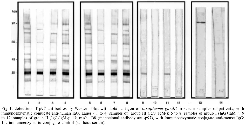
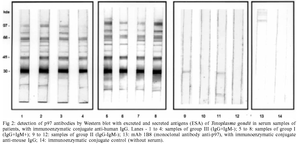
Western blot - Representative
data from Western blot with total antigen and ESA of T. gondii are demonstrated
in Figs 1 and 2.
In these assays, proteins from T. gondii recognized by human sera were
revelated with immunoenzymatic conjugate anti-human IgM and anti-human IgG-peroxidase.
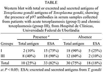
When the total antigen was used,
p97 was detected in only two samples from group I (recent phase) and eight samples
from group III (chronic phase) (Table).
However, it was recognized in these groups only by IgG antibodies. Results obtained
with Western blot and total antigens of T. gondii indicated that there
were significant differences between the frequencies relative to the presence
and absence of the band. The frequencies relative to the absence were higher
than the frequencies relative to the presence (P < 0.05).
With the use of ESA, p97 was detected
in 15 serum samples from group I and 18 serum samples from group III (Table).
Only one sample of group I identified the p97 through IgM antibodies. The other
samples identified this band only by IgG antibodies. Results obtained with Western
blot and ESA indicated that frequencies relative to the presence of antibodies
were higher than the frequencies relative to the absence of antibodies (P
< 0.05).
In group II (control group), p97
was not revealed with any antigenic extract.
DISCUSSION
It has already been reported by several
serologic studies that toxoplasmosis can be based on a classification that defines
the existence of three serologic profiles in T. gondii infection (Camargo
et al. 1978).
Profile I: the main marker is the
presence of specific IgM antibodies, however a rapid ascension of IgG antibodies
is observed (high titers detected by immunofluorescence and low titers by the
hemagglutination test); profile II: serological phase of transition in which
IgG antibodies were detected in high titers by immunofluorescence and hemagglutination
tests. IgM antibodies are absent or are found in low levels during this period
and tend to disappear; profile III: IgG antibodies are present in low titers
detected by immunofluorescence and hemagglutination tests and anti-Toxoplasma
IgM is absent.
In the present study, groups of serum
samples were established mainly based on the clinical symptoms and the results
of the ELISA assay for T. gondii. Group I (serum from IgG and IgM positive
patients) was made up of samples that could be classified as profile I or profile
II according to the titers of antibodies in each of the classes. The serum samples
in group II were negative controls of the reactions, while the samples in group
III corresponded to profile III infection by T. gondii.
The infection is generally diagnosed
by demonstration of specific antibodies to Toxoplasma in the serum samples
of infected patients. But in cases of acute toxoplasmosis, especially during
pregnancy, the serologic techniques currently used have been targeted with problems,
as persistent positive results for IgM antibodies, even one year more after
the primary infection with T. gondii (Giraldo et al. 2002).
Gross et al. (2000) studied an immunoblot
assay, which compares the early IgG profiles between the mother and her child
directed against a total cell lysate of T. gondii tachyzoites. The results
showed that this test is useful as an additional assay for the rapid diagnosis
of congenital toxoplasmosis. The use of alternative diagnostic techniques as
described in this work should be considered of great clinical relevance.
Martin et al. (1998) studied the
humoral response against Rop2, based on the detection of Toxoplasma-specific
IgG, IgM and IgA during human T. gondii infection. This antigen proved
to be a powerful tool for development of serological diagnostic systems to diagnose
either chronic or acute infections.
The IgG avidity determination is
another important serological marker that can be used to distinguish between
recent and chronic infections, allowing such a diagnosis of acute infection
to be made from a single serum sample. High-avidity index was characteristic
of the chronic phase, suggesting a progressive maturation of the affinity of
T. gondii specific IgG antibodies after the initial antigenic challenge.
Marcolino et al.(2000) observed that p97 was present among various antigenic
markers, being frequently recognized by low-avidity IgG in recent infection
and by high-avidity IgG in chronic toxoplasmosis.
In the present study, when we analyzed
human sera from acute and chronic phases of toxoplasmosis by Western blot, the
p97 antigenic fraction of T. gondii was detected more frequently by IgG
antibodies with the total antigens of T. gondii and ESA. When ESA was
used, the 97 kDa antigen was detected with high frequence and by IgG antibodies
in almost all samples. In contrast to this, another study previously showed
that the detection of IgM antibodies against an ESA 97 kDa antigen occurred
very soon after infection, and suggested the use of this antigen as a good marker
of early acute infection (Decoster et al. 1988). In the present study, however,
p97 could not be considered a good marker to distinguish acute from chronic
infections since this epitope was recognized in both phases of the infection,
when using the ESA or the total antigen extracts from T. gondii.
Taken all together, the present results
suggest that p97, an excreted and secreted component of T. gondii, stimulates
the immune system to produce both IgM and IgG antibodies in the beginning of
the infection, but with the synthesis of IgG remaining throughout the chronic
phase against this antigen. Therefore, further studies are necessary to carried
out an epitope mapping in the 97 kDa antigen in order to better select the components
that can behave as a good marker of acute toxoplasmosis.
REFERENCES

