Mem Inst Oswaldo Cruz, Rio de
Janeiro, Vol. 97(7), October
2002, pp. 1057-1061
Gregarine Cephaloidophora
communis Mawrodiadi, 1908 in the Barnacle Euraphia rhyzophorae, Oliveira,
1940 from Brazil
Dyrce Lacombe/+,
Sophie Jakowska*, Edalton Silva/++
Departamento de Entomologia, Instituto
Oswaldo Cruz-Fiocruz. Estrada da Covanca 56, 22735-200 Rio de Janeiro, RJ, Brasil
*Department of Biological Sciences, College of Staten Island, City University
of NewYork, New York, USA
+Corresponding author. Fax:
+55-21-3392.1216. E-mail: dlacombe@bol.com.br
++Post graduation student in Animal Biology
of Universidade Federal Rural do Rio de Janeiro, Seropédica, RJ, Brasil
Received 19 December 2001
Accepted 5 June 2002
Code Number: oc02233
The gregarine Cephaloidophora
communis was observed for the first time in Brazil in the barnacles Euraphia
rhyzophorae collected in Angra dos Reis, Rio de Janeiro, Brazil, between
1990 and 1996. Histological studies showed growth phases of the parasite in
specific parts of the digestive system. The intracellular forms occurred in
the vacuoles of the intestinal cells. Syzygy was frequent, and the most common
form following syzygy was cylindrical, with a single membrane. The cytoplasm
of the gregarines was always irregular, dense, and occasionally presenting a
dark stoch area.
Key words: Cephaloidophora communis
- gregarines - barnacles - histology - digestive tube - Brazil
The gregarines were arranged in the
traditional phylum Sporozoa, renamed Apicomplexa by Levine (1970). They are
parasitic protoctists, all endoparasites of invertebrate or vertebrate hosts.
They show different degrees of host specificity (Vivier & Desportes 1989).
Dusznski (in Vivier & Desportes
1989) estimates that there are about 500 species in the class Gregarinia. The
wide majority of gregarines are monoxenous, but some species may be heteroxenous
because their life cycles are still unknown.
Gregarines in the intestine of barnacles
were first reported by Kölliker (1848), and described in the intestine
of Balanus pallidus as Gregarina balani.
Mawrodiadi (1908) demonstrated C.
communis in B. eburneus, B. amphitrite, B. crenatus and Megabalanus
tintinnabulum.
Tregouboff (1912) and Kamm (1922)
pointed out that the members of the family Cephaloidophoridae are parasites
of crustaceans and develop within the cells, producing one or more oval spores.
Ball (1973) and Henry (1938) also studied gregarines in barnacles. Tuzet and
Ormières (1964) contributed to their morphology and Barnes (1953) to
the developmental physiology of barnacle larvae and their gregarines. Reger
(1966) and Reger et al. (1967) studied the spores of C. communis in the
cells of the intestine of M. tintinnabulum using electron microscopy.
Arvy and Nigrelli (1969), reported
the gregarines in intestine of B. nubilis and B. eburneus
collected on Coney Island, New York, near the Osborn Laboratory of Marine Science.
Gregarines in barnacles have been
studied by various authors in the United States of America, England, France
and Germany. In Brazil there has been relatively little interest in these sessile
crustaceans. Since, this is the first report of gregarine in the intestine of
the barnacle Euraphia rhyzophorae, Oliveira (1940) collected on the shore
in Angra dos Reis, near the city of Rio de Janeiro.
MATERIALS AND METHODS
Approximately 150 adult barnacles
of E. rhyzophorae were collected from the roots of Rhyzophora mangle
on the shore in Angra dos Reis, State of Rio de Janeiro, Brazil, between 1990
and 1996.
The barnacles, with or without substrate,
were immediately immersed in a fixative such as Bouin's (according to Duboscq-Brasil)
or Heidenhein's original of Susa.
In the laboratory the specimens were
separated for anatomical, microanatomical and histological studies. Dehydrated
in alcohol-benzol series, they were embedded in "histoseck" paraffin.
Serial sections cut at 5 and 7 µm were stained with Gallocyanin, Chromotrop
2R, Heidenhain Iron Hematoxylin and Mallory Hematoxylin, following the methods
presented by Pinto (1919), Romein (1928), Henry (1938), Pantin (1948), Barth
(1953) and Pearse (1960).
RESULTS AND DISCUSSION
C. communis gregarines were
observed in the mid and hind gut of the barnacles E. rhyzophorae (Fig.
1, GR, INT). This species of gregarine was described by Mawrodiadi (1908)
from other species of barnacles: B. improvisus, B. amphitrite,
B. eburneus and M. tintinnabulum, in which oval spores 50 to 60
µm in size were observed inside. In gregarines of the family Cephaloiphoridae
these organisms can be seen associated during their growth in groups of usually
two individuals, or may develop singly (Fig.
1), as reported by Vivier and Desportes (1989).
The gregarines in E. rhyzophorae
were always numerous in the digestive tube and exhibited different growth phases.
Although almost all organs of the host may be infected by apicomplexan zoites
or other forms, there is always a motile stage that may penetrate the target
organs, where the development may be completed (Vivier & Desportes 1989).
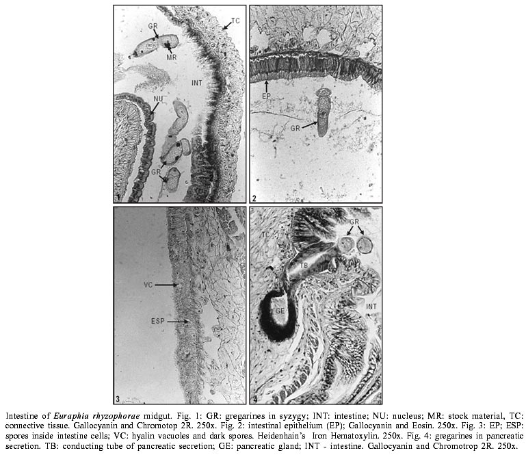
Fig.
1 shows deuteromites (GR) containing dark round bodies (MR) of stock material
in the lumen of the midgut (INT). NU identifies the nucleus of one of the epithelial
cells.
The forms seen next to the epithelial
cells (Fig. 2 EP) are limited to
the plasma membrane that lines the intestinal lumen. The epithelial wall of
the intestine in barnacles has extended narrow cells. Their cytoplasm is dense,
with a central nucleus, round and rich in chromatin. Within the cytoplasm clear
vacuoles are seen (Fig. 3, VC).
These contain one or more sporocysts measuring about 40 µm which stand
out as dark bodies in the hyaline vacuoles (ESP). They emerge into the lumen
throughout the entire intestine.
These gregarines of rounded forms
(Fig. 4, GR) emerge from the duct
(TB) of the pancreatic gland (GE). This picture shows where the duct of the
pancreatic gland comes in contact with the epithelium of the intestine and two
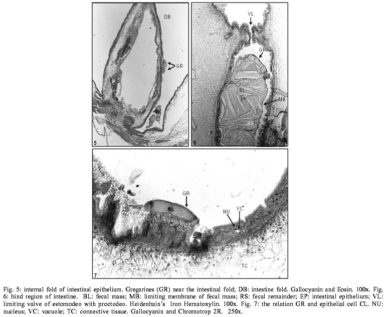
spherical gregarines. Some gregarines (Fig.
5, GR) are seen in the intestinal folds (DB). Others appear to be passing
from the midgut into the proctodeum, remaining in the posterior intestine close
to the fecal mass (Fig. 6, BL).
In this longitudinal section one sees the sphincter (VL) that separates the
midgut from the posterior intestine.
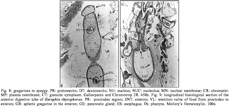
Fig.
7 shows a giant gregarine (GR), about 150 µm long, close to a row of
epithelial cells. In the mid part of the deutomerite (Fig.
8, DT) there is a disctinct nucleus (NU), spherical, poor in chromatin (CR)
but with an accentuated nuclear membrane. A dense nucleolus (NUC) is present.
The cytoplasm is granular (CT), almost alveolar but it may also show dark spots,
as in Fig. 1 (MR).
Some gregarines present matching
posterior parts, familiar figures of syzygy, as can be seen in Figs
2 and 8.
The gut parts of E. rhyzophorae,
can be observed in Fig. 9, where
within the enteron (ENT) a round gregarine (GR), the esophagean valve (VL),
a part of the esophagus (ES) and the pharynx (FA), which constitutes the foregut
are detected; the pancreatic gland (GE) is always lateral to the intestinal
wall.
C. communis found in the
barnacles E. rhyzophorae in Brazil may be another giant gregarine like
Porospora giganteal and Didymophyes gigantea that parasitizes
coleopterans larva (Grassé 1953). In barnacles there was no evidence
of cellular damage associated with the presence of these gregarines.
ACKNOWLEDGMENT
To Genilton Vieira and collaborators
of the Laboratory of Production and Handling of Images of the Instituto Oswaldo
Cruz-Fiocruz, for assistance in figures reproduction.
REFERENCES

