 |
| About Bioline | All Journals | Testimonials | Membership | News |
|
||||||
|
||||||
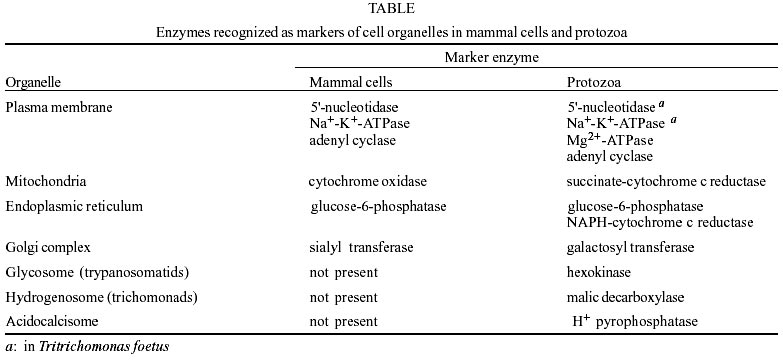
Mem Inst Oswaldo Cruz, Rio de Janeiro, Vol. 98, No. 2, March, 2003, pp. 151-170 Cell Fractionation of Parasitic Protozoa - A Review Wanderley de Souza+, Narcisa Leal da Cunha-e-Silva
Laboratório
de Ultraestrutura Celular Hertha Meyer, Instituto de Biofísica Carlos Chagas
Filho, Universidade Federal do Rio de Janeiro, CCS, Bloco G, Ilha do Fundão,
21941-900 Rio de Janeiro, RJ, Brasil. Received 30 October
2002 Code Number: oc03050 Cell fractionation, a methodological strategy for obtaining purified organelle preparations, has been applied successfully to parasitic protozoa by a number of investigators. Here we present and discuss the work of several groups that have obtained highly purified subcellular fractions from trypanosomatids, Apicomplexa and trichomonads, and whose work have added substantially to our knowledge of the cell biology of these parasites.
Key words: parasitic protozoa - cell fractionation - trypanosomatids - Apicomplexa - trichomonads
The protozoa comprise a large number of species, including some agents of human and animal diseases such as malaria, leishmaniasis, Chagas disease, African trypanosomiasis, amebiasis, giardiasis, toxoplasmosis, coccidiosis, theileriosis, and babesiosis, to mention only some of the more important examples. These protozoa are also of interest from a cell biological point of view since they contain unique cytoplasmic structures and organelles that have been the subject of investigation in some detail for many years. These studies have provided new information of general biological interest since some investigators assume that some of the protozoa form the roots of the evolutionary tree of eukaryotic cells. THE CELL FRACTIONATION TECHNIQUE
To disrupt tissue cells, separate their components by differential centrifugation and analyze the distribution of known enzymes among the different fractions obtained has been a powerful tool for exploring cell structure and composition. Transmission electron microscopy of thin sections has provided detailed views of cell structure. However, to address many questions concerning the function of subcellular organelles it has proven necessary to gently disrupt the cell and isolate its components. This has been accomplished by differential centrifugation, a methodology developed in the 1940s and 1950s mainly by the cell biology group coordinated by Albert Claude, Christian de Duve and George Palade at the Rockefeller University. Their reviews should be consulted for descriptions of the early studies (De Duve 1971, 1975, Claude 1975, Palade 1975 De Duve & Beaufay 1981). Their work established a baseline of important concepts: (a) polydispersity, which allowed the rules of sedimentation coefficients (Svedberg & Pedersen 1940), as defined for macromolecules, to be adapted for subcellular particle centrifugation; (b) biochemical homogeneity and unique localization, which resulted in the definition of marker enzymes; and (c) latency, which distinguished among enzymes confined in different membrane-bound compartments by the differences in time of release after membrane lyses (De Duve 1967). Subsequently, the knowledge obtained by analytical cell fractionation has been used to design preparative cell fractionation schemes aimed at isolating and purifying specific organelles for further studies. The cell fractionation process usually starts with disruption of the plasma membrane, using conditions that minimize damage to the membranes bounding intracellular organelles. Disruption can be achieved by procedures such as exposure to high-frequency sound (sonication), treatment with a high-speed blender, grinding in a mechanical homogenizer, nitrogen cavitation, etc. Usually, it is necessary to monitor the process by phase contrast microscopy in order to avoid rupture of the organelles or disorganization of cytoplasmic structures. Once the cells are broken open, the suspension can be separated into its main components using a series of centrifugations at increasing speeds. This causes cell components to move toward the bottom of the centrifuge tube, forming a pellet at a rate that depends on their size and density. The supernatant is then collected and subjected to further centrifugation at higher speed, and the process may be repeated several times depending on the cell type and the structures to be isolated. Usually differential centrifugation does not yield very pure material. Therefore, it is necessary to continue the isolation procedure by using density-gradient centrifugation, a procedure where the organelles and structures are separated by sedimentation through a gradient of a dense solution such as sucrose, Metrizamide, Percoll, etc. In this process, the fraction first obtained by differential centrifugation is layered on the top of a gradient composed of a non-ionic substance. Alternatively, sample can be positioned under the gradient solution or mixed with it, in the case of self-forming gradients. The tube is centrifuged at high speed for a extended period, allowing each structure to migrate to an equilibrium position where the density of the structure is equal to that of the surrounding liquid. Therefore, in this process separation is based on the buoyant density rather than on the size and shape of the particles. Following centrifugation, the fractions can be collected individually for further analysis and determination of the efficiency of separation. An important requirement for successful cell fractionation is the evaluation of the isolation procedure, usually accomplished using morphological and/or biochemical methods. The morphological approach applies light and electron microscopy of thin section to the various fractions obtained. The biochemical approach is based on the determination of the enrichment of enzymes or other molecules (a special type of lipid, an antigen, etc.) that can be considered as a marker for a given structure or organelle. To be considered a marker, an enzyme must be known as a component of the organelle in all cells studied (biochemical homogeneity) and absent from the rest of the cell (unique localization). These notions resulted from analytical cell fractionation studies (De Duve 1975) and defined a number of classical enzyme markers (Table). Based on experience accumulated over many years, we strongly recommend the simultaneous use of both approaches. There are relatively few reports on the isolation of structures and organelles from parasitic protozoa due to several factors, including difficulty in obtaining enough cells to start the fractionation procedure, and difficulty in breaking open the cells under conditions in which the structures are well preserved. This is especially important for those protozoa in which a layer of subpellicular microtubules renders the cells more resistant to rupture, as in the case of trypanosomatids and Apicomplexa. Finally, there is the absence of well-defined markers to characterize the isolated structure. Nevertheless, the discovery of two special organelles from protozoa came from analytical cell fractionation experiments: the glycosome of trypanosomatids (Opperdoes & Borst 1977) and the hydrogenosome of trichomonads (Lindmark & Muller 1973). The present review will describe attempts made to obtain structures and organelles from protozoa belonging to the Trypanosomatidae and Trichomonadidae families and the Apicomplexa phylum. They were selected because they are good examples of the use of the cell fractionation technique to obtain highly purified and well characterized fractions, which were further characterized using morphological and biochemical approaches. CELL FRACTIONATION OF TRYPANOSOMATIDS
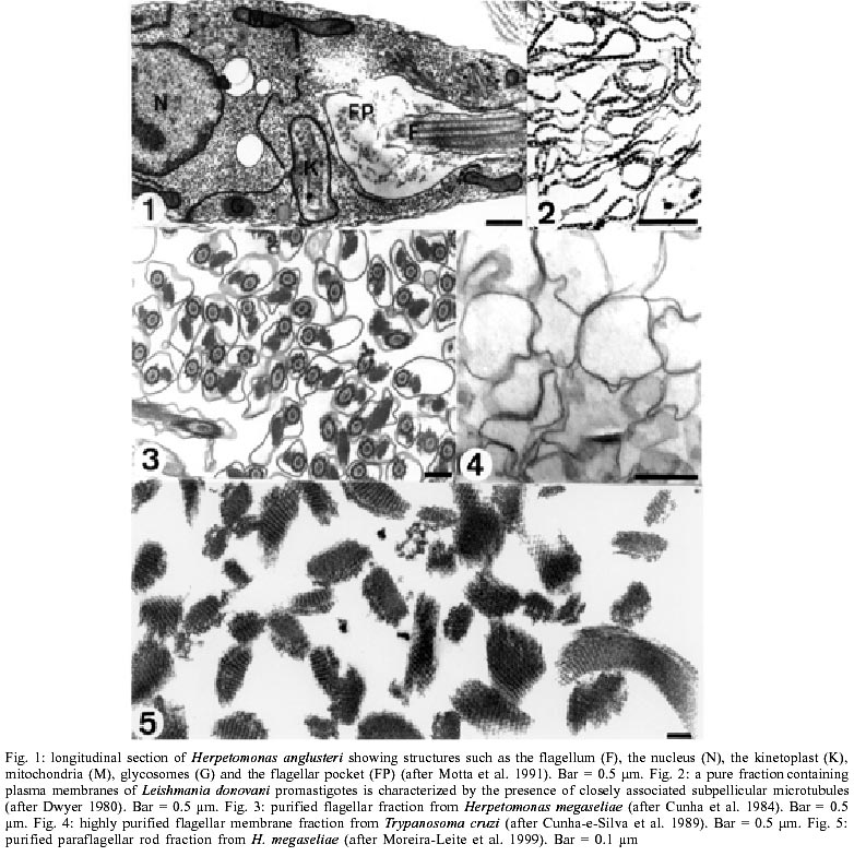
Fig. 1 shows a general view of a thin section of a trypanosomatid examined by transmission electron microscopy. The protozoan is surrounded by a typical plasma membrane, which envelops the cell body, invaginates at the anterior end of the cell forming the flagellar pocket, and continues over the flagellum. Therefore, we can distinguish at least three main domains of the plasma membrane: those surrounding the cell body, the flagellum, and the lining of the flagellar pocket. Below the plasma membrane, except in the region of the flagellar pocket, there is a layer of microtubules, known as subpellicular microtubules, which are in contact with each other, with the plasma membrane and with profiles of the endoplasmic reticulum through filamentous structures. The flagellum emerges from the basal body located in the anterior region of the cell and projects forward. It displays a typical flagellar structure with a 9+2 pattern of microtubule doublets. In addition, it exhibits a highly elaborated network of filamentous structures connected to the axoneme through small bridges forming a structure known as the paraflagellar (or paraxial) structure (or rod). The nucleus of the protozoan is centrally located. The cytoplasm contains randomly distributed ribosomes and profiles of endoplasmic reticulum. The Golgi complex is located in the anterior region, close to the flagellar pocket. Some organelles are specific for the trypanosomatids. The first one is the kinetoplast, which corresponds to a condensation of extranuclear DNA that lies within the unique ramified mitochondrion, and is localized in front of the basal body that gives rise to the flagellum. The second characteristic organelle, designated the glycosome, is usually round, is enveloped by a unit membrane, and contains a matrix denser than the cytoplasm. Most of the enzymes of the glycolytic pathway are localized in this organelle. Since in some species the glycosome contains catalase and enzymes involved in the b-oxidation of lipids, it corresponds to a peroxisome. The third characteristic organelle consists of several distinctive vesicles, which usually display an empty interior and an electron-dense material at their periphery. This organelle, designated acido-calcisome, has been the subject of intense investigation in recent years and is involved in the regulation of the cytoplasmic concentration of calcium, with the participation of several ATPases. Another important organelle is the reservosome, which is found in epimastigote forms of trypanosomatids of the genus Trypanosoma, of the sub-genus Schyzotrypanum, and is involved in the accumulation of macromolecules ingested through the endocytic pathway.
Isolation of the plasma membrane Attempts have been made to obtain a pure plasma membrane fraction from trypanosomatids. This is not an easy task, as the continuous layer of microtubules below the plasma membrane makes trypanosomatids resistant to conventional methods for cell breakage. Hunt and Ellar (1974) disrupted Leptomonas collosoma by blending the cell suspension with glass beads and were able to isolate a plasma membrane fraction. They found that the subpellicular microtubules remained attached to the plasma membrane, and could be used as a morphological criterion in assessing the purity of the fraction. This association of isolated membranes with microtubules has been confirmed for L. collosoma (Linder & Staehelin 1977), Trypanosoma cruzi (Meyer & De Souza 1976), Trypanosoma brucei (Voorheis et al. 1979), Leishmania mexicana (Timm et al. 1980), Leishmania donovani (Dwyer 1980), and Trypanosoma lewisi (Dywer & D'Alessandro 1980). In some cases, the microtubules did not remain associated with the plasma membrane (Pereira et al. 1978, Franco da Silveira et al. 1979, Zingales et al. 1979, Rovis & Baekkeskov 1980). In these cases the only way to assess the purity of the fraction is to use the membrane enzyme markers or the attachment of microtubules to the isolated membranes. Since different methods have been used to rupture the cells, we cannot exclude the possibility that, in some cases, the attachment of microtubules to the membrane depends on differences in the methods used. But even when the same method was used for the isolation of the membrane, it gave different results in T. cruzi (Pereira et al. 1978) and in L. mexicana (Timm et al. 1980). Three procedures have been used to obtain a purified plasma membrane fraction from epimastigotes of T. cruzi. Cells previously swollen by treatment with Triton X100 were disrupted using a Douncetype homogenizer. The membrane fraction was obtained through successive steps of differential centrifugation and its purity was evaluated by electron microscopy and by testing for enzyme markers. Activities of 5'nucleotidase and Na+K+ATPase, the two classical markers of the plasma membrane in mammals, were not found in T. cruzi. Acid phosphatase and succinate dehydrogenase were absent from the purified membrane fraction. The specific activities of Mg2+dependent ATPase and adenyl cyclase in the membrane fraction were two and five times higher, respectively, than in the whole homogenate. Electronmicroscopy showed slight contamination with ribosomes (Timm et al. 1980). In the second approach, cells were ruptured by sonication and the membrane fraction were isolated by differential centrifugation, followed by equilibrium centrifugation on sucrose gradients. The fractions were characterized by electron microscopy, enzyme markers (adenyl cyclase, succinate dehydrogenase, cytochrome c reductase, acid phosphatase, and glucose6phosphatase), and by distribution of surface-bound 131I activity in cells previously labelled using lactoperoxidase. A 10fold increase in the adenyl cyclase activity was obtained with this method. Na+K+ATPase and 5' nucleotidase activities were not found (Franco da Silveira & Colli 1981). In a third report, plasma membrane was induced to vesiculate by incubating the cells with aldehydes, N-ethylmaleimide, pchloromercuribenzoate, or acid buffers, followed by isolation of the vesicles by sucrosedensity centrifugation (Franco da Silveira et al. 1979). The fractions were characterized by distribution of 131I activity as in the second approach described above, and by electron microscopy. Unfortunately, the authors did not assay enzyme markers. Examination of the electron micrographs of T. cruzi treated with vesiculating agents showed that vesicles form only at certain points of the membrane which encloses the cell body and the flagellum. Therefore, the membrane fraction isolated by this procedure certainly does not contain all areas of the plasma membrane. The method is interesting as it shows that we can examine specific regions of the cell surface of T. cruzi that, for unknown reasons, are more likely to form vesicles. A 30fold to 50fold enrichment of some proteins was detected in the membrane fraction isolated from the vesicles (Franco da Silveira et al. 1979). These studies indicate that the plasma membrane of epimastigotes of T. cruzi does not have two of the enzymes that are typical of the plasma membrane of eukaryotic cells: Na+K+ATPase and 5'nucleotidase. It seems that adenyl cyclase is the best marker to determine the purity of a plasma membrane fraction of T. cruzi. In T. brucei, Na+K+ATPase was found and was used to establish the purity of the isolated plasma membrane fraction (Rovis & Baekkeskov 1980, Mancini et al. 1982). Three procedures have been used to obtain a plasma membrane fraction from T. brucei. First, cells were disrupted using nitrogen cavitation followed by differential centrifugation and successive centrifugations in gradients of dextran and sucrose (Rovis & Baekkeskov 1980). The fraction obtained was characterized by electron microscopy and by using enzyme markers (Na+K+ATPase and adenyl cyclase) and polyacrylamide gel electrophoresis. In the second approach, cells were broken by sonication, followed by differential centrifugation and centrifugation in a sucrose gradient (Mancini et al. 1982). The fraction obtained was characterized by electron microscopy, assay of enzyme markers (adenyl cyclase and Na+K+ATPase), analysis of iodinatable surface proteins, and the presence of the variable surface coat glycoprotein. Finally, cells were disrupted using a Dounce homogenizer followed by centrifugation in a sucrose gradient. The fraction obtained was characterized by electron microscopy and assay of the enzyme markers Na+K+ATPase and adenyl cyclase (Voorheis et al. 1979). L. donovani cells, previously treated with dilute ethanol in a hypotonic buffer, were disrupted on ice in a Dounce homogenizer and the membrane fraction was purified by centrifugation in a sucrose gradient. Characterized by electron microscopy, the fraction exhibited plasma membrane profiles with associated subpellicular microtubules (Fig. 2), which served as natural morphological markers to distinguish the plasma membrane from internal membranes profiles (Dwyer 1980). The freezefracture technique (reviewed in De Souza 1995) shows that the membrane that covers the various regions of the trypanosomatids (cell body, flagellar pocket and flagellum) is not homogeneous. Therefore, it would be of interest to obtain membrane fractions from each of the different regions. Some attempts have been made to isolate the flagellar membrane. On treating an isolated flagellar fraction of T. cruzi with Triton X100, Piras and co-workers (1981) obtained a soluble flagellar membrane preparation, but did not characterize it in detail. Some years later, Cunha-e-Silva and co-workers (1989) isolated a highly purified flagellar membrane fraction from two different species of trypanosomatids. By treating a flagellar fraction devoid of cell bodies with Triton X100 we separated the flagellar membrane by centrifugation in a sucrose gradient (Fig. 4; see the following section). Reviewing the work on the isolation of the plasma membrane of trypanosomatids has led to the important conclusion that the bridges of a protein nature connecting the inner portion of the plasma membrane to the subpellicular microtubules showed by Souto-Padrón and co-workers (1984) in quick-frozen and deep-etched replicas of T. cruzi are very stable structures.
Isolation of the flagellum The flagellum is responsible for the motility of trypanosomatids and participates in their interaction with the hosts, by adhering to the insect digestive tract (Vickerman & Tetley 1990), or initiating the contact with mammalian cells (De Souza 1984). It arises from a basal body at the bottom of a plasma membrane invagination, the flagellar pocket. The intraflagellar cytoskeleton comprises a canonical type 9+2 axoneme and a unique paraflagellar or paraxial structure, which is present in all trypanosomatids except for those that bear an endosymbiont (Freymuller & Camargo 1981). The initial attempts to isolate the flagellum of trypanosomatids were published in 1977. Segura and co-workers (1977) disrupted T. cruzi epimastigotes using cavitation. The flagellum-enriched fraction obtained by differential centrifugation was contaminated with membranes of flagellar and non-flagellar origins, due to the cell rupture method. The authors performed double diffusion tests against an antibody raised to the entire parasite and found five precipitin lines against the flagellar fraction. Flagella also showed good results (80 to 90%) in protective studies against a lethal challenge with trypomastigotes. The use of adjuvants raised this protection to 100% (Ruiz et al. 1986). It is noteworthy that the flagellar was the only subcellular fraction that did not cause cardiac lesions when inoculated into mice (Ruiz et al. 1986). In the same year, Pereira and co-workers (1977) designed another method for isolating flagella based on separating the flagellum from the undisrupted cell body. They treated Crithidia fasciculata, Herpetomonas samuelpessoai and Leishmania tarentolae with Lubrol-PX, a non-ionic detergent, in the presence of magnesium chloride, before deflagellation with a Dounce homogenizer. Flagellar fractions thus obtained were very pure, but consisted of demembranated flagella as a result of the detergent. Although devoid of membranes, isolated flagella from H. samuelpessoai were very effective in protecting mice against T. cruzi, indicating that highly conserved antigens were present inside the flagella. One year later, Pereira and co-workers (1978) obtained a similar flagellar fraction from T. cruzi epimastigotes. This fraction was also protective. In 1981, Piras and co-workers developed a protocol for the isolation of the flagellum of T. cruzi epimastigotes, based on sonication of the parasites in the presence of bovine serum albumin and calcium chloride to protect the cell body from rupture, followed by differential and sucrose gradient centrifugation. They obtained a highly purified and well-preserved flagellar fraction. By treating the flagellar fraction with Triton X-100, they also isolated an axoneme (plus paraxial rod) fraction and a soluble flagellar membrane fraction. Both flagellar cytoskeleton and flagellar membranes gave precipitin lines in double-diffusion tests against chagasic patients' sera. We have adapted and improved Piras' method to purify flagella from H. megaseliae (Cunha et al. 1984). This monoxenic trypanosomatid possesses a very big paraxial rod. The flagellar fraction from H. megaseliae was highly purified and we demonstrated by electron microscopy that the flagella were well preserved and had intact membranes (Fig. 3). In the same study, we obtained purified flagella from Crithidia deanei, a trypanosomatid that does not possess the paraxial structure. By comparing the electrophoretic profiles of purified flagella from both parasites, we could identify which protein were the major components of the paraxial structure of H. megaseliae. Since the first protection studies with flagellar fractions (see above), it was clear that an internal component of flagella (even from monogenetic trypanosomatid species) could confer a high degree of protection against lethal challenges with T. cruzi trypomastigotes (Segura et al. 1974, 1977, Pereira et al. 1977, Ruiz et al. 1985). Considering that the components of the axoneme are highly conserved in eukaryotes, the paraflagellar structure had the potential to be used as an immunogen for vaccination, with the advantage that it is different from any cytoskeletal structure of the mammalian host cell. Pursuing this lead, a research group developed immunization protocols using purified major paraxial proteins with excellent results (Segura et al. 1974, Wrightsman et al. 1995, Miller et al. 1996, 1997). The paraxial structure is an intricate lattice of protein filaments that parallels the axoneme for almost all its length and is connected to its microtubule doublets 4 to 7 (Fuge 1969, De Souza & Souto-Padrón 1980). Three portions form the lattice: one proximal to the axoneme, one distal and one intermediary portion connecting the other two. Layers of thin (7 nm) and thick (25 nm) filaments crossing at an angle of 100o form the basic organization of the proximal (two layers) and distal (eleven layers) portions. Alternating Y- and I-shaped thin filaments form the intermediary portion (Farina et al. 1986). The biochemical composition of the paraxial structure was analyzed by different approaches and shown to contain two major proteins in all trypanosomatids: a slowly migrating protein, PFR1, ranging from 70 to 78 kDa and a faster one, PFR2, ranging from 68 to 73 kDa (reviewed in Bastin et al. 1996). It was recently demonstrated that at least in T. cruzi there are four major proteins, two of 70 kDa - PAR1 and PAR 3 - and two of 68 kDa - PAR2 and PAR4 (Fouts et al. 1998). There is no correlation between paraxial major proteins and paraxial filaments. Furthermore, it is evident that such an intricate structure must have minor components. For T. brucei, those minor components were suggested to be spectrin-type proteins (Schneider et al. 1988, Woods et al. 1989, Müller et al. 1992, Woodward et al. 1994, Imboden et al. 1995). The best way of investigating the paraflagellar rod composition was to obtain a subcellular fraction containing purified and intact paraxial rods. We decided to try to improve a protocol described by Russell and co-workers (1983), who used trypsin to disrupt the connections between paraxial rod and axoneme. Purified and detergent-demembranated flagella from H. megaseliae were treated with trypsin in a very careful manner and applied on top of a 1.8-2.2 M continuous sucrose gradient. After centrifugation, six fractions of equal volume were collected from top to bottom. The third fraction was a highly purified paraflagellar rod fraction (Fig. 5) (Moreira-Leite et al. 1999). Using tubulin as an internal marker it was demonstrated by SDS-PAGE and Western blots that the fraction was devoid of axonemes. Fractions from higher sucrose densities were progressively richer in axonemes. The purified paraflagellar fraction allowed us to identify minor components in the range of 120 to 190 kDa. The flagellar membrane is also of great interest, as many trypanosomatids adhere to their insect hosts by the flagellum (Steiger 1973, Vickerman 1973, reviewed in De Souza 1989) and depend on this adhesion to survive and differentiate. Ultrastructural studies have demonstrated some interesting features concerning the flagellar membrane: freeze-fracture replicas of L. samueli, H. megaseliae and T. cruzi, (Souto-Padrón et al. 1980, 1984) showed that the membrane that surrounds the flagellum has far fewer intramembranous particles than the membrane enclosing the cell body of the same cell, suggesting a low protein/lipid ratio. Special particle arrays are present at the membrane of the flagellar base and flagellum-cell body attachment region (Linder & Staehelin 1977, De Souza et al. 1978b, 1979). Although poor in protein, flagellar membrane is immunogenic, since it is recognized by chagasic serum (see above, Piras et al. 1981). We managed to get a highly purified flagellar membrane fraction from H. megaseliae and T. cruzi epimastigotes (Fig. 4) (Cunha-e-Silva et al. 1989) by combining detergent treatment on ice, vortexing and separation on a sucrose equilibrium gradient. Analyzing the purified fraction by using filipin cytochemistry for sterols combined with freeze-fracture, we showed that the flagellar membrane is almost devoid of intramembranous particles and rich in sterols. Electrophoretic analysis showed few bands, some of which bound Concanavalin A after being transferred to nitrocellulose. The identification of these protein bands as receptors or adhesins still awaits further investigation.
Isolation of the mitochondrion-kinetoplast complex Trypanosomatids possess only one ramified mitochondrion, which spans the parasite's entire body. In the portion of the mitochondrion localized below the basal body, where the flagellum arises, there is a special concentration of DNA molecules that form a slightly concave disc-like structure, the kinetoplast. Kinetoplast DNA is composed of about 25,000 concatenated minicircles and about 30 maxicircles in a single network per cell and represents up to 25% of the total DNA. It would be important to obtain a purified subcellular fraction containing the mitochondrion-kinetoplast complex, as it would allow studies concerning the molecular characteristics of kinetoplast DNA and its processing, and a better understanding of parasite metabolism, including the balance between aerobic and anaerobic metabolism in bloodstream and procyclic forms of T. brucei. Hill and co-workers (1968) obtained the first mitochondrion-kinetoplast enriched fraction from C. fasciculata and characterized the cytochromes from this protozoan. Braly and co-workers (1974) isolated the mitochondrion-kinetoplast complex from L. tarentolae, obtaining a purified fraction, but the mitochondrion was fragmented. Nichols and Cross (1977) used digitonin under hypotonic conditions to disrupt C. fasciculata, and purified the mitochondrion-kinetoplast complex using differential centrifugation and density gradients, analyzing its biochemical properties and RNA content. Another mitochondrial fraction from C. fasciculata was prepared at the same time by Edwards and Lloyd (1977) using rate zonal centrifugation. By evaluating the activity of the enzymes NADH-cytochrome c oxidoreductase and p-nitrophenyl phosphatase in each fraction, they demonstrated the separation of mitochondria from lysosomal vacuoles. Several years later, Opperdoes and co-workers (1981) established a cell fractionation method for procyclic forms of T. brucei. They obtained a glycosome fraction equilibrating at 1.23 g/cm3 and a mitochondrial fraction equilibrating at 1.16 to 1.18 g/cm3. They showed the presence in the latter of succinate dehydrogenase and oligomycin-sensitive ATPase activity as well as L-threonine 3-dehydrogenase and carnitine acetyl transferase. The latter two enzymes, involved in threonine metabolism, were 15-fold higher in procyclic mitochondrial fraction than in that of bloodstream forms. Cell disruption using silicon carbide and grinding in a mortar was applied to T. cruzi, followed by isopycnic centrifugation, in order to determine the subcellular localization of phosphoenolpyruvate carboxykinase and malic enzyme I (Cannata et al. 1982). They were found in two different compartments, glycosomes and mitochondrion, respectively. Subcellular fractions containing the mitochondrion-kinetoplast complex have been used as starting material to purify macromolecules whose origin can be ascribed undoubtedly to the complex. Schneider and co-workers (1994) demonstrated that T. brucei mitochondrial tRNAs are encoded in the nucleus and imported into the mitochondrion, where they are spliced. The authors modified tRNA genes by site-directed mutagenesis and found an accumulation of unspliced mutant tRNA in the mitochondrial fraction, thereby demonstrating that the tRNA had been imported. Using cell fractionation and immunofluorescence microscopy, Sullivan and co-workers (1994) localized a heat-shock protein of 60 kDa (hsp60) that acts on mitochondrial protein folding. Unlike hsp70, which is associated with the kinetoplast, hsp60 is distributed all over the mitochondrial matrix. Recently, Heise and Opperdoes (2000) used a cell fractionation, isopycnic centrifugation and digitonin-titration protocol to purify the enzyme 3-hydroxy-3-methylglutaryl-coenzyme A reductase from procyclic T. brucei, and demonstrated that it is localized in the mitochondrion. This is the first description of an enzyme from the ergosterol synthesis pathway of T. brucei. The authors suggested that the mitochondrial localization of this enzyme, which is resident in the endoplasmic reticulum in mammalian cells (Lecureux & Wattenberg 1994), is due to the use of 2-carbon molecules from the metabolism of threonine and/or leucine as a source of acetyl groups for incorporation into sterols.
Isolation of the Golgi complex The Golgi complex of trypanosomatids is formed by 4 to 10 stacked cisternae localized in the anterior region of the cell, close to the flagellar pocket. Biochemical studies have shown that in members of the Trypanosomatidae family the pathway of N-linked oligosaccharide processing of protein is similar to that found in other eukaryotic cells (Parodi et al. 1981, 1989, Parodi 1993). However, a novel series of unique O-linked N-acetylglucosamine-containing oligosaccharides have been found in si-aloglycoproteins (Previato et al. 1994, 1995). It has been assumed, based on the results obtained with mammalian cells, that the Golgi complex of trypanosomatids is involved in the glycosylation process. Indeed, a few cytochemical studies in which thin sections of Lowicryl-embedded trypanosomatids were incubated with gold-labeled lectins showed labeling of the Golgi complex. Two attempts have been made to purify the Golgi complex of trypanosomatids. The first one was carried out with African trypanosomes (Grab et al. 1984). Transmission electron microscopy showed that the fraction consisted predominantly of smooth surface vesicles and flattened cisternae rather than stacked Golgi cisternae. The second attempt was carried out on epimastigote forms of T. cruzi (Morgardo-Díaz et al. 2001). The parasites were suspended in a hypotonic solution containing protease inhibitors and then disrupted by controlled sonication followed by centrifugation at 95,000x g for 90 min in a discontinuous sucrose density gradient (1.2, 1 and 0.8 M sucrose). Transmission electron microscopy of a band recovered at a position corresponding to the 1-1.2 M sucrose interfaces was highly enriched in stacked cisternae and vesicles. The fraction was characterized biochemically as significantly enriched in galactosyl transferase, O-a-GlcNAc transferase and acid phosphatase (Morgado-Díaz et al. 2001). A protein of 58 kDa that has been shown to be involved in the association of the Golgi complex with microtubules in mammalian cells was found by immunofluorescence microscopy and Western blotting in the Golgi complex of T. cruzi (Morgado-Díaz et al. 2001).
Isolation of the glycosome (peroxisome) Membrane-bound cytoplasmic structures that resemble those initially designated as microbodies and later on as peroxisomes in mammalian cells, have been described in trypanosomatids since the initial studies on their fine structure (Vickerman & Preston 1976, reviewed in De Souza 1984). The peroxisomes usually appear as spherical organelles with a diameter of about 0.7 µm and are randomly distributed throughout the cell. In some cells, as in Leptomonas samueli, they appear as elongated structures that can reach a length of 2.8 µm (Souto-Padrón & De Souza 1982). In some Phytomonas species, the peroxisomes may be packed arranged and even associated with cytoskeletal structures (Attias & De Souza 1985). In trypanosomatids that harbour an endosymbiont, the peroxisomes concentrate around the symbiont (Motta et al. 1997). Usually the glycosome contains a homogeneous and slightly dense matrix, although a crystalloid core can be seen in some protozoa such as T. brucei and L. mexicana (J Alexander, cited by Vickerman & Preston 1976). Peroxisomes have been defined as organelles that are bounded by a single membrane and that contain catalase and H2O2-producing oxidases. Catalase is easily localized using the alkaline diaminobenzidine incubation medium and has been used as an enzyme marker, to identify an organelle as a peroxisome. The application of this approach in trypanosomatids led to the identification of two groups. One includes digenetic trypanosomatids such as T. brucei, T. cruzi and Leishmania, where no significant enzyme activity could be detected by either enzyme assay or cytochemistry. The other includes the monogenetic trypanosomatids such as Crithidia, Leptomonas, etc. where the enzyme is easily detected (Souto-Padrón & De Souza 1982). Initial cell fractionation studies showed that the first seven enzymes of the glycolytic pathway are localized in the peroxisomes, rather than in the cytoplasm as in all other eukaryotes. The compartmentalization of glycolysis, first described in Trypanosoma brucei (Opperdoes & Borst 1977), was observed in all members of the Try-panosomatidae family (Cannata et al. 1982, Coombs et al. 1982, Taylor et al. 1979, Opperdoes et al. 1977) and the organelle was thus called a glycosome. Biochemical studies carried out on isolated fractions have shown that the glycosomes of trypanosomatids are involved in other metabolic pathways such as carbon dioxide fixation (Opperdoes & Cottem 1982), purine salvage and de novo pyrimidine biosynthesis (reviewed in Opperdoes 1987, Shih et al. 1998, Michels et al. 2000, Parsons et al. 2001). Opperdoes and co-workers obtained a highly purified subcellular fraction containing glycosomes from bloodstream-form trypomastigotes of T. brucei: grinding with silicon carbide disrupted the parasites and a glycosome-enriched fraction was recovered from a Percoll gradient. The organelle was further purified on a sucrose gradient, equilibrating at 1.23 g/cm3 (Opperdoes et al. 1984). The authors achieved the separation of the glycosome membrane from its matrix by incubating the purified fraction on ice, at pH 11.5, in hypotonic buffer. Electron microscopy and biochemical analysis were used to evaluate the purity of the fraction and analyze the isolated organelle. Morphometric analysis in situ showed that glycosomes are round or ellipsoid compartments with a mean diameter of 0.27 µm, present in an average number of 230 per cell, representing 4.3% of the volume of the parasite. Biochemical data demonstrated the enrichment of several glycolytic enzymes and the absence of mitochondrial contamination and DNA. The same group applied the glycosome purification scheme to procyclics, the insect stage of T. brucei (Hart et al. 1984). Comparing the enzymatic activities of purified glycosomes from bloodstream and procyclic forms, the authors found lower glycolytic activities in the latter. On the other hand, they found adenyl kinase, malate dehydrogenase and phosphoenolpyruvate carboxykinase in procyclic glycosomes, enzymes that are also present in the mitochondrion (Opperdoes et al. 1981). The enzymes involved in glycerol metabolism had equivalent activities in both forms. A few years later, Aman and Wang (1986) compared glycosome fractions that had been isolated using three different gradients: organelles obtained from two sequential sucrose gradients were more purified than those obtained from gradients of Percoll or Nicodenz. In this paper, the authors compared enzymatic activities from purified glycosomes of procyclic and bloodstream form trypomastigotes, and found that glycolytic enzyme activities from procyclic forms were reduced, whereas activities of glycerol-modifying enzymes were maintained. Purified glycosome fractions were permeabilized with toluene (McLaughlin 1985) or Triton X-100 after treatment with cross-linking agents (Aman et al. 1985), to test the activity of core enzymes. These experiments demonstrated that glycolytic enzymes are not bound to the membrane, but are closely arranged spatially. Besides glycolysis, glucose can be metabolized via the pentose phosphate pathway. NADP+-dependent glucose-6-phosphate dehydrogenase is an enzyme of this latter pathway that converts glucose-6-phosphate to ribose-5-phosphate to be used for nucleotide biosynthesis. Recently, it was found to be associated with glycosomes and with the cytoplasm of T. brucei procyclic promastigotes by testing enzyme latency with a digitonin titration experiment (Heise & Opperdoes 1999).
Isolation of the acidocalcisome Acidocalcisomes are membrane-bounded organelles with an electron-dense content, acidic character and calcium storage capacity (reviewed in Docampo & Moreno 1999). The ultrastructural appearance of these organelles varies, as their inorganic content is not well preserved by chemical fixatives (Miranda et al. 2000), so that fixation results in an apparently empty vacuole with a peripheral dense deposit, previously known as volutin granules. The best preservation was obtained with high-pressure freezing followed by freeze substitution, revealing a very electron-dense aspect. Acidocalcisomes were detected in several protozoa: T. brucei (Vercesi et al. 1994), T. cruzi (Docampo et al. 1995), L. amazonensis (Lu et al. 1997), Toxoplasma gondii (Moreno & Zhong 1996) and Plasmodium falciparum (Marchesini et al. 2000). Biochemical data obtained from digitonin-permeabilized cells indicated the presence of a bafilomycin A-sensitive H+-ATPase and a vanadate-sensitive Ca2+ pump in the acidocalcisome. Immunochemical approaches have detected some enzymes that could be responsible for these transport activities: a vacuolar H+ ATPase (Moreno et al. 1998, Benchimol et al. 1998), a Ca2+ - H+ translocating ATPase (Lu et al. 1998) and a vacuolar-type H+-pyrophosphatase. This last enzyme was considered similar to plant pyrophosphatases, as its activity was stimulated by K+ and inhibited by Na+ (Scott et al. 1998, Luo et al. 1999, Rodrigues et al. 1999a, 1999b, 2000). The function of the acidocalcisome is still not completely clarified. Docampo and Moreno have recently (1999) suggested some possibilities, such as a storage role in calcium homeostasis and in energy metabolism, in view of the large accumulation of inorganic pyrophosphate (Urbina et al. 1999) Other possibilities include pH control and osmoregulation, and the presence of Na+-H+ exchange activity (Rodrigues et al. 1999b). To properly assess the function of acidocalcisomes, it is necessary to study the membrane transport events listed above. A suitable preparation for such studies is a sub-cellular fraction containing well-preserved acidocalcisomes and no other cell membranes. The first attempt to isolate acidocalcisomes was made in 1997 (Scott et al. 1997): T. cruzi epimastigotes were treated with Triton WR-1339, to decrease the density of intracellular vacuoles, and lysed by grinding with silicon carbide. The organelles were then separated on a Percoll gradient. In a subsequent paper from Scott and Docampo (1998), an improvement was obtained by omitting the detergent treatment. The organelles were denser than glycosomes and were concentrated at the bottom of the gradient together with Percoll particles. This led to great loss of acidocalcisomes when Percoll was washed out. Similar methods were adopted to isolate acidocalcisomes from L. donovani (Rodrigues et al. 1999a) and T. brucei (Ro-drigues et al. 1999b). Recently, a new protocol was established by Scott and Docampo (2000) using iodixinol, a density gradient solute that does not precipitate, allowing acidocalcisome isolation in good yield (Fig. 6). The presence of a H+-pyrophosphatase was confirmed using isolated acidocalcisomes. However, different from plants, vacuolar H+-ATPase was not found in the same compartment, although it has been detected in the same Ca2+-containing compartment in permeabilized cells (Lu et al. 1998) and by immunocytochemistry (Benchimol et al. 1998). Isolated acidocalcisomes were probed for V-type H+-ATPase using an antibody against a membrane subunit that should be detected even if peripheral subunits had been lost during isolation. These data leave the possibility that two populations of acidocalcisomes are present in epimastigotes: one less dense that bears both enzymes and another, denser form that was purified and lacks the ATPase (Scott & Docampo 2000).
Isolation of endocytic compartments In trypanosomatids, few data concerning an endocytic pathway are available. Due to a subpellicular array of the parallel, closely arranged, stable microtubules, the only possible sites for the uptake of macromolecules are the flagellar pocket and the cytostome, in those forms where it is present. In T. brucei, coated vesicles bud from the flagellar pocket (Langreth & Balber 1975, Coppens et al. 1987, Webster 1989). Endocytosed proteins are subsequently found concentrated in a tubular network, which the authors named the endosome, and in round lysosome-like organelles (Webster 1989, Webster & Fish 1989). Early endosomes, often closely associated with recycling compartments, were only recently characterized, using the small GTPases TbRab4 and TbRab5 as markers (Field et al. 1998). Although transferrin and its receptor are not recycled in T. brucei, as in other eukaryotic cells (Grab et al. 1992), the variant surface glycoprotein (VSG) is internalized and recycled. An early attempt to isolate endosomal compartments in T. brucei bloodstream forms was made in 1980, as part of a comprehensive fractionation scheme (Steiger et al. 1980). Hydrolase activities and latency were used as criteria to identify isolated fractions. In this way, a-mannosidase and acid proteinase, recovered in a gradient at a density of about 1.20 g/cm3, were associated with lysosomes, while acid phosphatase, totally accessible in intact cells, was associated with the flagellar pocket. Some years later, coated vesicles were purified from T. brucei bloodstream trypomastigotes (Shapiro & Webster 1989). Parasites were lysed by freezing and thawing and coated vesicles were purified by subsequent sucrose and Percoll gradients. The analysis of the fraction showed that the vesicles contained VSG, which formed a coat on the inner face of the vesicle itself. They also contained IgG, IgM and serum albumin. Their cytoplasmic coat does not contain clathrin as a major component, as found in coated vesicles of mammalian cells. Recently, preparative free-flow electrophoresis was used to separate and analyze endocytic compartments from T. brucei that had taken up a fluid phase marker, horseradish peroxidase (Grab et al. 1998). At low temperatures, peroxidase was found in positively charged compartments, but did not co-localize with lysosomal proteases. When compared to isopycnic density gradient centrifugation, preparative free-flow electrophoresis proved to be more suitable to discriminate between only slightly different compartments. In T. cruzi epimastigotes, the endocytic pathway is very polarized: proteins are captured mainly in the cytostome, in the cell's anterior region, close to the flagellar pocket (Porto-Carreiro et al. 2000) and stored in reservosomes, uncommon organelles always present at the posterior region of epimastigotes (De Souza et al. 1978a, Soares & De Souza 1988, 1991). We have obtained a highly purified subcellular fraction (Fig. 7) containing reservosomes from epimastigotes (Cunha-e-Silva et al. 2002). The protein profile of the organelle analyzed by SDS-PAGE revealed a wide range of protein bands, predominantly those corresponding to 85 kDa, a triplet of 67-57 kDa and a doublet of 24-23 kDa. Protease activity in substrate-containing gels, in the presence or absence of protease inhibitors, showed that cysteine proteinase is enriched and very active in the purified fraction. Enzymatic assays demonstrated the absence of pyrophosphatase, an acidocalcisome marker, and hexokinase, a glycosomal marker, although both enzymes were detected in other regions of the gradient. Western blot analysis using specific antibodies showed an enrichment of TcRab11, a GTPase present in the organelle in the intact epimastigote (Mendonça et al. 2000). Thin-layer chromatographic neutral lipid analysis of purified reservosomes demonstrated that the organelle stores large amounts of cholesterol and triacylglycerol. Phospholipid analysis indicated phosphatidylcholine and phosphatidylethanolamine as the major constituents of reservosome membranes.
CELL FRACTIONATION OF APICOMPLEXA
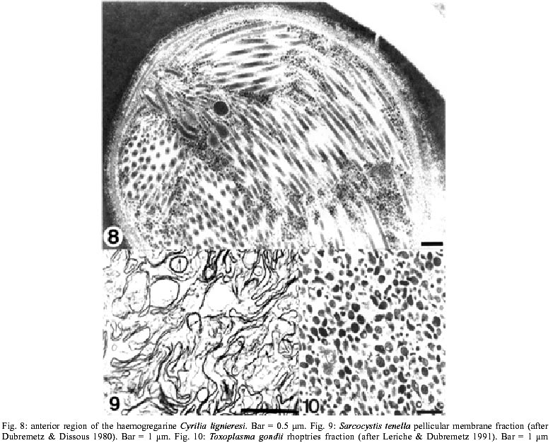
Fig. 8 shows a thin section of a trophozoite form of Apicomplexa represented by the haemogregarine Cyrilia lignieresi. The protozoan is surrounded by a complex membrane system formed by an outer plasma membrane and two closely apposed unit membranes, which form the inner membrane complex, which is interrupted at some points of the cell body and at the very anterior region of the cell. A layer of microtubules is localized under the inner membrane complex. These microtubules originate from the inner ring of the conoid, a structure located at the apical pole, and project toward the posterior but do not reach the most posterior portion of the cell. The nucleus is located in the central portion. Four special organelles are found in the cytoplasm of the protozoan. The first is a group of small, cigar-shaped organelles that are restricted to the apical third of the protozoan body and known as micronemes. Their number varies according to the species and the developmental stages. In some they are hardly seen while in others they are so numerous that they are the most abundant organelle found in the cell (Fig. 8). The organelle is surrounded by a typical unit membrane and presents an electron-dense matrix due to its high protein content. Indeed, the organelle was intensely stained when the protozoa were subjected to the ethanolic phosphotungstic acid technique, which reveals basic proteins (De Souza & Souto-Padrón 1978). It has been shown that when the infective forms of Apicomplexan parasites touch the host cell surface a process of Ca2+ release is triggered with the subsequent discharge of the content of the micronemes at the junction between the parasite and the host cell (Carruthers et al. 1999). This material then mediates parasite attachment (Carruthers et al. 1999, Carruthers & Sibley 1999). The second group of special organelles in Apicomplexa is formed by long, club-shaped organelles connected by thin necks to the extreme apical pole of the parasite and known as rhoptries (Fig. 8). At their basal portion the matrix of the organelle shows a spongy appearance, while the neck region is uniformly electron dense, making it difficult to distinguish from the micronemes. Their number varies according to the species. Several rhoptries can be seen in T. gondii whereas only two, often designated as the paired organelle, are found in Plasmodium. A typical unit membrane surrounds the organelle. Secretion of rhoptry proteins takes place immediately after adhesion of the parasites to the host cell surface. In the case of T. gondii kinetic studies showed that release of the proteins is completed in about 1 minute and that the proteins are internalized and will form part of the membrane lining the parasitophorous vacuole (Sam-Yellowe et al. 1988, Carruthers & Sibley 1997). A third group comprises spherical organelles distributed throughout the cell rather than localized at the apical complex, with a mean diameter of about 0.7 µm, known as dense granules. Their matrix is uniformly electron dense due to the high concentration of protein. Kinetic studies have shown that secretion of the dense-granule content takes place after parasite invasion and localization within the parasitophorous vacuole, persisting for several minutes (Carruthers & Sibley 1997). In contrast to secretion of micronemes and rhoptries, which takes place in the apical region, dense granule secretion occurs at the lateral regions of the protozoan. The secreted proteins associate with the membrane of the parasitophorous vacuole and with the parasite-derived intravacuolar membranous network. The fourth organelle is surrounded by four membranes (reviewed in Gleeson 2000) and localized in the anterior region, next to the nucleus. Based on data showing that it contains a 35 kb DNA molecule similar to that found in the chloroplast genome, the term apicoplast has been suggested for this organelle (Kohler et al. 1997). The presence of the four unit membranes surrounding the apicoplast has been interpreted as evidence that it originated as a secondary endosymbiont derived by ingestion of a eukaryote that itself harbored a plastid, analogous to the plastids of chlorarachniophytes and cryptomonads.
Isolation of the membrane complex Several attempts have been made to obtain isolated fractions containing the plasma membrane of protozoa of the Apicomplexa phylum. Ideally we should get a single fraction containing the plasma membrane, the inner membrane complex and the subpellicular microtubules. Subsequently, these components could be separated. The most successful approach was that used by Dubremetz and co-workers (Foussard et al. 1991, Rabjeau et al. 1997) using tachyzoites of T. gondii. The parasites were disrupted by sonication and the homogenate subjected to differential centrifugation to pellet unbroken cells and the nuclear fraction. The 600 x g supernatant was then collected and sedimented at 30,000 x g for 30 min and the resultant pellet applied to discontinuous sucrose gradients. In a first attempt a 5-step sucrose gradient was used and a membrane enriched fraction was observed at the 0.25-1.0 M sucrose interface (Foussard et al. 1990). The purification was significantly improved by applying the post 600 x g supernatant to the bottom of a 4-step sucrose gradient (1.6, 1.4, 1.2 and 1.0 M), followed by centrifugation at 120,000 x g for 120 min. A layer at the 1.0-1.2 M sucrose interface was highly enriched in the pellicle containing the outer plasma membrane, the inner membrane complex and, in some cell fragments, even the subpellicular microtubules (Rabjeau et al. 1997). Two criteria were used to assess the purification: the morphology of the fraction as seen by transmission electron microscopy of thin sections (Fig. 9) and the enrichment in SAG 1, a well characterized plasma membrane protein detected by SDS-PAGE followed by Western blot. The 1.0-1.2 M interface fraction showed a 15-20-fold enrichment in SAG. Subsequently, the plasma membrane was separated from the whole pellicle by high salt-glycerol treatment followed by one cycle of freezing and thawing and sonication. The suspension was then centrifuged at 10,000 x g to sediment the undisrupted pellicles and the supernatant layered on top of sucrose gradient and centrifuged at 140,000 x g for 10 h. The centrifuge tube showed a pellet and a very faint low-density band close to the top of the gradient at buoyant density of 1.085 g/cm3. This band contained well-conserved vesicles with diameters ranging from 20 to 100 nm. The nature of the vesicles was not determined.
Isolation of the rhoptries The rhoptries of tachyzoites of T. gondii have been purified following rupture of the cells with a French press operated at 28 kg/cm3. Then, unbroken cells were pelleted by low-speed centrifugation and the homogenate was centrifuged at 20,000 x g for 15 min. The resulting pellet was then resuspended and adjusted to 30% Percoll and centrifuged at 50,000 x g for 25 min. Rhoptries were found in the bottom and then centrifuged again at 100,000 x g for 90 min to remove the Percoll particles yielding a highly enriched rhoptry fraction, as evaluated by transmission electron microscopy (Fig. 10) (Leriche & Dubremetz 1991). The fraction was, however, contaminated with dense granules and all attempts to separate the two organelles were unsuccessful. Only the lipids of the fractions were analyzed. Phospholipids were about two times lower while cholesterol was about five times higher in the fraction as compared to the total protozoan. The rhoptries of P. falciparum were isolated starting with schizont-infected erythrocytes which were disrupted by nitrogen cavitation using a nitrogen-pressure chamber at 50 kg/cm2 for 18 min followed by centrifugation at 8,000 x g for 10 min. The supernatant was collected and layered onto a linear 0.4-1.6 M sucrose gradient placed over a cushion of 2.2 M sucrose and centrifuged at 72,000 x g for 30 min. The layer above the gradient was removed and the gradient further centrifuged at 130,000 x g for 2.5 h after which fractions were collected and analyzed. Transmission electron microscopy of the fractions showed that the rhoptries were localized in fractions with densities of 1.155 and 1.166 g/cm3. These two fractions were also enriched in the 110 and 140 kDa proteins previously identified as markers of the rhoptry (Holder et al. 1985, Sam-Yellowe et al. 1988). SDS-PAGE of the rhoptry fraction showed the presence of a large number of proteins indicating the complexity of the organelle. A rhoptry-containing fraction was also obtained from sporozoites of Eimeria tenella (Kawazoe et al. 1992). Oocysts were induced to sporulate and sporozoites purified by anion-exchange chromatography were disrupted by sonication and subjected to differential centrifugation. The 8,500 x g supernatant was centrifuged at 58,000 x g for 30 min in a sucrose gradient. The white layer at the top of each gradient was removed and re-centrifuged at 70,000 x g for 72 hours. The combined pellets from 1.0-1.6 M gradients were resuspended and layered onto a preformed gradient of 1.9-2.5 M sucrose and centrifuged at 62,000 x g for 2 h. A fraction collected at 2.3 M sucrose contained organelles with a diameter of 500-700 nm, which probably correspond to rhoptries. A large number of polypeptides were detected in this fraction by SDS-PAGE, including several of high molecular weights (118, 122, 124, 132, 180, and 220 kDa).
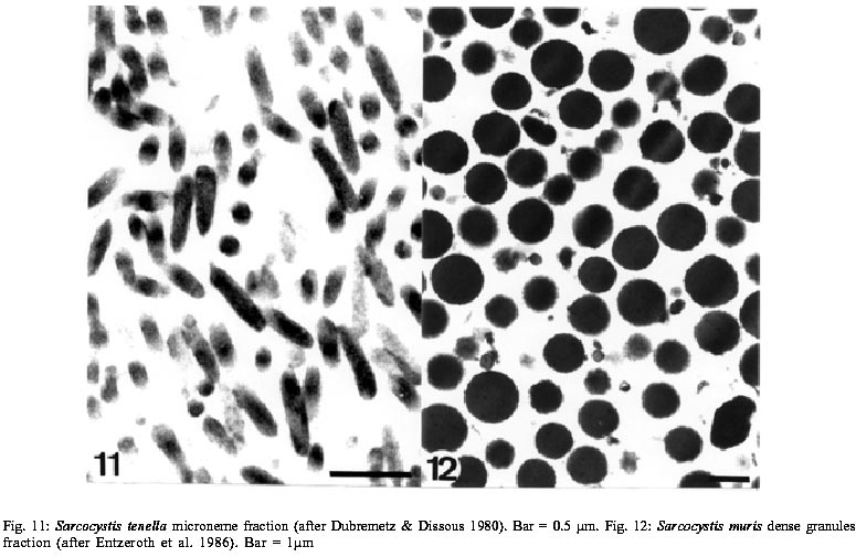
Isolation of the micronemes In the attempts to obtain dense granules and rhoptries from Sarcocystis and Eimeria, as described above, a fraction containing structures similar to the micronemes was detected around 1.4 M sucrose (Fig. 11). SDS-PAGE showed that it had a relatively simple polypeptide content, with enrichment of bands at 26, 28, 31, 47, 130 kDa and one of > 200 kDa (Dubremetz & Dissous 1980, Dubremetz et al. 1989).
Isolation of the dense granules Entzeroth and co-workers (1986) reported a procedure for the isolation of dense granules from zoites of Sarcocystis muris. The parasites were disrupted in a French pressure cell operated at 50 kg/cm2 and the homogenate centrifuged at 500 x g for 10 min to eliminate intact or incompletely emptied cells. The supernatant was then submitted to a centrifugation at 12,000 x g for 10 min. The obtained pellet was re-suspended and adjusted to 1.4 M sucrose with 2.2 M sucrose, layered on a discontinuous sucrose gradient (2.0, 1.6, 1.4 and 0.25 M) and centrifuged for 1 h at 112,000 x g. The material detected at the 1.6-2 M interface was collected and sedimented for 45 min at 113,000 x g. The fraction obtained showed a large number of organelles, which by their size and shape correspond to the dense granules seen in intact cells (Fig. 12). CELL FRACTIONATION OF TRICHOMONADIDAE
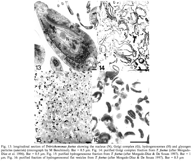
Fig. 13 shows a thin section of Tritrichomonas foetus, where the main structures and organelles observed in members of the Trichomonadidae family are depicted (reviewed in Honigberg 1978). The protozoan is surrounded by a typical plasma membrane that envelops the cell body as well as the group of anterior flagella and the recurrent flagellum. The area of contact between the recurrent flagellum and the cell body is specialized and known as the undulating membrane. The cytoplasm of the protozoan shows a large number of free ribosomes, glycogen particles and vacuoles. This protozoan has a remarkable cytoskeleton, composed of several structures: (a) a sheet of microtubules that originate in the anterior end and project towards the posterior end, forming the pelta-axostylar complex; (b) a large striated structure known as the costa, also originating at the anterior end projecting towards the posterior region, usually subjacent to the region of contact of the recurrent flagellum with the cell body; and (c) a series of striated structures that originate close to the basal bodies and radiate in various directions, even establishing contact with the cis face of the Golgi complex. One characteristic feature of this group of protozoa is the presence of a large number of spherical organelles concentrated around the axostyle and the costa, known as hydrogenosomes. The hydrogenosome is a spherical or slightly elongated organelle of 0.5 to 1.0 mm in diameter, surrounded by two closely apposed membranes, which separate at certain points to form a peripheral vesicle (reviewed in Benchimol 1999) The hydrogenosome plays a fundamental role in the metabolism of the protozoan and is the site of production of ATP and molecular hydrogen (Muller 1993). The organelle has been the subject of intense investigation in recent years, especially by those interested in the process of evolution of cell organelles, since there are several similarities and differences between hydrogenosomes and mitochondria (Biagini et al. 1997, Muller 1997, Martin & Muller 1998, Hackstein et al. 1999).
Isolation of the plasma membrane There is only one description of cell fractionation of trichomonads aimed at isolating the plasma membrane of T. foetus (Morgado-Díaz et al. 1996). The protozoa were collected and resuspended in buffer and disrupted with a Potter-type homogenizer. The homogenate was subjected to differential centrifugation and a microsomal fraction obtained as sediment after centrifugation at 80,000 x g. The resuspended pellet was applied to a discontinuous sucrose gradient (1.0, 1.3, 1.6 and 1.9 M) and centrifuged at 130,000 x g for 3 h. Several bands were collected and that localized at the sample-1.0 M sucrose interface was characterized as highly enriched in plasma membrane, as judged by electron microscopy and the assay of membrane marker enzymes. The fraction contained a population of small and large vesicles, some of which showed a multivesicular appearance. Enzyme assays showed an enrichment in the fraction of 5'-nucleotidase and Na+-K+-ATPase, two well characterized markers of the plasma membrane of eukaryotic cells and previously shown, using a cytochemical approach (Queiroz et al. 1991), to be localized in the plasma membrane of T. foetus.
Isolation of the Golgi complex The Golgi complex of trichomonads is a large organelle consisting of 8-12 parallel cisternae. It has been isolated from T. foetus (Morgado-Díaz et al. 1996). The cells were disrupted with a Potter-type homogenizer and the homogenate centrifuged at 2,500 x g for 10 min to sediment unbroken cells, nuclei and other dense structures. The supernatant was mixed with 2.3 M sucrose to make a 1.4 M sucrose solution that was loaded into the bottom of a centrifuge tube, then overlaid in succession with 1.2, 1.0 and 0.8 M sucrose, and then centrifuged at 95,000 x g for 2.5 h. Two bands, corresponding to 0.8-1.0 and 1.0-1.2 M interfaces, designated as GF1 and GF2, respectively, were seen to contain cisternae and vesicles resembling the components of the Golgi complex. The GF2 fraction was significantly enriched in cisternae of the Golgi complex (Fig. 14) and highly enriched in galactosyltrans-ferase, a well-known marker of the Golgi cisternae of eukaryotic cells. A preparation of GF1 and GF2 pooled together was sub-fractionated by sodium carbonate treatment and alkaline pH in order to separate the Golgi content and membrane fractions. About 15 protein bands were identified in the total Golgi purified fraction by SDS-PAGE (Morgado-Díaz & De Souza 1998).
Isolation of the hydrogenosome The hydrogenosome has been the subject of excellent reviews published in the last years (Muller 1993, Biagini et al. 1997, Benchimol 1999). It appears as a spherical or slightly elongated structure with a mean diameter of 0.5 µm, reaching 2 µm in dividing organelles. The hydro-genosome is surrounded by two closely apposed unit membranes. Invaginations of the hydrogenosomal membrane forming inner compartments are occasionally seen. A characteristic feature of most of the hydrogenosomes is the presence of structures designated as peripheral vesicles. In some species only one or two vesicles are seen. In others, as is the case of T. vaginalis, several peripheral vesicles are found. The vesicle corresponds to about 8% of the volume of the hydrogenosome. Usually the vesicle is flat; however, its size and shape may vary significantly. When the cells are fixed in solutions containing calcium an electron dense product is seen within the vesicle. Cytochemical studies have shown that the peripheral vesicle is a site for the accumulation of calcium. The hydrogenosomal matrix usually appears as granular and homogeneous. The hydrogenosome does not contain nucleic acids. The hydrogenosomal proteins are synthesized in free cytoplasmic ribosomes and post-translationally inserted into the organelle via leader sequences which are absent from the mature protein and which are similar to mitochondrial pre-sequences. The hydrogenosome compartmentalizes the fermentative metabolism of pyruvate, leading to the production of molecular hydrogen. Pyruvate generated by glycolysis or by conversion from malate is decarboxylated by pyruvate:ferredoxin oxidoreductase to the iron sulfur protein ferredoxin and subsequently to protons, forming molecular hydrogen. The enzyme hydrogenase participates in this step. Acetyl-CoA is converted to acetate with concomitant conversion of succinate into succinyl-CoA synthetase. The generation of acetyl CoA is coupled to ATP production via substrate-level phosphorylation. Interconversion of malate and pyruvate is catalyzed by a malate dehydrogenase which can utilize either NAD+ or NADP+. The hydrogenosome does not contain enzymes of the tricarboxylic acid cycle and does not carry out oxidative phosphorylation. The hydrogenosome has been the subject of intense investigation from an evolutionary perspective. The absence of mitochondria in trichomonads and the fact that the hydrogenosome is surrounded by two membranes and participates in some metabolic pathways that take place in the mitochondria of other cells strengthen this interest. In view of the important role played by the hydro-genosome in the cell biochemistry and physiology of trichomonads, it has been the subject of intense investigation, including the isolation by cell fractionation techniques (Muller 1973). We present here one procedure, which yielded the purest fraction obtained for hydrogenosomes of T. foetus (Morgado-Díaz & De Souza 1997). The cells were disrupted in a Potter-type homogenizer. The homogenization process was controlled by observation of the cells under a phase contrast microscope and stopped while some unbroken cells were still present. The resultant homogenate was subjected to differential centrifugation at 1,500 and 4,000 x g. The 4,000 x g pellet, which contained the hydrogenosomes, was layered on top of a gradient of 53, 45, 24 and 18% Percoll and then centrifuged at 36,000 x g for 30 min. A pure fraction containing well-preserved hydrogenosomes was recovered from the Percoll gradient. The purity of the fraction was assessed by electron microscopy (Fig. 15) with an enrichment of about 12 fold in malic decarboxylase, a marker enzyme of the hydrogenosome. As previously mentioned a flattened membrane-bound vesicle is observed at the periphery of the hydro-genosome. A pure hydrogenosomal fraction, as shown in Fig. 15, was resuspended in buffer containing 2% Triton X-100 and 50 mM 3-(N-morpholine) propanesulfonic acid (MOP-3)-NaOH, pH 7.0, and incubated on ice for 1 h. This treatment led to the complete solubilization of the membrane complex surrounding most of the organelle, but not the membrane lining the peripheral vesicle. In addition, components of the matrix remained attached to the vesicle. Further incubation of the fraction for 45 min at room temperature in the presence of proteinase K and subsequent centrifugation at 350,000 x g for 30 min gave a highly purified fraction containing only the peripheral vesicles (Fig. 16). This observation shows unequivocally that the peripheral vesicle is a special domain of the hydrogenosome. It is possible that it is involved in processes of regulation of cytoplasmic calcium concentration since cytochemical studies indicate that this cation accumulates in the peripheral vesicles (Benchimol et al. 1982a, Benchimol & De Souza 1983, De Souza & Benchimol 1988). The fractionation procedure was also monitored by analysis of the proteins using SDS-PAGE. Enrichment in about 12 polypeptide bands with molecular masses between 10 and 120 kDa was observed in the hydrogenosomal fraction. The peripheral vesicle showed few protein bands, with a major one of 45 kDa. Interestingly, this protein is an N-acetylglucosamine-containing glycoprotein, as demonstrated by Western blots probed with alkaline phosphatase-conjugated wheat germ agglutinin (WGA). This observation is in agreement with previous studies showing the presence of WGA-labeling sites only in the peripheral vesicle of hydrogenosomes found in intact cells (Benchimol et al. 1996). These results raise interesting questions concerning the mechanisms involved in the sorting of glycoproteins to a well-defined domain of the hydrogenosome, an organelle whose proteins are synthesized in free polyribosomes, released into the cytoplasm and subsequently translocated into the organelle (Lahti & Johnson 1991, Johnson et al. 1993). We cannot exclude the possibility of a close relationship between the peripheral vesicle and the endoplasmic reticulum.
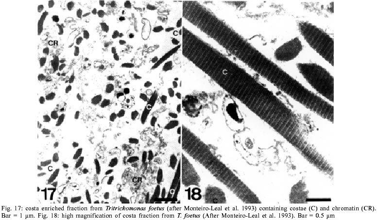
Isolation of the costa The costa is a cytoskeletal structure characteristic of members of the Trichomonadidae family, appearing as a striated root that originates near the basal bodies of the flagellum, and extends towards the posterior region of the protozoan. Some attempts have been made to isolate the costae. Taking advantage of the size of the costae of large trichomonads found in the gut of termites, and using micromanipulation it was possible to purify the structure from T. gigantea (Amos et al. 1979). The first attempt made to isolate the costae of T. foetus yielded a cell fraction predominantly composed of carbohydrates (Lemoine et al. 1983, Sledge et al. 1978). However, cytochemical studies of intact cells showed that the costae were composed of proteins and did not contain carbohydrates (Benchimol et al. 1982b). The second cell fractionation approach yielded a highly enriched costa fraction from T. foetus (Monteiro-Leal et al. 1993). For this the protozoa were suspended in a medium containing 0.05% Lubrol PX detergent, washed in buffer and disrupted with a Dounce-type homogenizer. Cell disruption was monitored by light microscopy in an effort to obtain cell rupture without breaking the nuclear membrane. The homogenate was then vortexed and centrifuged for 15 min at 1000 x g over a 2 M sucrose cushion. The supernatant and the interface were pooled and layered on the top of a second cushion of 1.5 M sucrose and centrifuged for 15 min at 10,000 x g. The pellet was collected, layered on a four-step sucrose gradient (1, 1.5, 2 and 2.5 M sucrose from the top to the bottom) and centrifuged at 19,000 x g for 1 h. The material localized at the interface of 2/2.5 M was collected, resuspended in buffer and centrifuged at 10,000 x g for 10 min, yielding a highly enriched costa fraction (Figs 16, 17). SDS-PAGE analysis of the isolated fraction showed a group of polypeptide bands of 122, 115, 108, 93, 87 and 82 kDa. Further studies are necessary to determine the localization of each protein in the complex structural organization of the costae. PERSPECTIVES
Cell fractionation is still the only approach that enables us to determine directly the chemical composition of an organelle, a cell structure or a region of the plasma membrane. As shown in this review, in some situations it is also possible to sub-fractionate an organelle into its components for further analysis. There also exists the possibility of isolating a given organelle in its different physiological states, which has been relatively little explored. Studies carried out in mammalian cells have shown that it is possible to use cell fractionation techniques to isolate special domains of the plasma membrane. Such an approach should be used with parasitic protozoa, especially with those where there is evidence for the presence of specialized domains such as (a) the flagellar pocket of trypanosomatids, (b) the region of attachment of the flagellum to the cell body in trichomonads and try-panosomatids.
ACKNOWLEDGEMENTS
To Dr Martha Sorenson for a critical reading of the manuscript. To Dr R Docampo from University of Illinois, USA, Dr JF Dubremetz from INSERM, France, Dr M Benchimol from Universidade Santa Úrsula, Brazil, Dr MCM Motta from Universidade Federal do Rio de Janeiro, Brazil, and JA Morgado-Díaz from the Instituto Nacional de Cancer, Brazil, for kindly supplying photographs. REFERENCES
The following images related to this document are available:Photo images[oc03050f17-18.jpg] [oc03050f11-12.jpg] [oc03050t1.jpg] [oc03050f13-16.jpg] [oc03050f8-10.jpg] [oc03050f6-7.jpg] [oc03050f1-5.jpg] |
| |||||||||
