 |
| About Bioline | All Journals | Testimonials | Membership | News |
|
||||||
|
||||||
Mem Inst Oswaldo Cruz, Rio de Janeiro, Vol. 98, No. 2, March, 2003, pp. 265-268 Use of Polymerase Chain Reaction and Enzymatic Cleavage in the Identification of Helicobacter spp. in Gastric Mucosa of Human Beings from North Paraná, Brazil PL Camargo/+, AA Alfieri/++, APFRL Bracarense*, R Menoli, SR Spinosa**, MK Hagiwara*** Departamento
de Clínicas Veterinárias *Departamento de Medicina Veterinária
Preventiva **Departamento de Clínica Médica, Faculdade de Medicina,
Universidade Estadual de Londrina, Campus Universitário, 86051-970 Londrina,
PR, Brasil ***Departamento de Clínica Médica, Faculdade de Medicina
Veterinária e Zootecnia de São Paulo, São Paulo, SP, Brasil
Received
28 June 2002 Code Number: oc03065
Helicobacter pylori is the most common gastric bacteria of human beings. Animal-borne helicobacter have been associated with gastritis, ulceration, and gastric mucosa-associated lymphoid-tissue lymphoma in people. We attempted to identify the species of Helicobacter spp. that infect human beings in north Paraná, Brazil. Samples of gastric mucosa from 38 dyspeptic patients were analyzed by optic microscopy on silver stained slides, polimerase chain reaction (PCR), and enzymatic cleavage. Genus and species-specific primers to H. pylori, H. heilmannii, H. felis, and consensual primers to H. bizzozeronii or H. salomonis were used. The PCR products were submitted to enzymatic cleavage by VspI (Helicobacter spp. product) and HinfI (species products) enzymes. Thirty-two out of 38 patients evaluated had 3.2 to 5 µm long bacteria that resembled H. pylori in Warthin-Starry stained slides and were positive to the genus Helicobacter by PCR. In 30 of these patients the bacteria were identified as H. pylori. Two samples positive by silver stain were negative to all species tested by PCR. None of the 38 samples was positive to animal-origin helicobacter species. These results show that PCR and enzymatic restriction are practical methods to identify the species of helicobacters present in gastric mucosa of human beings. People in north Paraná appear to be infected mostly with H. pylori.
Key words: Helicobacter spp. - dyspeptic patients - diagnosis - polymerase chain reaction - Paraná - Brazil
Helicobacter pylori is the most prevalent helicobacter species in human beings. Helicobacter species, usually found in dogs, cats, and pigs, have been associated in different parts of the world with gastric inflammation, ulcers and neoplasia, especially mucosal associated-lymphoid tissue lymphoma (MALT) in humans (Morris et al. 1990, Solnick et al. 1994, Dieterich et al. 1998, Morgner et al. 2000). Dogs, cats, and pigs may have been the source of infection to humans (Lavelle et al. 1994, Stolte et al. 1994, Dieterich et al. 1998, Meining et al. 1998). Infection rates in humans for helicobacters other than H. pylori are usually low, ranging from 0.01% in Italy (Foschini et al. 1999) to 6.2% in Thailand (Yali et al. 1998), a developing country. The prevalence of the less common helicobacters in human gastric mucosa is unknown. H. heilmannii type 1 apparently is the most prevalent bacteria (78.5%), moreover there are infections by H. salomonis (2.4%) and non-identified species (Trebesius et al. 2001). H. heilmannii may cause MALT lymphoma, because spontaneous regression of gastric lymphoma type MALT occurs after its eradication (Regimbeau et al. 1997, Morgner et al. 2000). It has been suggested that the best therapeutic approach to eradicate H. heilmannii may not be the same used to eradicate H. pylori in humans (Foschini et al. 1999). Tightly spiraled bacteria have been found occasionally in the gastric mucosa of dyspeptic patients in Brazil (Queiroz et al. 1990, Nogueira et al. 1993), but the species or the source of infection were not determined. In a swine population in the State of Minas Gerais, H. heilmannii was present in 77% (54 out 70) of the stomachs (Queiroz et al. 1996). In the northern region of the State of Paraná, 64% (32 out 50) of the cats were infected by H. heilmannii, whereas H. felis was identified in 62% (31 out 50) of the cats and 51% (36 out 70) of the dogs (Camargo 2002). Since the species that infect humans in our region have not been determined, we attempted to verify the adequacy of PCR associated to enzymatic restriction in order to determine which Helicobacter species inhabit the gastric mucosa in dyspeptic patients from the north of Paraná, Brazil. MATERIALS AND METHODS
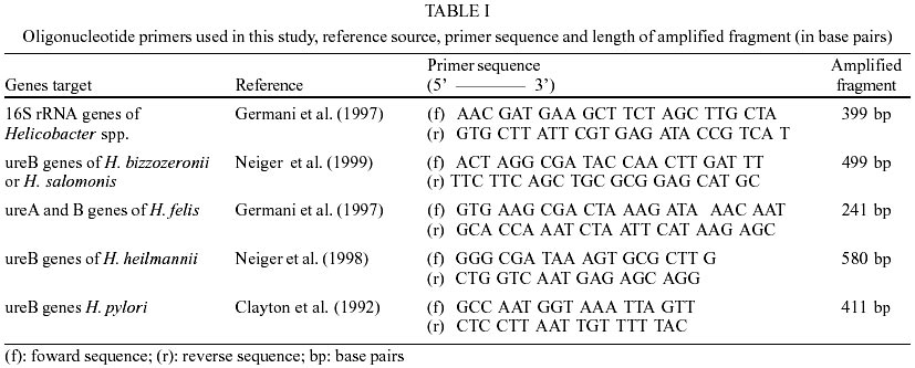
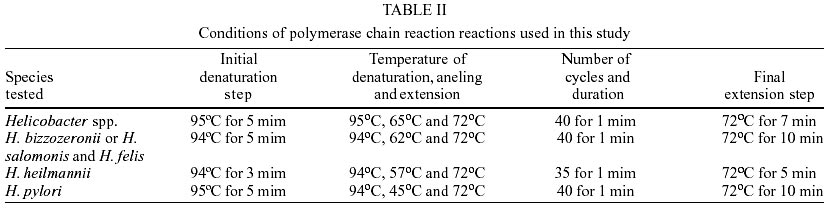
Patients - Samples from gastric mucosa were collected from 38 dyspeptic patients admitted in the Gastroenterology section, Hospital Universitário, Universidade Estadual de Londrina, in Londrina, PR, Brazil. Men and women, aged from 26 to 89 years, were included in the study. Only patients with clinical indication for upper endoscopy and mucosa collection were admitted in the study. Mucosa biopsy samples from the stomach body and antrum were obtained for histological and molecular biology analysis. Morphological analysis of bacteria - Mucosal fragments from each patient were fixed in 10% phosphate-buffered formalin. Sections of paraffin-embedded specimens were applied onto glass slides, and stained by the Warthin-Starry technique (Michalany 1980). The slides were scanned for presence of bacteria and morphological analysis. DNA extraction - Mucosal specimens from each patient were sealed into Eppendorf vials and mixed to 50 µl of proteinase K (20 mg/ml) and 200 µl of lyses buffer pH 8 (100 mM of NaCl, 100 mM of EDTA and 0.5% of dodecyl sodium sulphate), agitated for 30 sec and then incubated at 56oC for 3 h. DNA was extracted using phenol/chloroform (Sambrook et al. 1989), followed by guanidine/isothiocyanate silica (Boom et al. 1990). PCR amplification - PCR was performed in a final volumes of 50 µl solutions composed of 10 µl of DNA mix [(4 µl of extraction product, 4 µl of ultra pure water, and 20 pM of each of the oligonucleotide primers (Table I)], plus 40 µl of Taq PCR mix containing 23.5 µl of ultra pure water, 1.6 mM of dNTP, 4 µl of PCR buffer (20 mM of Tris HCl pH 8.4 and 50 mM of KCl), 2.5 U of Taq DNA polymerase recombinant, and 1.5 mM of MgCl2. Amplification were performed in a thermocycler (PTC-100TM - MJ Research Inc.) under PCR conditions described in Table II. DNA of H. pylori and H. felis standard strains were used as positive controls. Positive controls for the other species were developed by presumptive identification by transmission electron microscopy of bacteria present in gastric mucosa from dogs and cats. Presumptive identification was confirmed by PCR using species-specific primers and enzymatic restriction. All gastric mucosal samples, including those non-reactive to the genus primer, were tested for the species H. pylori, H. heilmannii, H. felis, and H. bizzozeronii or H. salomonis. PCR products were electrophoresed in 1.5% (w/v) agarose gel with 0.3% ethidium bromide in a 10% Tris-Borate-EDTA buffer (TBE). Readings were performed in UV light illumination. Photographic registration was done using a digital Kodak system (Kodak EDAS 290TM). DNA enzymatic restriction - Products amplified with genus primers were cleaved by VspI enzyme (Germani et al. 1997). H. pylori amplified products were cleaved by HinfI (Clayton et al. 1992). DNA restriction digestion and electrophoresis were performed by standard procedures (Sambrook et al. 1989). The sequences of amplified products from H. heilmannii, H. bizzozeronii or H. salomonis and H. felis primers were estimated based on sequences present in the GenBank. The length of fragments generated with HinfI cleavage was established using the Gene RunTM software. RESULTS
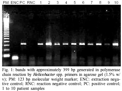
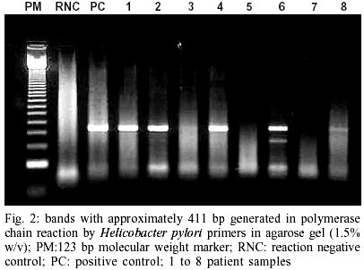
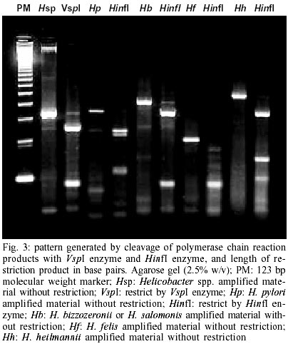
In 32 out of 38 patients small slightly curved or S-shaped bacteria with size ranging from 3.2 to 5 µm in length were visualized on Warthin-Starry stained slides. The bacteria were distributed on mucosal surface and in the glandular pits. The same 32 patients were positive by PCR for Helicobacter spp. (Fig. 1). Of the 32 patients with Helicobacter spp., 30 were identified by PCR as having H. pylori (Fig. 2). The two remaining patients were negative to all species tested. Cleavage of the products amplified by genus specific primer (399 bp) using VspI enzyme resulted in fragments of 295 bp and 104 bp in length. Cleavage using HinfI enzyme resulted in fragments with 277 bp and 134 bp of the H. pylori (411 bp) amplified products, 408 and 172 bp for H. heilmannii; 100 bp for H. felis, and 395 and 100 bp length for H. bizzozeronii or H. salomonis (Fig. 3). DISCUSSION
The bacteria visualized with the Warthin-Starry staining showed size and morphology compatible with H. pylori. When compared to the bacteria found on slides of gastric mucosa from dogs and cats, stained by the same technique, remarkable differences in both morphology and size could be observed. H. pylori can be differentiated from animal-infecting species by optical microscopy if adequate staining is used (Queiroz et al. 1990, Lavelle et al. 1994, Goddard et al. 1997, Foschini et al. 1999). The Helicobacter genus-specific primers to PCR amplification of 16S rRNA genes, to H. felis urea A and urea B genes (Germani et al. 1997), and urea A to H. pylori (Clayton et al. 1992), and urea B genes to H. heilmannii and H. bizzozeronii or H. salominis (Neiger et al. 1998, 1999) amplified products of expected sizes. Enzyme cleavage of the PCR-generated products with VspI enzyme (Germani et al. 1997) and HinfI enzyme (Clayton et al. 1992) was a rapid and practical method to confirm that the genetic material amplified was from Helicobacter spp. and H. pylori. The restriction of the PCR-generated products with primers to the species H. bizzozeronii or H. salomonis and H. heilmannii generated fragments of expected length. In the H. felis amplified material however, two bands of 100 bp were superposed and the other expected band (41 bp) could not be seen in agarose gel due to its small size. The use of polyacrylamide gel might have allowed visualization of the smaller bands. The association of specific primers with enzymatic cleavage allowed species identification in a large number of samples, showing it to be a practical and reliable tool for routine laboratory use. Samples from two patients were positive by PCR for the Helicobacter genus, but negative to all tested species. Technical errors and defective reaction with the primers are unlikely, based on the presence of positive controls. These patients may have hosted a Helicobacter species different from those tested. The low infection rate of human beings with animal specific Helicobacter spp. probably results from a host-specificity among these bacteria. Nevertheless small infection rates with animal-origin helicobacter were reported in Europe (Foschini et al. 1999) higher rates (6.2%) were reported in Thailand (Yali et al. 1998). In the State of Minas Gerais, Brazil, one out of 315 dyspeptic patients (0.4%) was infected with both, H. pylori and "Gastrospirillum hominis" (Queiroz et al. 1990), an animal-origin helicobacter. Also in Minas Gerais 2.5% of low socioeconomic level patients (one out of 40) with gastric cancer also had mixed infection (Nogueira et al. 1993). All samples tested with species-specific primers for helicobacters other than H. pylori yielded, negative results despite the fact that these patients have low socioeconomic level, and live in a region with a high incidence of H. felis and H. heilmannii in dogs and cats (Camargo 2002). Lack of close contact with animals could be a plausible hypothesis to explain the absence of animal-origin helicobacters in the patients. Pets may still be a source of infection to humans. Organisms similar to ones found in humans with gastric diseases were also found in their pets (Lavelle et al. 1994, Dieterich et al. 1998). In one case clinical remission of gastric signs in a human patient was only achieved after eradicating the bacteria from both patient and his two pet dogs (Thomson et al. 1994). Despite questions about the zoonotic potential of these microorganisms, species identification within the Helicobacter genus is important. There is an association between infection by H. heilmannii and gastric lymphoma type MALT. Remission is achieved when the bacteria are eradicated (Regimbeau et al. 1997, Morgner et al. 2000). Gastric ulceration related with H. heilmannii improved after eradication of these bacteria (Goddard et al. 1997). If it is necessary to eradicate the bacteria in order to achieve cure in human patients, the precise species identification and known animal sources of infection become even more important. Therapeutic strategies designed to eradicate animal-born helicobacter may differ from the ones used to eradicate H. pylori in human patients.
ACKNOWLEDGMENTS
To Drs Andrey A Lage and Kenneth William Simpson for providing the DNA of standard H. pylori and H. felis strains. To Dr Idércio Luiz Sinhorini, for the transmission electron microscopy of the bacteria used with positive controls, and to Dr Maria de Lourdes Estrela Faria and Dr Helio Autran for reviewing the manuscript. REFERENCES
Copyright 2003 Instituto Oswaldo Cruz - Fiocruz The following images related to this document are available:Photo images[oc03065f2.jpg] [oc03065f3.jpg] [oc03065f1.jpg] [oc03065t2.jpg] [oc03065t1.jpg] |
| |||||||||
