 |
| About Bioline | All Journals | Testimonials | Membership | News |
|
||||||
|
||||||
Mem Inst Oswaldo Cruz, Rio de Janeiro, Vol. 98, No. 2, March, 2003, pp. 273-276 SHORT COMMUNICATION
Perspective of a New Diagnostic for Human Trichomonosis Sheila B Lecke, Tiana Tasca*, André A Souto**, Geraldo A De Carli/+
Laboratório de Parasitologia
Clínica, Faculdade de Farmácia **Laboratório de Síntese
Orgânica, Faculdade de Química, Pontifícia Universidade Católica
do Rio Grande do Sul, Av. Ipiranga 6681, 90619-900 Porto Alegre, RS, Brasil *Departamento
de Bioquímica, Instituto de Ciências Básicas da Saúde,
Universidade Federal do Rio Grande do Sul, Porto Alegre, RS, Brasil This work was supported by Faculdade de Farmácia, PUCRS, and CNPq (# 470148/01-2). SBL is recipient of a fellowship (# 00501323) from Fundação de Amparo à Pesquisa do Estado do Rio Grande do Sul, and TT from the Conselho Nacional de Desenvolvimento Científico e Tecnológico. Received
20 August 2002 Code Number: oc03067
Several diagnostic techniques have been employed for the detection of Trichomonas vaginalis. Microtubules constitute the cytoskeleton in eukaryotic cells and are sensitive to antimitotic drugs, such as Taxol (paclitaxel). We used FLUTAX a fluorescent taxoid - to analyze the microtubule distribution in living trophozoites of T. vaginalis in urine and in vaginal discharge. A high intensity of fluorescence was observed in living T. vaginalis, epithelial cells and leukocytes present in urine and vaginal discharge. Our preliminary results show the perspective of a new diagnostic technique for trichomonosis and will contribute to the understanding of the cytoskeleton of T. vaginalis.
Key words: Trichomonas vaginalis - diagnostic techniques - FLUTAX
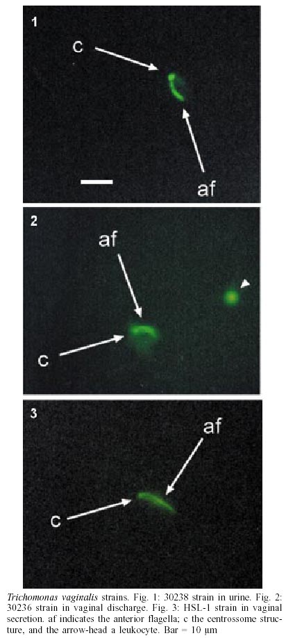
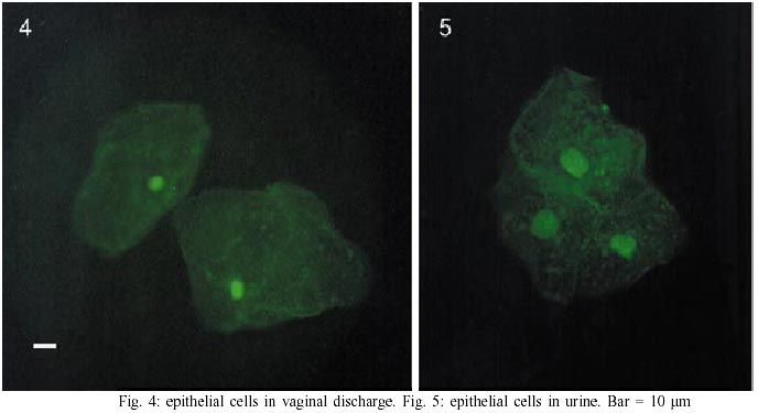
Trichomonas vaginalis is a flagellated parasitic protist of the human urogenital tract. The parasite is a common cause of infection in the female tract, and its clinical presentation ranges from a totally asymptomatic infection to a severe vaginitis. In men, the infection is mostly asymptomatic, but in some cases it can lead to a mild urethritis which usually resolves spontaneously within two weeks. Recent publications indicate that this parasite's impact is not only limited to vaginitis but also a major factor in promoting transmission of HIV (Sorvillo & Kerndt 1998, Fleming & Wasserheit 1999), in causing low-weight and premature birth (Lehker & Alderete 2000), and in predisposing women to atypical pelvic inflammatory disease (Heine & McGregor 1993), cervical cancer (Gram et al. 1992, Kharsany et al. 1993, Zhang & Begg 1994), and infertility (Grodstein et al. 1993). Like others trichomonads, T. vaginalis does not present a cystic form, only the trophozoitic one. The organism is ellipsoidal and presents four anterior flagella unequal in size, an undulating membrane, an axostyle, a nucleus, a Golgi apparatus and hydrogenosomes, instead of mitochondria. Trichomonosis is the most common non-viral sexually-transmitted disease (STD) in the world (Gerbase et al. 1998). Donné described the protozoan for the first time in 1836, however diagnosis of trichomonosis still presents some difficulties. The diagnosis of this infection, based only on clinical aspects, such as characteristics of vaginal discharge, may be erroneous (Madico et al. 1998). Various laboratory techniques have been employed for the detection of T. vaginalis. Positive diagnosis of trichomonosis is generally established by wet-mount examination, but this method is only 30-80% sensitive when compared to the gold standard of culture (Lehker & Alderete 2000). Conversely, culture is laborious, sometimes inaccessible and often cost prohibitive for many clinical settings, and is not routinely used (Heine & McGregor 1993, Heine et al. 1997). Different stains or cytochemical reactions including acridine orange, Giemsa, Leishman, Diff-Quick, Fontana, Gram, periodic acid-Schiff, iron hematoxilin and Papanicolau (De Carli 2001) have also been used in smears. A cost effective and sensitive alternative, with a similar clinical efficacy as traditional culture, is the InPoucht TV culture system (Ohlemeyer et al. 1998). This is a disposable culture system for the maintenance, transport, and detection of T. vaginalis in clinical specimens (Borchardt & Smith 1991). Other diagnostic techniques such as enzyme-linked immunosorbent assay (ELISA) (Sharma et al. 1991), hybridization (DeMeo et al. 1996) and fluorescent antibody test (Krieger 1988) have been used to detect the parasite. These procedures contribute to increase the certainty of diagnosis, but do not substitute the culture exams (Honigberg & Burgess 1994, De Carli 2001). The advent of the polymerase chain reaction (PCR), however, has opened a new avenue for diagnosis of T. vaginalis (Shaio et al. 1997, Madico et al. 1998). In this study, we present a perspective of a new diagnostic technique for trichomonosis, based on the analysis of microtubule distribution in living trophozoites of T. vaginalis in urine and vaginal discharge, using the fluorescent taxoid Flutax-2. Flutax-2, described by Souto et al. (1995), is an active fluorescent derivative of Taxol (paclitaxel). Like Taxol, FLUTAX-2 binds to the polymerized αβ-tubulin dimer, allowing direct and rapid observation by fluorescence microscopy (Diaz et al. 2000). T. vaginalis presents a poorly known cytoskeleton formed by an axostyle, which is a trunk that courses from one end of the cell to the other oriented by microtubules (Benchimol et al. 2000), and a pelta, which like the axostyle, is also formed by stable structures such as microtubules (Ribeiro et al. 2000). Microtubules are essential for the maintenance of cell shape and its organization; for cytoplasm transport, motility and division in all eukaryotic cells. Urine and vaginal secretions were observed in an hemocytometer to determine number of trophozoites, epithelial cells, leukocytes, artifacts and the presence of some erythrocytes and yeast. We used three strains in this study: HSL-1, recently isolated from women with trichomonosis in a State medical unit, and two American Type Culture Collection (ATCC) strains, 30236 (sensitive to metronidazole) and 30238 (resistant to metronidazole). The ATCC strains were cultivated axenically in vitro at 37°C, in trypticase-yeast extract-maltose (TYM), Diamond medium (Diamond 1957) without agar, pH 6.0, supplemented with 10% heat inactivated bovine serum, penicillin (1000 IU/ml) and streptomycin sulfate (1 mg/ml). Both strains were counted in an hemocytometer to obtain 106 cells/ml. The positive urine and vaginal discharge from the patient (HSL-1), and ATCC strains were incubated with FLUTAX-2 (1 µM, final concentration) at 37°C, for 40 min, and observed with an Axiolab MC 80 DX (Zeiss) fluorescence microscope. The photomicrographs showed a high intensity of fluorescence in the axostyle, centrossome and flagella of living cells of T. vaginalis, strongly suggesting the microtubule composition of these structures (Figs 1-3). Epithelial cells showed an homogenous fluorescence throughout the cytoplasm and surrounding the nucleus (Figs 4, 5). Prominent plasma membranes in the epithelial cells were particularly noted, suggesting that they are superimposed. Leukocytes were observed as small spheres with intensive fluorescence. No erythrocytes or yeast were observed. Under fluorescence microscope T. vaginalis was easily differentiated from other structures, since the parasitic trophozoite is alive, presenting flagella mobility, while all other cells are dead. In addition, the size of each structure represents another feature for identification of each organism. A difference in fluorescence intensity, size and shape between living T. vaginalis and other cells was also observed. Our data are in accordance with Lopes et al. (2001), who showed, using specific monoclonal antibodies, that T. vaginalis presents a-tubulin at the flagella, basal bodies and the axostyle and β-tubulin at the axostyle and flagella, since FLUTAX-2 binds to αβ-tubulin dimers. Although no positive results were found, it is possible that trichomonads present the γ-tubulin isoform (Lopes et al. 2001). Although 30236 and 30238 T. vaginalis strains present different sensitivities to metronidazole, our results demonstrated no differences in the binding profile of FLUTAX-2. The same was observed with the analyzed isolated strain, demonstrating that diagnosis of human trichomonosis with FLUTAX-2 may become a new alternative. Our new technique is still being tested on vaginal discharges and urine and must be improved for use as a routine laboratory method. Furthermore, an analysis of the composition of the cytoskeleton of T. vaginalis will contribute to the understanding of the biochemical aspects involved in cellular morphology and behavior, as well as the mechanisms related to host-parasite interactions.
ACKNOWLEDGEMENTS
To Dr Sérgio De Meda Lamb, Pharmacy School Chairman for his support; to Dr Denise C Machado, IPB, PUCRS, for microscope assistance and to Rafael V Michel and Fernanda S Meira for technical assistance. REFERENCES
Copyright 2003 Instituto Oswaldo Cruz - Fiocruz The following images related to this document are available:Photo images[oc03067f4-5.jpg] [oc03067f1-3.jpg] |
| |||||||||
