 |
| About Bioline | All Journals | Testimonials | Membership | News |
|
||||||
|
||||||
Mem Inst Oswaldo Cruz, Rio de Janeiro, Vol. 98, No. 2, March, 2003, pp. 287-290 SHORT COMMUNICATION
Preparation of Sand Fly (Diptera: Psychodidae: Phlebotominae) Specimens for Histological Studies Beatriz Gomes Brazil/+, José Lino-Neto*, Reginaldo Peçanha Brazil
Centro de Pesquisas René
Rachou-Fiocruz, Av. Augusto de Lima 1715, 300190-002 Belo Horizonte, MG, Brasil
*Departamento de Biologia Geral, UFV, Viçosa, MG, Brasil This work received financial support from Capes, CNPq (Project 52307/95-6), Pronex and Fiocruz. Received
18 June 2002 Code Number: oc03070
Phlebotominae sand fly specimens were prepared for histological and physiological studies. Different fixatives were tested on sectioned and whole bodied adult females in order to obtain good fixation and provide satisfactory penetration of the embedding media. All fixed specimens were infiltrated (up to seven days under 5ºC) and embedded in hydroxyethyl metacrylate. Two-three µm sections were stained, mounted in Canada balsam and observed by light microscopy. Best results were achieved when whole bodied insects were double fixed in Bouin's and Carnoy's fluids (4 h/2 h) and stained in Hematoxilin/Eosin or fixed in calcium formaldehyde and stained in mercury bromophenol blue.
Key words: Phlebotominae - histology - historesin preparations
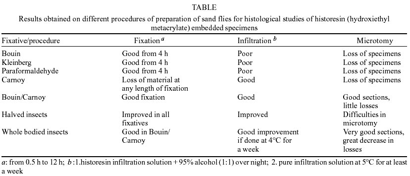
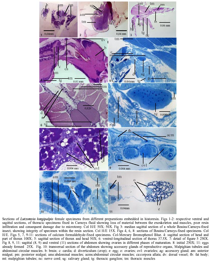
There are few morphological studies on the internal organs of phlebotomine sand flies, vectors of the Leishmania spp. causing cutaneous and visceral leishmaniasis. Most published studies were done several years ago and involved predominantly Old World species (Adler & Theodor 1926, Christophers et al. 1926, Perfilev 1928a,b, Lewis & Minter 1960, Lewis 1965, Davis 1967). More recent publications on this subject refer to parasite-vector interactions or peritrophic membrane formation and blood digestion (Gemetchu 1974, Walters et al. 1987, 1989, 1995, Blackburn et al 1988). Guzman et al. (1994) and Abassy et al. (1995 a, b, c) using histological methods studied the physiology of feeding and embryonic development on Phlebotomus duboscqi and P. papatasi respectively under the light microscope obtaining satisfactory results what indicate these methods as practical tools for physiological studies of sand flies. The most recent literature on histological techniques suggests that historesin is a good alternative to paraffin for obtaining sections as thin as 0.5 µm, eliciting a better visualization of the tissue and cell structures (Junqueira 1995). Based on this and aiming to study the functional morphology of the salivary glands of adult phlebotomine sand flies, we developed a procedure in which specimens of different chronological and physiological ages could be obtained and prepared for histological studies. Specimens of Lutzomyia longipalpis from a colony of the Laboratory of Leishmaniasis (CPqRR- Fiocruz) maintained in the laboratory according to the already preconized techniques (Brazil et al. 1997) were used. When pupation occurred the specimens were sexed as described by Brazil and Brazil (2000) and transferred individually to emergence vials. The vials were numbered and emergence of the insects monitored at hourly intervals. Unfed adult specimens were fixed at pre-determined times from the first hour after emergence until the fifth day. Five day-old sand flies were fed and fixed in the same way. Prior to fixation the insects were aspirated from the emergence containers, anaesthetized by cooling at 4°C and placed on a glass slide in a drop of fixative under a stereoscopic microscope. After removing the appendages each specimen was transferred to glass vials containing the fixative of choice. The following fixatives were evaluated for histology: Bouin's fluid, Carnoy's fluid, paraformaldeyde, Kleinberg's fluid and double fixation in Bouin's and Carnoy's fluids. Different fixation times were tested, ranging from 0.5-12 h. Specimens either had the scutellum removed, the thorax separated from the abdomen or where left entire. At the end of fixation, the specimens were rinsed several times in distilled water for 45 min until no more yellowish color could be seen. They were than transferred to 70% alcohol. When necessary the insects were stored in alcohol at 4°C to complete dehydration later. For protein detection the insects were fixed by immersion in calcium formaldehyde for 24 h, and then quickly rinsed in distilled water to remove excess. Following, the specimens were rinsed three more times for 15 min being posteriorly transferred to 70% alcohol. In all cases dehydration was performed by passing the specimen three times for 10 min through each dilution of an alcoholic series (70%, 90% and 95% ethanol). As embedding media we used historesin (hydroxyethyl metacrylate-Leica historesin embedding Kit-Leica instruments GmbH). After several timing experiments, the following schedule was established to be used on the supplier's two-step recommended infiltration: (i) pre-infiltration in a 1:1 mixture of historesin infiltrating solution/95% alcohol, kept overnight in the refrigerator; (ii) slow infiltration in the pure infiltrating resin at 5°C for at least a week. During the embedding the vessel containing the resin preparation was kept on ice inside a Styrofoam box to prevent early polymerization. At the end of the embedding procedures the molds containing the specimens were kept at room temperature under cold light for about 2 h or until polymerization was completed. As this resin is somewhat hygroscopic and the RH in the laboratory was high, at the end of polymerization the molds containing the specimens were taken to an oven and kept at 60ºC for one night. The specimens were then stored in a dessicator containing a dehumidifying agent (silica gel), until microtomy could be carried out. Thin sections (2-3 µm) of the specimens were obtained using a rotary microtome (Microm HM 340 E) with glass knives. The sections were recovered one by one in drops of distilled water on a clean microscopy slide and left on a hot plate for few hours to ensure adherence. Prior to utilization and at no more than a week the slides were cleaned for 1 h in a sulfochromic solution, rinsed in distilled water and left to dry at 70ºC for one night (Chandler & Schowenwolf 1987). The sections were rehydrated and stained in Harris hematoxylin/eosin for general morphology studies. Mercury bromophenol blue was used for protein detection according to (Pearse 1972). After staining and drying, the slides were diaphanized in xylene and mounted in Canada balsam. The methodology detailed above allowed preparations of various sand fly organs to be obtained. Good results were achieved for the observation of salivary glands, digestive tract, Malpighian tubules, ovaries, accessory glands, nerve cord, muscles and fat body and the results are presented in the Table and in the Figs 1 to11. Studies of the functional morphology of the salivary glands as of other organs of adult insects require the exact age of the specimen to be known, since this could reveal age-related differences. Individual confinement of pupae and hourly monitoring of adult emergence ensured this in the present study. Phlebotomine sand flies are small insects and specimens can easily suffer mechanical damage, so that excessive manipulation should be avoided in morphological studies. Sexing the pupae and individualizing them in emergence vials, from which specimens can be obtained directly for fixation, can avoid such damage. Despite this fragility, the chitinous exoskeleton of the insects presents a barrier to the penetration of plastic infiltrating solutions and embedding media. This results in the loss of suitable material during microtomy, due to separation of the specimen from the resin block. To avoid this problem, various methods of fixation were tried. In spite of their good fixative properties, paraformaldehyde, Kleinberg's and Bouin's fixatives did not permit good infiltration of the resin. Carnoy's fluid reacted with chitin softening the insect body in such a way that it could be damaged during the subsequent procedures. It also leached material from the epidermis and consequently provided very poor histological preparations. Better results were achieved through the double fixation of whole insects in Bouin's and Carnoy's fixatives. These specimens were immersed individually in the former for 4 h and than transferred to the latter for 2 h. Fixation in calcium formaldehyde was satisfactory and did not cause any problems with infiltration. However, the most satisfactory infiltration was achieved for insects fixed either in Bouin/Carnoy's or calcium formaldehyde fixatives if the specimens were left infiltrating for at least a week in the refrigerator. Loss of material was also reduced. Historesin is the best embedding medium for the histological preparation of sand fly-sized insects (2-3 mm) as infiltration may be carried on inside small glass or plastic vials using little quantities of resin. This process also avoids excessive heat that would be harmful to the integrity of such small specimens. The possibility to obtain sections less than 5 µm thick is interesting since it allows detailed examination of the tissues and cells under the light microscope, as well as permitting larger number of sections to be made from the same small specimen. Microtomy is undoubtedly the more difficult step in this procedure due to the dimensions of the object to be sectioned and the thickness of the sections required. These are very difficult to see implying the use of high magnification lenses. Obtaining a ribbon of sections is too difficult and each section must be transferred to drops of water placed on slides using small surgical forceps. The common histological procedure of placing sections on a water bath and collecting them with a glass slide is difficult since they may not be seen on the surface of the water. Although somewhat time-consuming and laborious, this methodology is reliable and provided good preparations of different organs and tissues of sand flies and could be used for future studies of the internal morphology of these insects.
ACKNOWLEDGEMENTS
To Patricia Monteiro de Freitas Teixeira, Jeane Cristina Menezes Alves and Mariana Junqueira Pedras for their help in sand fly collections and maintenance of the insects in the laboratory; to José Eduardo Serrão, Federal University of Viçosa for critical reading of the manuscript, Bruce Alexander for revision of the English text and Heloiza Diniz, Laboratory of Image Treatment, Instituto Oswaldo Cruz for helping in preparation of the plate. REFERENCES
Copyright 2003 Instituto Oswaldo Cruz - Fiocruz The following images related to this document are available:Photo images[oc03070t1.jpg] [oc03070f1-11.jpg] |
| |||||||||
