 |
| About Bioline | All Journals | Testimonials | Membership | News |
|
||||||
|
||||||
Mem Inst Oswaldo Cruz, Rio de Janeiro, Vol. 98, No. 3, April, 2003, pp. 335-344 Comparative Study of the Stridulatory Sulcus, Buccula and Rostrum of the Nymphs of Triatoma guazu Lent & Wygodzinsky, 1979 and Triatoma jurbergi Carcavallo, Galvão & Lent, 1998 by Scanning Electron Microscopy (Hemiptera, Reduviidae) Maria Beatriz Araújo Silva, Helene Santos Barbosa*, Cleber Galvão/+, José Jurberg, Rodolfo U Carcavallo Laboratório Nacional e Internacional de Referência em Taxonomia
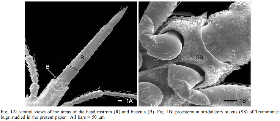
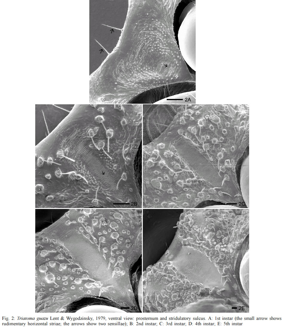
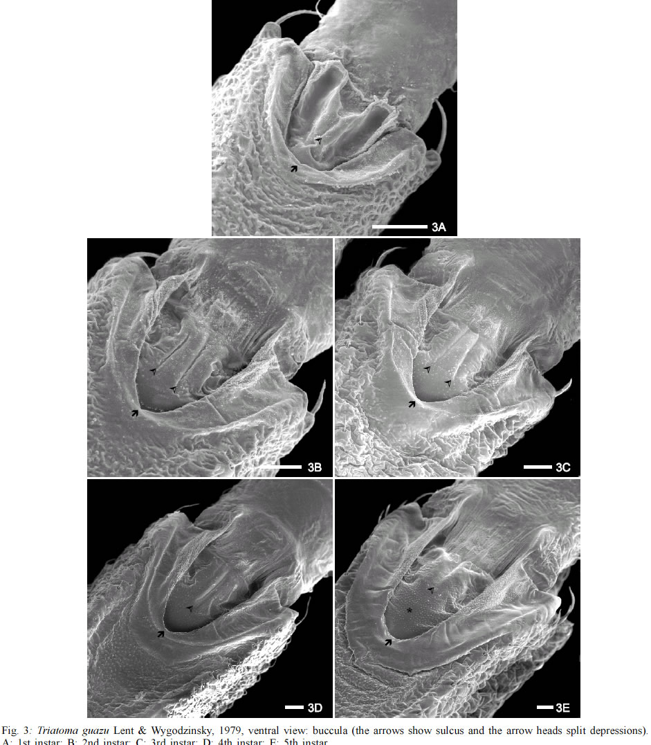
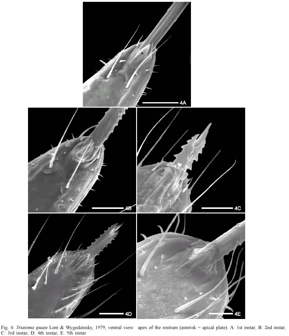
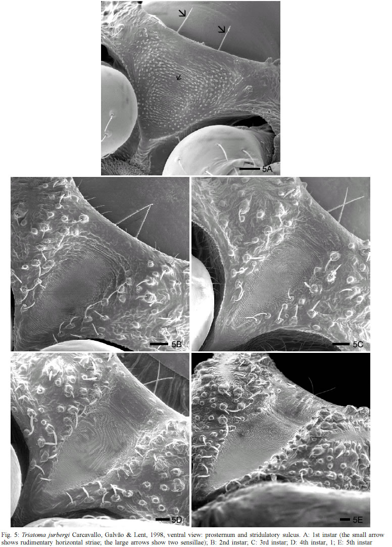
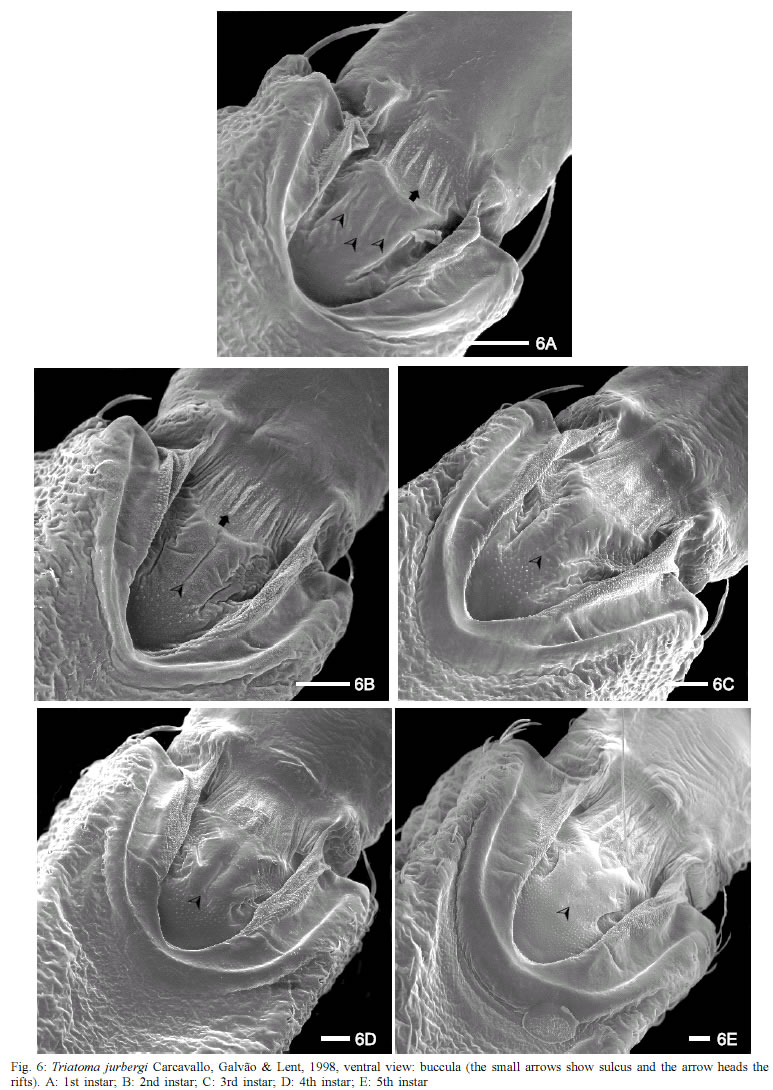
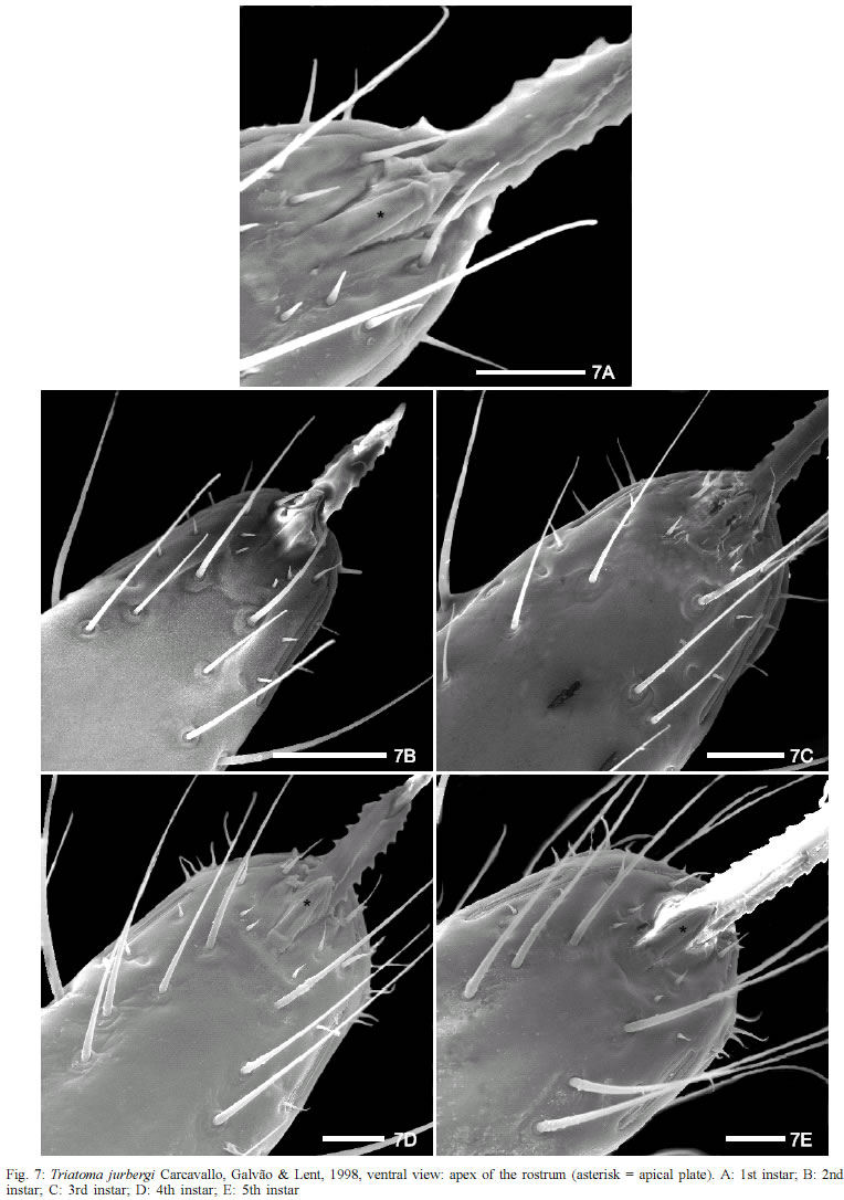
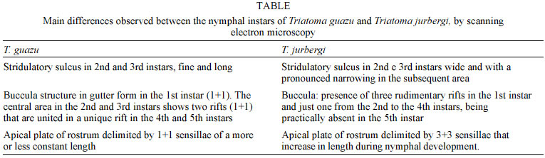
de Triatomíneos, Departamento de Entomologia Supported by CNPq, Funasa/Fiocruz and Faperj. Received 17 September 2002 Code Number: oc03078 The ultrastructural morphology of the ventral region of the head (rostrum and buccula) and proesternum (stridulatory sulcus) of nymphs from the 1st to 5th instars of Triatoma guazu Lent & Wygodzinsky, 1979 and Triatoma jurbergi Carcavallo, Galvão & Lent, 1998 was described. Morphological differences between the two species and of the five nymphal stages development of each species were observed. These structures showed systematic differential characteristics of the studied species and may be used to increase their taxonomic range. Key words: Triatominae - Chagas disease vectors - buccula - stridulatory sulcus - rostrum Chagas disease constitutes a major public health in Latin America. It is caused by infection with the protozoan parasite Trypanosoma cruzi (Chagas, 1909), mainly transmitted in the faecal droppings of triatomine bugs. Actually the subfamily Triatominae is composed of 138 species, all characterized by their obligate haematophagy (Carcavallo et al. 2002, Galvão et al. 2002). These insects are distributed in the Americas and some species are found in the Old World. Its geographic distribution is wide, being found in the Neartic Region between the latitudes 42ºN, in Salt Lake City in the North of America (USA) until the Neotropical region 46ºS in the Patagonia, with occurrences of some species in the Oriental region (Jurberg 1996). Triatoma guazu Lent & Wygodzinsky, 1979 and Triatoma jurbergi Carcavallo, Galvão & Lent, 1998 are related species present in the state of Mato Grosso, Brazil, between the latitudes of 15º49'01" and 16º43'10"S. These species belong to the oliverai complex, considered to be a very homogeneous group according to morphological characteristics (Carcavallo et al. 2000). The Triatominae taxonomy is based on the external morphological characters (Lent & Wygodzinsky 1979). During the last decade, the scanning electron microscopy (SEM) has been used as an important tool for Triatominae systematics, clearing the status of the cryptic species and their complexes. Studies by Pinto (1931) reveal the importance of the rostrum in the characterization of the genera of Tria-tominae, considering that the length of the articles of the rostrum in the Hemiptera in general, especially in the hae-matophagous families is important for the systematic of these insects. Carcavallo et al. (1994) studied 11 species of Panstrongylus genus using SEM, considering P. herreri, P. humeralis and P. lignarius as related species. Later, Carcavallo et al. (1995), comparing various anatomic zones of genus Psammolestes Bergroth, 1911, found important differences between the species. This methodology was the principal tool used to study 97 of 110 species of Triatominae (Carcavallo et al. 1998), which has been employed by Ferro et al. (1997) to analyze structures of buccula and gula of nymphs of 5th instar of Triatoma williami Galvão, Souza & Lima, 1965 and Triatoma gerstaeckeri (Stål, 1859). Our group has developed a series of papers in the study of the comparative morphologic characterization between T. guazu and T. jurbergi demonstrating little known structures of 1st instar nymphs of T. guazu such as proesternum, antenniferous tubercle and the articulations between the antennal segments (Silva et al. 1998), partial results of apical plate morphology of first instar of both species (Silva et al. 1999), description of the five instars nymphs and in the exochorion of eggs through optical and scanning electron microscopy (Silva et al. 2000, Jurberg et al. 2002) and more recently, a comparative ultrastructural analysis of the antenna of T. guazu and T. jurbergi during the nymphal stage describing characteristics that allowed the taxonomic separation of the nymphs of these two species (Silva et al. 2002). In the present study, morphological structures of head and proesternum as the stridulatory sulcus, buccula and rostrum, of nymphs from the 1st to 5th instar of T. guazu and T. jurbergi, were compared using scanning electron microscopy. MATERIALS AND METHODS Insects - The five nymph stages of T. guazu and T. jurbergi were obtained from acclimatized colonies that were respectively started in 1996 and 1997, at the insectary of the Laboratório Nacional e Internacional de Refe-rência em Taxonomia de Triatomíneos of the Departamento de Entomologia of the Instituto Oswaldo Cruz, Rio de Janeiro, Brazil. These colonies were established with insects from Rondonópolis and Barra do Garças, Mato Grosso, sent by the Fundação Nacional de Saúde. All material analyzed belongs to the Rodolfo Carcavallo Collection at the Departamento de Entomologia, Instituto Oswaldo Cruz. SEM - Ventral views of the head and prosternum of three nymphs of each instar (1st to 5th instar) of the two species were analyzed. The 1st instar nymphs were collected soon after hatching, killed by freezing and adhered to metal stubs with 1.2 cm of diameter using double-faced sticky tape. Due to the small diameter of the stubs, the cephalic region of the nymphs of 4th and 5th instars were cut with a scalpel and adhered to the stubs exposing either the dorsal or the ventral surface. The mounted specimens were sputter-coated with gold and observed under a Zeiss DSM 940 scanning electron microscope, operated at 15 kV. The images were either photographed with Neopan SS 120 Fuji film or digitally captured by IBM-PC computer and processed using the Adobe Photo Shop 5.0 software. RESULTS In order to get more informations about the morphology of the of 1st to 5th instars of T. guazu and T. jurbergi, the ventral views of the head (rostrum and buculla) and the proesternum (stridulatory sulcus) of Triatominae bugs were examined in the present paper. Triatoma guazu - The structure of the prosternum region of nymphs from the 1st instar of T. guazu shows a central area with rudimentary horizontal striae, the future stridulatory sulcus. A granular area surrounding the central region was observed, where papillae and sensilla shall be present in the 2nd to 5th instar (Fig. 2 A). The anterior edge presents in prominence two wide sensilla, observed in all the instars (Figs 2 A-E). From the 2nd to 4th instar, the sulcus is fine and long with well defined parallel striae. Along the development, the anterior edge of the sulcus becomes straighter, the posterior edge was gauged and the striae condensed (Figs 2 B-E). The 5th instar nymphs show a wider "V" shaped stridulatory sulcus (Fig. 2 E). Papillae and sensilla are present in setiferous tubercles, limiting the depression of stridulatory sulcus insertion, increasing in number during the nymphal development (Figs 2 A-E). The buccula, localized in the protero-ventral region of the rostrum, is "U" shaped with thick edges. During the nymphal development, the edges of the buccula seem like a depression with two lateral groves, central rifts and a granular texture in the antero-central region (Figs 3 A-D). Nymphs of 5th instar loose the depression in this region, which differentiates it of the other instars (Fig. 3 E). A finger-like structure with flat boards is observed in the posterior region of each edge (Figs 3 A-E). This structure is rounded in the 1st instar. Starting from the 2nd instar, it is more elongated, with flat and well delimited boards. Internally, an area with a flat and glabrous surface with granulations in its superior region can be seen, which presents longitudinal striations on the first segment of the rostrum, more evident during the nymphal development. The central region of the buccula of the 1st instar has a split depression in the anterior area 1+1 and two longitudinal lateral depressions, forming a gutter, which is internally limited by granular pleats (Fig. 3 A). This intern structure modifies in the 2nd and 3rd instars and in the 4th instar forms an unique structure with a long central sulcus, which is short, apical and with granular texture in the 5th instar (Figs 3 B-E). The rostrum is inserted in an arched area of the apex of the labium. It presents at the terminal region a pair of structures localized on the ventral and medial region (Fig. 4A). The basal part is welded with the labium and the free apex is constituted of 1+1 lamellae to foliate one upon the other, which receive the name of "apical plate" (Figs 4 A-E). The proximal lamella is wide and the distal lamella is narrower, limited laterally by two short and gauged sensillae 1+1 (Figs 4 A-E). Below the apical plate is a pair of long sensillae (Figs 4 A-E) and short sensillae are in parallel aligned position, already visible in the 1st and 2nd instars (Figs 4 A-B). During the nymphal development a considerable increase in the number, type and sensilla length is observed below the apical plate and in the lateral area of the rostrum (Figs 4 C-E). The presence of two lateral rifts (1+ 1) is observed close to the apex of the rostrum (Figs 4 A-E). Triatoma jurbergi - The stridulatory sulcus is "V" shaped and large up to the 1st to 5th instar, with a deep central concavity (Figs 5 A-E). Laterally, it is delimited by papillae and sensillae, inserted in setiferous tubercula and it presents central parallel grooves that are compressed during the development of the instars. The posterior edge of this structure narrowed gradually during the nymphal development. The buccula of T. jurbergi is "U" shaped with thick edges and presents pleats and a granular surface. The anterior region shows accentuated straight pleats, present in all instars (Figs 6 A-E). The central region presents three rudimentary rifts in the 1st instar (Fig. 6 A). Small granulations appear up and one longitudinal rift is observed from the 2nd to the 4th instar (Figs 6 B-D) that disappeared in the 5th instar (Fig. 6 E). The rostrum in T. jurbergi is similar to T. guazu (Figs 7 A-E). The apical plate has a lozenge formed inferior lamella and the superior one has a digit form. This structure is laterally delimited by 3+3 sensillae, which gradually increase in length, during the nymphal development. Two very long and fine sensillae were inserted in tubercles of a short and circular basis in the 1st instar (Fig. 7 A). The number and length of sensillae in the third segment of the rostrum increase during the nymphal development presenting a parallel distribution. Two lateral rifts 1+1 were observed at the apex of the rostrum. DISCUSSION Considering that T. jurbergi has been found naturally infected with Trypanosoma cruzi, a comparative morphological study is related between T. guazu and T. jurbergi. We have analyzed the buccula, rostrum and the stridulatory sulcus from the 1st to the 5th instars of T. guazu and T. jurbergi. They demonstrated particular characteristics during the nymphal development that can be used as taxonomical data (Table). The studies developed by Lent and Wygodzinsky (1979) point out the taxonomic importance of the stridulatory sulcus for characterization of the species, which varies in form, length, number and spacing of grooves. These characteristics can aid in the determination of the species. In the present study it was possible to verify that nymphs of T. guazu and T. jurbergi present different morphology of the stridulatory sulcus in all the instars. In contrast, Costa et al. (1991) studying the nymphs and adults of Cavernicola lenti Barrett & Arias, 1985, observed that this structure is absent in nymphs and vestigial in adults. In the species studied here, the stridulatory sulcus narrowed and prolonged in T. guazu in the 2nd and 3rd instars, while, in T. jurbergi, this structure is enlarged and the posterior region is very gauged. Ferro et al. (1997) studied the buccula and the gula of nymphs of the 1st and 5th instars of Triatoma williami Galvão, Souza & Lima, 1965 and Triatoma gerstaeckeri (Stål, 1859), and described differences in the structures of the two species. The buccula was studied by Barth (1953) and Galíndez-Girón et al. (1998), who suggested that the function of the buccula in addition to the genas is to support the labium. The buccula of T. guazu and T. jurbergi studied here in all instars showed differences of boards in the central region, which presents two structures in gutter form only in the 1st instar of T. guazu, and two rifts 1+1 in the 2nd and 3rd instars, which melt in only one rift in the 4th and 5th instars. This structure in T. jurbergi showed peculiar characteristics: in the 1st instar, the central region shows three rudimental rifts and only one in the 2nd until the 4th instars, being almost absent in the 5th instar. Catalá (1996) studying ten species of the rostrum sensillae of eight species of Triatominae, observed that the sensilla type chaetica is more abundant along the surface of the rostrum, distributed in six columns along most of the 2nd and 3rd segments, increasing in number on the last segment. In the present study, columns of sensillae along the principal axis on the ventral surface of the 3rd segment of the rostrum in both species were observed. The sensillae increased in size and number during the development of nymphs. The main difference between the species was observed in the 1st e 2nd instars of T. guazu, which showed two long sensillae below of the apical plate along the longitudinal axis and two pairs of short sensillae, while in T. jurbergi these pairs of sensillae showed medium length in the two first instars. The labial apical plate (Quadri 1951, Parsons 1966, Cobben 1978, Silva et al. 1999) was another parameter to differentiate species. We showed that T. guazu has this pair of structures limited by 1+1 sensillae which present a large base, while, in T. jurbergi these structures are localized within a rectangular depression, have a lozenge form and the extern structure is digit form, limited by 3+3 sensillae, which increase gradually in length. Two lateral rifts 1+1 were observed close to the apex of the rostrum, that were modified during the nymphal development in both species, as described by Lent and Wygodzinsky (1979) in the genus Dipetalogaster Usinger, 1939 using optical microscopy. The data described here point out that these structures in T. guazu and T. jurbergi showed differences that may be used to distinguishing these species. In conclusion, the ultrastructure of buccula, rostrum and the stridulatory sulcus presented in this study, associated to the morphologic characteristics of the antennae described previously by Silva et al. (2002), the exochorion of eggs and the morphometry of three characteristics of the eggs and of every nymphal stage (Silva et al. 2000, Jurberg et al. 2002) present different patterns of systematic value that allowed the taxonomic diagnosis of T. guazu and T. jurbergi nymph's. These data can be used as a tool to identify the two species both found in peridomiciliary habitats and one, T. jurbergi, found naturally infected with T. cruzi. ACKNOWLEDGMENTS To Bruno Ávila from Departamento de Ultra-estrutura e Biologia Celular, Instituto Oswaldo Cruz-Fiocruz, for the image processing. Two anonymous reviewers helped to improve the manuscript. REFERENCES
Copyright 2003 Instituto Oswaldo Cruz - Fiocruz The following images related to this document are available:Photo images[oc03078f7.jpg] [oc03078f2.jpg] [oc03078f6.jpg] [oc03078f3.jpg] [oc03078f5.jpg] [oc03078t1.jpg] [oc03078f1.jpg] [oc03078f4.jpg] |
| |||||||||
