 |
| About Bioline | All Journals | Testimonials | Membership | News |
|
||||||
|
||||||
Mem Inst Oswaldo Cruz, Rio de Janeiro, Vol. 98, No. 4, June, 2003, pp. 513-517 Physa acuta Draparnaud, 1805 (Gastropoda: Physidae): a Study of Topotypic Specimens W Lobato Paraense/+, Jean-Pierre Pointier* Departamento de Malacologia, Instituto
Oswaldo Cruz-Fiocruz, Av. Brasil 4365, 21045-900 Rio de Janeiro, RJ, Brasil
*Laboratoire de Biologie Marine et Malacologie, EPHE, UMR 5555 du CNRS, Université de
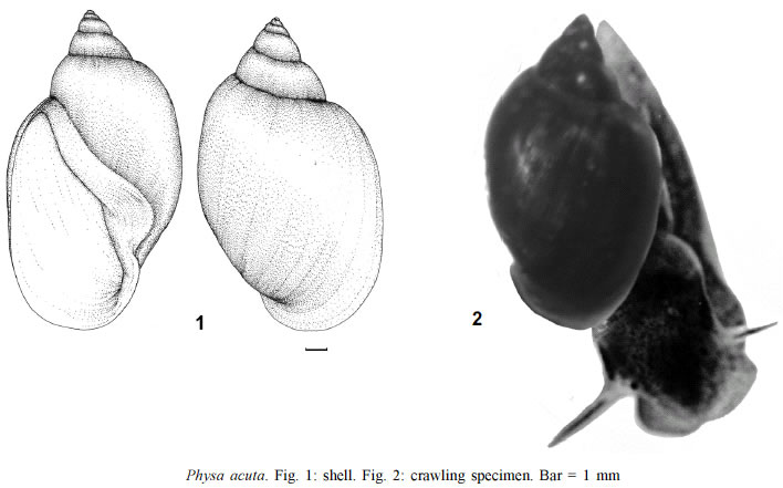
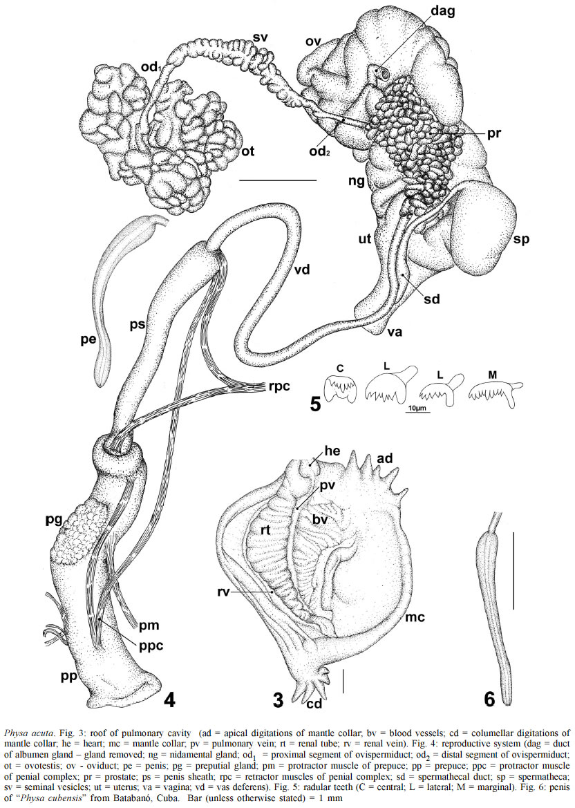
Perpignan, Perpignan, France Received 11 December 2002 Code Number: oc03112 A description is given of Physa acuta Draparnaud, 1805, based on topotypic specimens from the Garonne river basin, and additional samples from the environs of the French cities of Montpellier and Perpignan. It proved indistinguishable, in shell and anatomy, from topotypic Physa cubensis Pfeiffer, 1839, thus leading the authors to admit the synonymy of the two nominal species under the older name, P. acuta. Key words: Physa acuta - Physa cubensis - synonymy This species was described by Draparnaud (1805 : 55, Pl. III, Figs. 10, 11) as follows: 2. P. aiguë. P. acuta. P. testâ sinistrosâ ovatâ; spirâ brevissimâ acutâ; peristomate marginato. Desc. L'animal n'a point de digitations au manteau. Coquille assez semblable à la précédente [Physa fontinalis], mais plus grande, plus ventrue, plus épaisse, moins transparente, moins luisante, de couleur un peu cendrée, striée longitudinalement. Quelquefois même les stries forment des bandes pâles longitudinales sur le dernier tour qui est très-grand. La base de la columelle est profondément sinuée avec un bord blanc. Ouverture grande et rétrécie supérieurement; péristome bordé en dedans d'un bourrelet blanc. La spire a cinq tours et est aiguë à son sommet. Habite dans la Garonne et les rivières qui s'y jettent. MATERIALS AND METHODS The snails were collected from four places in the Garonne basin: Saint Léger (on Garonne river), Viale (on Baïse river), Clairac (on Lot river), tank at Saint Étienne de Fougères. Additional samples were collected from an artificial pond at Montpellier and a canal at Perpignan. The specimens to be dissected were relaxed for 8 h in a 0.05% solution of nembutal. Then they were immersed for 40 sec in water heated at 70oC, from which they were transferred to water at room temperature. The animals (under water) were drawn from the shell with a small forceps applied to the cephalopedal mass, and fixed in slightly modified Railliet-Henry's fluid (distilled water 930 ml, sodium chloride 6 g, formalin 50 ml, glacial acetic acid 20 ml). Voucher specimens were deposited in the Museum National d'Histoire Naturelle de Paris (Laboratoire de Biologie des Invertébrés Marins), the École Pratique des Hautes Études of Perpignan (Laboratoire de Biologie Marine et Malacologie), and the Malacological Collection of Instituto Oswaldo Cruz. DESCRIPTION Shell (Fig. 1) elongate-ovate, thin, smooth, moderately lustrous and translucent, light fawn colored; fine, close-set lines of growth, non-perceptible spiral lines. Protoconch distinct, rounded exserted. Whorls 5, regularly and rapidly increasing, the first minute, the last very large, roundly shouldered. Spire short. Suture slightly impressed. Aperture large, ear-shaped, about ¾ total length of shell; outer lip thin, sharp; inner lip closely appressed to the columellar region completely closing the umbilical region; columellar plait somewhat twisted; parietal callus wide, columellar fold well marked. Largest shell 16 mm in length, 9 mm in width. Cephalopedal mass uniformly dark gray. Fig. 2 shows some features of a crawling specimen: labial palps fan-like, slightly bilobed anteriorly, roundly angled laterally; foot tapering to a pointed caudal tip that does not overreach the shell apex. Roof of pulmonary cavity deeply pigmented and flecked with small circular to oval unpigmented spots. Renal tube (Fig. 3, rt), as usual in physids, tightly folded into a zig-zag course, ending by a short ureter which opens through a subterminal meatus just behind the pneumostome. Mantle collar (Fig. 3, mc) with about 3-5 digitations on the apical (ad) and 5-10 on the columellar (cd) region, reflected _ the latter more extendedly _ over the edge of the shell. By the way, in the original description of P. acuta it is stated that "the animal has no mantle digitations at all", whereas seven digitations over the columella are mentioned by Moquin-Tandon (1855). Reproductive system (Fig. 4) - Ovotestis (ot) embedded in the digestive gland. Ovotestis follicles (about 80 in a minutely dissected specimen) emptying into a collecting canal which continues into the ovispermiduct. Between the proximal (od1) and distal (od2) segments of the latter are the seminal vesicles (sv). The ovispermiduct empties into the carrefour, as well as the albumen gland (removed in the figured specimen). From the carrefour the oviduct (ov) emerges as a narrow tube which runs leftwards, gradually widens, becomes bosselated and highly convoluted, proceeding into the nidamental gland (ng). This latter shows no remarkable features and narrows to the uterus (ut) and vagina (va), which receives the spermathecal duct (sd), about twice as long as the spermatheca (sp). Immediately after emerging from the carrefour the spermiduct gives off a series of short prostatic diverticula (pr). They are closely packed, so that their numbers are difficult to determine; in one thoroughly dissected specimen about 45 units were recorded. They may be simple or divided into usually 2-4 short branches. After giving off the last diverticulum the spermiduct continues into the vas deferens (vd), which opens into the caudal end of the penis. The penis sheath (ps) is somewhat swollen proximally, tapering gradually distalward and expanding into a sarcobellum at its junction with the prepuce. The penis, free within the penis sheath (ps), decreases in width toward its extremity, and has a terminal outlet (Fig. 4, pe). The prepuce (pp), much wider than the penis sheath, is from about as long to twice as long as the latter; a lenticular gland (pg) is present on the proximal half of the preputial wall. A set of retractor (rpc) and protractor (pm) muscles are inserted into the walls of the penial complex. Some radular teeth are shown in Fig. 5. DISCUSSION Many records have been published of P. acuta ouside its type locality, and only in a few of them was its identification based on anatomic characters: Moquin-Tandon (1855), from France, Slugocka (1913), from Switzerland, and Aboul-Ela and Beddiny (1969), from Egypt. The following citations, although not exhaustive, give an idea of its ubiquity: In Europe: Austria (Strouhal 1934); Belgium (Adam 1960); Bulgaria (Dragneva & Kanev 1983); Czechoslovakia (Wohlgemuth 1987); England (Cooper 1918); Germany (Martens 1902); Greece (Eleutheriades et al. 1993); Hungary (Soós 1917); Italy (Clessin 1886); Malta (Mienis 1987); Netherlands (Meeuse & Hubert 1949); Northern Ireland (Dillon et al. 2002); Poland (Feliksiat 1939); Portugal (Azevedo et al. 1967); Romania (Iordau et al. 1964); Russia (Kazannikov 1978); Scotland (Jenkins 1890); Sicily (Sowerby 1873-74); Spain (Clessin 1886); Ukraine (Stadnishenko 1972); Yugoslavia (Krkac 1982); In Asia: Azerbaijan (Aliev 1960); Bangladesh (Begum & Nazneens 1992); China (Dudgeon & Lam 1985); Georgia (Kurashvili et al. 1986); India (Raut et al. 1995); Iran (Massoud & Hedayeti-Far 1979); Iraq (Altaif et al. 1978); Israel (Mienis 1983); Japan (Gotoh & Kawata 2000); Jordan (Burch et al. 1989); Korea (Ha et al. 1981); Malaysia (Ali 1993); Oman (Brown & Gallagher 1985); Pakistan (Nazneen et al. 1992); Saudi Arabia (Magzoule & Kasim 1980); Tadzhikistan (Izzatullajev 1978); Turkestan (Lindholm 1929); In Africa: Algeria (Bourguignat 1864); Botswana, Ethiopia (Brown 1965); Kenya, Mauritius, Rhodesia, Transvaal (Mandahl-Barth, after Verdcourt 1971); Madagascar (Brygoo 1968); Mauritania, Reunion, Zimbabwe (Madsen & Frandsen 1989); Morocco (Mohamed & Najat 1998); Namibia (Brown et al. 1985); Nigeria (Fashuyi 1990); South Africa and Uganda (Bruggen 1966); Sudan (Brown et al. 1984); Tunisia (Pallary 1923); Zaire (Mandahl-Barth et al. 1974); In Australia and Hawaii: Burch and Tottenham (1980); In North America: USA (Beetle 1973 in Virginia), Clench (1934 in Massachusetts). Such ubiquity was expressed by Smith (1989), who also discusses the methods of dispersal and colonization of non-marine molluscs: "Two of the six species considered ecologically insignificant are found in every region of the world. These are the snail Physella acuta (Draparnaud) and the minute bivalve Pisidium casertaneum Poli, both of which may have spread with only limited assistance from man". The likeness in shell characters of P. acuta to P. cubensis is mentioned by Pfeiffer (1839) in his description of the latter: "Sehr ähnlich unsrer europäischen Ph. acuta Dr." Such similarity is also observed in the anatomic characters (see Paraense 1987), pointing to the synonymy of the two nominal species under the older name, Physa acuta. By the way, in Paraense's (1987) paper it was erroneously stated that "the penis has an axial canal with a subterminal outlet"; as seen in Fig. 5 of that paper and Fig. 6 of the present one, the outlet is really terminal. ACKNOWLEDGEMENT To the technologist JE Prado for the graphic documentation. REFERENCES
Copyright 2003 Instituto Oswaldo Cruz - Fiocruz The following images related to this document are available:Photo images[oc03112f3-6.jpg] [oc03112f1-2.jpg] |
| |||||||||
