 |
| About Bioline | All Journals | Testimonials | Membership | News |
|
||||||
|
||||||
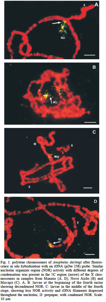
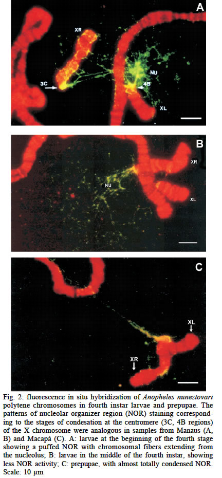
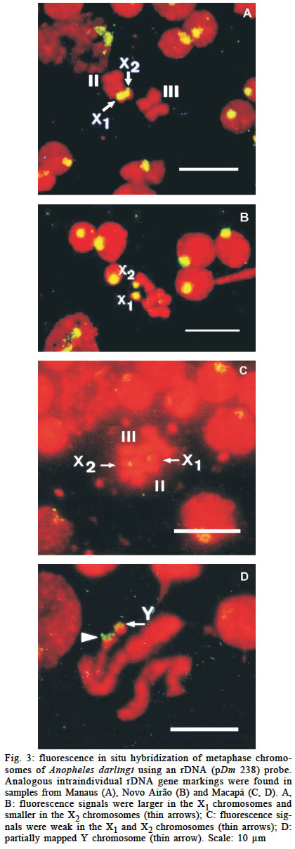
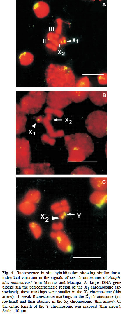
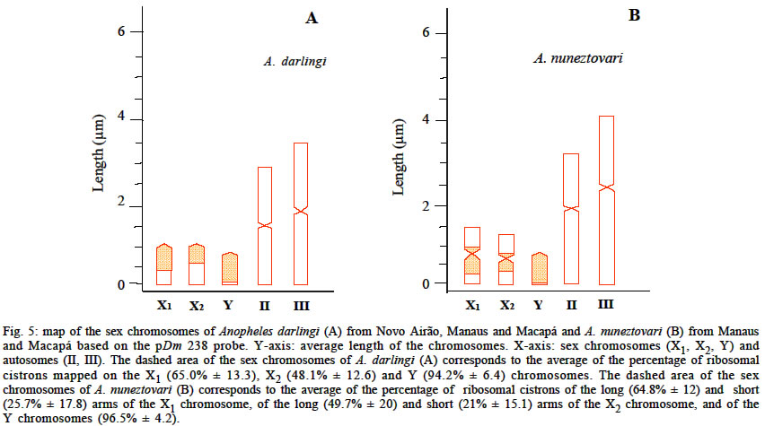
Mem Inst Oswaldo Cruz, Rio de Janeiro, Vol. 98, No. 5, July, 2003, pp. 629-635 Location of Ribosomal Genes in the Chromosomes of Anopheles darlingi and Anopheles nuneztovari (Diptera, Culicidae) from the Brazilian Amazon Míriam Silva Rafael/+, Wanderli Pedro Tadei, Shirlei Maria Recco-Pimentel* Coordenação de Pesquisas
em Ciências da Saúde, Instituto Nacional de Pesquisas da Amazônia,
Av. André Araújo 2936,
69083-000 Manaus, AM, Brasil *Departamento de Biologia Celular, Instituto de
Biologia, Universidade Estadual de Campinas, Campinas, SP, Brasil This research was supported by PPG-7/Finep, Ministério da Ciência e Tecnologia, Brasília, DF, Brazil and Projeto Institucional do Inpa-PPI 3190, Manaus, AM, Brasil. Recived 8 March 2003 Code Number: oc03132 Fluorescence in situ hybridization of Anopheles darlingi and A. nuneztovari demonstrated nucleolar organizer region activity at the end of the fourth larval instar, when the nucleolar organizer regions underwent gradual condensation. The heteromorphic sex chromosomes showed intraindividual size variation in the rDNA blocks located in the pericentromeric region and this coincided with the location of constitutive heterochromatin (C-banding). Key words: Anopheles darlingi - Anopheles nuneztovari - fluorescence in situ hybridization - rDNA genes - nucleolar organizer region - sex chromosomes Anopheles (Nyssorhynchus) darlingi Root, 1926 and A. (N.) nuneztovari Gabaldón, 1940 are important vectors of human malaria parasites. In Brazil, A. darlingi is the main vector of malaria, especially in the Amazon region, where more than 97% of all cases in the country occur (Tadei & Dutary-Thatcher 2000). In Colombia and Venezuela, the main vector is A. nuneztovari (Kitzmiller et al. 1973). Intraspecific variation involving constitutive heterochromatin in mitotic chromosomes is a general phenomenon in many groups of animals. Such structural changes result in the loss or gain of heterochromatin, as occurs in the chromosomes of Anopheles species in Thailand and Southeast Asia (Baimai et al. 1996, Baimai 1998). In A. nuneztovari and A. darlingi, which have karyotypes with 2n = 6 chromosomes, with a pair of submetacentric autosomes (III), one pair of metacentric (II) and one pair of sex heteromorphic (XX/XY) chromosomes (Rafael & Tadei 1998), there is intraspecific variation in the heterochromatic blocks located around the centromeric region in the sex chromosomes and autosomes (Rafael & Tadei 2000). Based on morphological variations (Faran & Linthicum 1981), geographic distribution, isoenzymes patterns (Rosa-Freitas et al. 1992, Freitas-Sibajev et al. 1995, Santos et al. 1999, Manguin et al. 1999), behavior, mitochondrial DNA sequences (Rosa-Freitas et al. 1992, Freitas-Sibajev et al. 1995, Conn et al. 1999), random amplified polymorphic DNA (RAPD) patterns and internal transcribed spacer 2 (ITS2) region profiles from rDNA markers (Manguin et al. 1999) of different populations, A. darlingi is considered to be a single species. However, comparisons of chromosomal variability among A. darlingi populations from Northern and Southern Brazil have revealed a higher frequency of heterozygous inversions in the northern populations (Kreutzer et al. 1972, Tadei et al. 1982). In contrast, environmental and isoenzymatic variations (Fritz et al. 1995, Scarpassa et al. 1999), ITS2 (Fritz et al. 1994, Onyabe & Conn 1999) and mitochondrial DNA marker (Conn et al. 1998, Scarpassa et al. 2000) analyses have shown important geographic interpopulational differences in A. nuneztovari, and it is still not clear whether A. nuneztovari is a single variable species or a complex of species. The fixed inversion on the X-chromosome in Brazilian population of A. nuneztovari differed from that of Vene-zuelan and Colombian populations (Kitzmiller et al. 1973). This fixed inversion, the frequencies of inversions in autosome II, and the presence of a chromocenter were used by Conn (1990) and Conn et al. (1993) to identify the cytotypes A, B and C for A. nuneztovari. Cytotype A showed one fixed inversion on the X-chromosome (Brazilian Amazon), cytotype B had inversion 2La (Venezuela) and cytotype C had inversions 2b, 2Lc and 2Ld (Colombia and Western Venezuela). Molecular biology and in situ hybridization techniques have provided powerful tools for gene mapping and for establishing physical gene maps of polytene and me-taphase chromosomes in mosquitoes species. As in other eukaryotes, the ribosomal DNA (rDNA) of mosquitoes is useful for studying genetic variability and divergences within and among species. The rDNA consists of tandemly repeated transcriptional units with highly conserved genes, and occurs with approximately 500 copies in the genome of mosquitoes (Collins et al. 1987). The rRNA genes, which are located in the nucleolar organizer regions (NORs), have been used as a probe in in situ hybridization methods to map the NORs in many insects, including Drosophila of the mulleri/D. arizonensis complex (Bicudo 1982), Ceratitis capitata (Bedo & Webb 1989), Sciara ocellaris (Dessen & Perondini 1991), and the blow flies Chrysomyia megacephala and C. putoria (Parise-Maltempi & Avancini 2001). In mosquitoes, the rRNA genes are located within and around the heterochromatic regions, especially on the sex chromosomes in the subfamily Culicinae and in some species of the subfamily Anophelinae (Marchi & Pili 1994). However, there are no in situ hybridization studies for A. darlingi and A. nuneztovari (both in the subgenus Nyssorhynchus). In the present study of A. darlingi and A. nuneztovari from Manaus (AM) and Macapá (AP) in the Brazilian Amazon, we investigated the NOR in the polytene chromosomes and the relationship of the NOR to the constitutive heterochromatin (C-banding) in the heteromorphic sex pair. We also compared the rDNA genes of both species using an rDNA probe (pDm 238 _ D. melanogaster) in fluorescence in situ hybridization. The use of this method to map the ribosomal genes in the chromosomes of these mosquitoes should allow the identification of chromosomal signals which may be useful in differentiating populations of both species. The FISH method should also be useful to separate the A. albitarsis complex, such as A. marajoara, which was incriminated as a primary malaria vector in Macapá (Conn et al. 2002). MATERIALS AND METHODS Mosquitoes rearing - From July 1998 to August 2000, individuals of A. darlingi and A. nuneztovari were collected at three locations in the Amazon region: Manaus (3o08'S, 60o01'W), and Novo Airão (1o56'S, 61o22'W) at Manairão locality, both in the state of Amazonas, and Macapá (0o02'S, 51o03'W) at Manuarum locality, in the state of Amapá. Adult females were captured from 18:30 to 20:30 h using oral aspirators while they were feeding on cattle, resting on stable walls or biting humans. Wild-caught adults were transported in moist chambers to the laboratory of malaria vectors at the Instituto Nacional de Pesquisas da Amazônia. Females were confined individually in plastic cups for egg laying. The offspring were reared to the fourth instar larvae and prepupal stage. Morphological identification of the specimens was according to Forattini (1962) and Faran and Linthicum (1981). Preparations of polytene chromosomes - Fourth instar (early and middle stages) larvae and prepupae of females A. darlingi and A. nuneztovari were used to prepare polytene chromosomes as described by French et al. (1962) and Kumar and Collins (1994). In some cases, during squashing of the salivary glands, the nucleolar contents and granular and fibrous compounds became dissociated and were lost. This loss of material, together with the fact that there were fewer specimens from Novo Airão than from Manaus, meant that there were fewer slide preparations from the former location. In all 71 slides containing well-spread polytene chromosomes were selected for in situ hybridization. In A. darlingi we analyzed 5, 21 and 18 slides from Novo Airão, Manaus and Macapá, respectively. In A. nuneztovari we analyzed 15 and 12 slides from Manaus and Macapá, respectively. Preparations of mitotic chromosomes - Mitotic chromosomes in air-dried neuroblast preparations from fourth instar larvae (early and middle stages) and prepupae (F1) were examined using the techniques described by Imai et al. (1988). Thirty-nine treated and fixed slide preparations containing 4 to 5 well-spread metaphase plates of A. darlingi and A. nuneztovari were used in the fluorescence in situ hybridization assays. We analyzed 2, 11 and 9 slides of A. darlingi from Novo Airão, Manaus and Macapá, respectively; and 7 and 10 slides of A. nuneztovari from Manaus and Macapá. Fluorescence in situ hybridization - FISH of polytene and mitotic chromosomes was done according to Viegas-Péquignot (1992), using a cloned rDNA probe (pDm 238) from D. melanogaster. Polytene and mitotic chromosome preparations were pretreated with RNase (100 µg/ml) at 37oC for 1 h. The slides were then dipped in 2X SSC for 2 min, dehydrated in an ethanol series for 2 min in each concentration, air-dried, denatured in 70% formamide (in 2X SSC) at 70oC for 2 min, and dehydrated in cold 50%, 75% and absolute ethanol. The rDNA probe was labelled with biotin-14-dATP using standard nick translation (GIBCO BRL) procedures. The labelled probe was denatured at 100oC for 10 min and added to the slides, which were then incubated in a humid chamber at 37o C for 36 h. Excess probe was removed by washing the slides twice in 50% formamide (in 2X SSC) and twice in 2X SSC (5 min per wash). The slides were subsequently blocked with the first antibody (antibiotin) in the presence of bovine serum albumin in a moist chamber at 37oC for 45 min. To detect the fluorescence signals, the slides were washed in PBT (0.4% of BSA 30% w/v, 0.1% of tween 20 and PBS buffer 1X) and then incubated with the second antibody (IgG-FITC - 1:100 v/v in PBT) in a moist chamber at 37oC for 45 min. After washing in PBT, the slides were counter-stained with propidium iodide (2 µg/ml), which was then removed by a quick wash. The slides were mounted with anti-fading (Vectashield) and examined using Zeiss Axioplan and Olympus fluorescence microscopes. Photographs were obtained using 400-ASA color negative Kodak film. RESULTS Polytene chromosomes - The rDNA probe (pDm 238 - D. melanogaster) physically mapped the NOR at the proximal end (5C region) of 58 salivary X-chromosomes of A. darlingi from Novo Airão, Manaus and Macapá. In A. nuneztovari, the genes were located in the 3C region of the right arm and the 4B region of the left arm in 67 X-chromosomes from Manaus and Macapá. In both species, the NOR was associated with the nucleolus through the presence of chromatin fibers and ribosomal cistrons protruding from the NOR, the activity of which revealed a gradual condensation on the X-chromosomes. The rDNA probe did not map to any other chromosome. The early fourth instar larvae of A. darlingi from Novo Airão, Manaus and Macapá showed analogous, decondensed NOR with thin filaments of rDNA (Fig. 1A, B), whereas in the middle of fourth instar larvae, the NOR became less active (Fig. 1C). When the larvae reached the prepupal stage, the NOR became condensed (Fig. 1D). In early fourth instar larvae of A. nuneztovari from Macapá and Manaus, the NOR was decondensed and there were thin rDNA filaments (Fig. 2A). By the middle of the fourth instar, the NOR had already started to condense (Fig. 2B), and in prepupae, the NOR was almost totally condensed (Fig. 2C). Mitotic chromosomes - The NOR was detected by FISH in the pericentromeric region of 63 sex chromosomes of A. darlingi from Novo Airão, Manaus and Macapá. Fifty-one nuclei of A. nuneztovari from Manaus and Macapá. The location of the rRNA genes showed similar intraindividual variation on the sex pairs and coincided with the centromeric heterochromatic blocks on these chromosomes. In both species, the fluorescence signals were greater in the X1 chromosome and smaller in the X2 chromosome. The X1 chromosome showed strong signals in approximately 80% of the nuclei whereas 70% of the X2 chromosome had weak fluorescence signals (Figs 3, 4). The Y-chromosome was partially or almost totally mapped (Figs 3C, 4C). All interphase nuclei exhibited one or two nucleoli with fluorescence. Fig. 5 shows the percentage of ribosomal cistrons mapped on the sex chromosomes of A. darlingi (A) from Novo Airão, Manaus and Macapá, and A. nuneztovari (B) from Manaus and Macapá. DISCUSSION Active NOR were detected on the polytene X-chromosome of A. darlingi and A. nuneztovari in early fourth instar larvae. In prepupae, the NOR became condensed and the nucleolus disappeared. The longest period in the development of both species, from eggs to adults, corresponds to the transformation of the fourth instar larvae into pupae (Santos et al. 1981, Scarpassa & Tadei 1990). According to these authors, this period may be related to the levels of the insect hormone ecdysone. The effects of this moulting hormone on protein and rRNA synthesis in insects have been shown in cytological studies such as in Rynchosciara (Amabis 1972). Salivary gland chromosomes from fourth instar larvae of S. ocellaris treated with ecdysone showed very active NOR that were abruptly repressed when pupation was induced (Dessen & Perondini 1985). The structural regulatory mechanism of NOR may be in-fluenced by the functional stage of cells, and this can affect chromatin condensation and nucleolus size, as occurs in Drosophila (Bicudo 1982), Rynchosciara (Amabis 1972) and S. ocellaris (Dessen & Perondini 1991). A similar phenomenon probably occurs in Anopheles species, since the degree of NOR condensation in the salivary gland chromosomes of A. subpictus, the vector of malaria in India (Chaudhry 1986), varies in fourth instar larvae and pre-pupae. An analogous situation was reported by Tiepolo et al. (1974) who observed divergent DNA synthesis in hybrids of A. atroparvus and A. labranchiae. In A. darlingi and A. nuneztovari, the differences in the intensity of protein and RNA synthesis probably reflect the functional stage of the cells and the effect of hormones such as ecdysone on NOR transcriptional activity. The presence of NOR in a single chromosome was detected using rRNA gene probes in Xenopus (Pardue et al. 1970), Drosophila of the mulleri/D. arizonensis complex (Bicudo 1982), S. occelaris (Dessen & Perondini 1991), A. labranchiae and Orthopodomyia pulcripalpis (Marchi & Pilli 1994). Centromeric heterochromatin occurs at a single locus in X-chromosomes of A. gambiae and A. arabiensis (Collins et al. 1987). A. merus, A. melas, and A. quadrimaculatus have a second rDNA locus, probably located on the Y chromosome (Collins et al. 1989). Likewise, the D. melanogaster rDNA probe mapped both the NOR in a single X chromosome (5C region) of A. darlingi and the centromere (3C and 4B regions) of A. nuneztovari. In A. darlingi and A. nuneztovari, the NOR was puffed and exhibited chromatin fibers and fluorescence grains protruding from within the nucleolar mass. The transcriptional activity of RNA genes and variations in the intensity of NOR staining have been observed in the X chromosomes of D. hydei (Pardue et al. 1970), D. mulleri/D. arizonensis (Bicudo 1982) and the medfly C. capitata (Bedo & Webb 1989). In O. pulcripalpis, 18S and 28S rRNA probes showed the NOR in the 4C band of polytene chromosome I and in the nucleolus of polytene chromosomes of A. labranchiae. The 4C band is associated with the nucleolus through fibers and grains that can be seen extending from the band into the nucleolus. This association may be related to the transcriptional activity in the NOR (Marchi & Pili 1994). A similar pattern of hybridization was observed in A. darlingi from Novo Airão, Manaus and Macapá, and A. nuneztovari from Manaus and Macapá. The internal transcribed 2 (ITS2) variant, a spacer which separates the 18S from 5.8S rDNA sub-units, was analyzed in A. nuneztovari mosquitoes by Onyabe and Conn (1999). According to these authors, multiple variants may be present on a single chromosome of this species, regardless of sex. These authors also suggested that the rDNA of A. nuneztovari may not be exclusively X-linked, thus resembling that of A. melas, A. merus, A. quadri-annulatus (Collins et al. 1989) and A. quadrimaculatus (Kumar & Rai 1990). These findings could be tested in the polytene chromosomes of A. nuneztovari by in situ hybridization, using rDNA-specific probes. An rDNA (pDm 238 _ D. melanogaster) probe was used to determine the relationship between the NOR and constitutive heterochromatin (C-banding) in A. darlingi and A. nuneztovari. This probe mapped the X (X1 and X2) and Y chromosomes, whose gene sites coincided with the constitutive heterochromatin (C-banding) in the pericentromeric region and showed a conspicuous association with the NOR of both species. This gene sites agree with the data for X1 acrocentric chromosomes of A. darlingi from Manaus and Macapá (Rafael & Tadei 2000). In these chromosomes, the constitutive heterochromatin was located in the centromeric region which extended to 1/3 of this chromosome whereas the X2 chromosomes showed fewer signals. In A. nuneztovari from Manaus, the intraspecific variations in the heterochromatic block signals in the submetacentric X1 (longer) and the X2 (shorter) chromosomes were the same as those of A. nuneztovari from Macapá. Identical ribosomal gene locations were obtained in the centromeric X chromosome and in the Y chromosome of D. melanogaster, D. simulans and D. hydei (reviewed in Bicudo 1985). In 20 species of mosquitoes belonging to eight genera of the subfamily Culicinae, the rDNA was located on a single mitotic chromosome which had rRNA genes dispersed along the length of all three pairs of theese chromosomes (Kumar & Rai 1990, 1991). In contrast, in A. petragnani, A. hispaniola (Marchi & Pili 1994), A. gambiae (Kumar & Collins 1994) and A. stephensi (Redfern 1981), the rRNA genes were located on the sex pairs, mainly within heterochromatic regions (C-banding) or adjacent to them. As in individuals with different rDNA amounts detected in populations of Anopheles, Culex, Aedes and Orthopodomyia (Marchi & Pili 1994), the oriental Anopheles species are prone to variations in the content of constitutive heterochromatin in their mitotic chromosomes, especially in the sex pairs with consequent differences in size and shape (Baimai et al. 1996, Baimai 1998). In dipteran insects, constitutive heterochromatin may account for more than 60% of the length of the X chromosome (Baimai 1998). The fluorescence signals of rDNA around the centromeric heterochromatic blocks of the sex pairs of A. darlingi and A. nuneztovari indicate that the rRNA genes are more dispersed in X1 than in X2 chromosomes. The differences of the fluorescence signals of the sex pairs of both species suggest intraindividual variation in the distribution of rDNA copies associated with constitutive heterochromatin. Intraspecific variation in the number of copies of rRNA genes has been reported in mosquitoes (Kumar & Rai 1990, 1991). This polymorphism probably results from recombination and unequal crossing-over during the meiosis. In Anopheles, the partially homologous X and Y chromosomes may undergo recombination and unequal crossing-over, and this may lead to differences in the arm length and heterochromatin content of these chromosomes (Baimai & Traikpavasin 1987, Marchi & Mezzanotte 1990). The heterochromatin on eukaryotic chromosomes has a significant role in the regulation and concerted evolution of the genome and may serve similar functions in the chromosomes of A. darlingi and A. nuneztovari. It is uncertain whether the acrocentric X chromosome of A. darlingi originated from the X metacentric chromosome. The metacentric-acrocentric chromosomes may originate via heterochromatinization of the metacentric-acrocentric arm and their successive loss. In A. nuneztovari, which shows polymorphism in the size of the X1 (longer) and X2 (shorter) submetacentric chromosomes, similar hete-rochromatinization may occur. Detailed investigations of the dynamics of heterochromatin loss, particularly at the molecular level, and of its evolutionary significance in the genome of A. darlingi and A. nuneztovari, remain to be undertaken. ACKNOWLEDGMENTS To Klélia Aparecida de Carvalho and Luciana Bolsoni Lourenço (Departamento de Biologia Celular, Unicamp, SP, Brazil) for technical assistance. REFERENCES
Copyright 2003 Instituto Oswaldo Cruz - Fiocruz. Free, full-text also available from http://www.memorias.ioc.fiocruz.br The following images related to this document are available:Photo images[oc03132f3.jpg] [oc03132f4.jpg] [oc03132f1.jpg] [oc03132f5.jpg] [oc03132f2.jpg] |
| |||||||||
