 |
| About Bioline | All Journals | Testimonials | Membership | News |
|
||||||
|
||||||
Mem Inst Oswaldo Cruz, Rio de Janeiro, Vol. 98, No. 5, July, 2003, pp. 709-712 Eleutherinone, a Novel Fungitoxic Naphthoquinone from Eleutherine bulbosa (Iridaceae) Tânia Maria Almeida Alves, Helmut Kloos*, Carlos Leomar Zani/+ Laboratório de Química
de Produtos Naturais, Centro de Pesquisas René Rachou-Fiocruz, Av.
Augusto de Lima 1715, 30190-002 Belo Horizonte, MG, Brasil *Department
of Epidemiology and Biostatistics, University of California, San Francisco
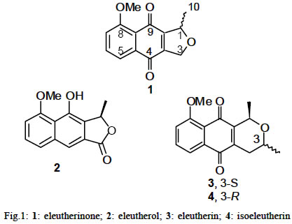
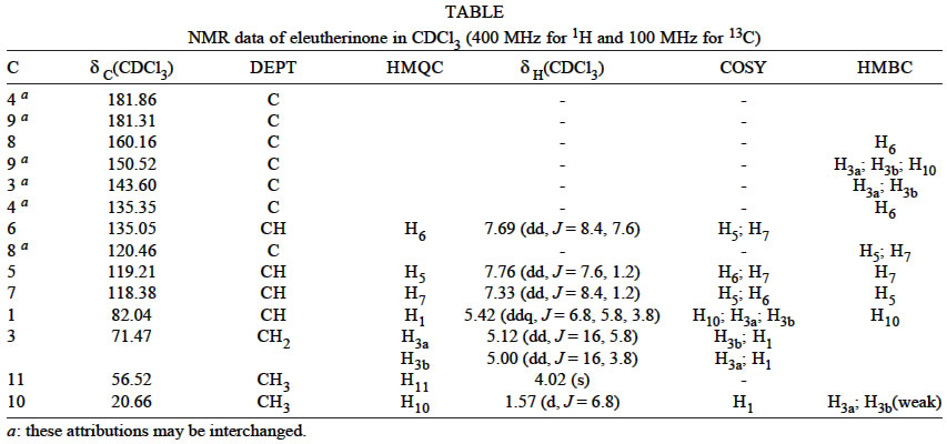
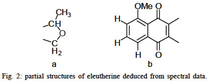
Medical Center, San Francisco, CA, US Sponsorship Fiocruz/CNPq/Pronex Received 20 December 2002 Code Number: oc03145 The dichloromethane extract prepared from the underground parts of Eleutherine bulbosa (Miller) Urban (Iridaceae) showed strong activity in the direct bioautography assay with the phytopathogenic fungus Cladosporium sphaerospermum. This assay was used to guide the fractionation of this extract and allowed the isolation of four compounds: the new naphthoquinone eleutherinone [8-methoxy-1-methyl-1,3-dihydro-naphtho(2,3-c)furan-4,9 -dione] and the known compounds, previously isolated from this species, eleutherin [9-methoxy-1(R),3(S)-dimethyl-3,4-dihydro-1H-benzo(g)isochromene-5,10-dione], isoeleutherin [9-methoxy-1(R),3(R)-dimethyl-3,4-dihydro-1H-benzo(g)isochromene-5,10-dione], and eleutherol [4-hydroxy-5-methoxy-3(R)-methyl-3H-naphtho(2,3-c)furan-1 -one]. All quinonoid compounds showed strong antifungal activity in the bioautography assay at 100 µg/spot, while eleutherol was inactive. Key words: naphthoquinone - Eleutherine bulbosa - fungicide - medicinal plant - Cladosporium sphaerospermum Eleutherine bulbosa (Miller) Urban is an herb from Iridaceae, a botanical family that comprises 90 genera and about 1 200 species (Schultes & Raffauf 1990). This plant is used by some populations as a vermifuge (Schultes & Raffauf 1990), for painful and irregular menstruation (Hodge & Taylor 1956), intestinal disorders (Van den Berg 1984, Lin et al. 2002) and as an abortive and antifertility agent (Weniger et al. 1982). In the state of Minas Gerais, some populations near the "Rio Doce" valley make infusions of the underground bulbs of this plant to treat intestinal infections (Kloos, unpublished data). Eleutherine americana Merr. ex K. Heyne is a related herb and its rhizome is used as a folk medicine for the treatment of coronary disorders (Ding & Huang 1983). The rhizomes of Eleutherine subaphylla Gagnep. are used as haemostatic and antibacterial agents (Dam & Mai 1990) . Several compounds, including elecanicin, eleutherol, isoeleutherol, eleutherin, isoeleutherin (Hara et al. 1997), hongconin (Zhengxiong et al. 1984) as well as anthraquinones (Komura et al. 1983) and their glycosides (Shibuya et al. 1997) have already been isolated and identified from bulbs of plants belonging to this genus. Some compounds present in the genus display important biological activities. Thus, Hara et al. (1997) described eleutherin as forming a type of "noncleavable complex" with topoisomerase II with stereospecific and selective inhibitory activity. These authors also described the inhibitory activity of isoeleutherin and isoeleutherol against HIV replication in H9 lymphocytes. The direct bioautographic assay is a rapid and sensitive method to detect fungitoxic compounds (Homans & Fuchs 1977). The use of this assay with Cladosporium sphaerospermum was demonstrated by the detection and isolation of antifungal amides from Piper arboreum (Silva et al. 1998) and polygodial from Polygonum punctatum (Alves et al. 2001). This fungus is pathogenic to plants such as Lutjanus campechanus (Blaylock et al. 2001) and other organisms including the invertebrate marine species Hippodiplosia insculpta (Sterflinger et al. 2001). With the aim of detecting bioactive natural products we evaluated hundreds of plant extracts using this assay. As a result the fungitoxic activity of the dichloromethane extract from bulbs of E. bulbosa was detected. Although previous study showed the presence of the xanthones mangiferin and isomangiferin in the leaves of E. bulbosa (Williams & Harborne 1985) no report on the chemical investigation of its bulbs could be found. We report herein the isolation and identification of the fungitoxic components of the bulbs of this plant extract by a bioassay-guided chemical fractionation protocol. MATERIALS AND METHODS The bulbs (170 g) of E. bulbosa were collected on February 1998 at Nova União (municipality of Itabirinha de Mantena), state of Minas Gerais, Brazil. A voucher specimen was deposited at the Federal University of Minas Gerais Herbarium, under the code BHCB 41473. The plant material was oven-dried at 45°C, crushed, and macerated (3 x 24 h) with dichloromethane to produce 2.5 g of crude extract. This extract was fractionated on a MPLC column (490 X 50 mm) of silica gel (230-400 mesh) at a flow rate of 60 ml/min using a step gradient of hexane-ethyl acetate 25% (35 min); 50% (15 min); 75% (25 min) and 100% (10 min). Nineteen fractions (270 ml) were collected and the solvents removed in a rotary evaporator under vacuum, at temperatures under 45°C. Fraction 11 (Rt 51 min; 110 mg) was subjected to a semi-preparative HPLC separation on a DIOL column (20 x 250 mm, 7 µm particle size) using a mixture of hexane-dichloromethane-acetonitrile (187:7:6) at 10 ml/min to yield 5 mg of eleutherinone (1). Under similar conditions, the fraction 10 (Rt 45 min; 78 mg) was eluted with a mixture of hexane-dichloromethane-acetonitrile (79:19:2) and yielded 2 (eleutherol, 29 mg), 3 (eleutherin, 4.7 mg), and 4 (isoeleutherin, 12 mg) (Fig. 1). Compound 1 crystallized from ethanol as yellow needles with mp 156-158oC; [a]25D +60 (c 0.13, CH2Cl2); UV (CH3OH) lmax (log e) 228 (4.29) ; 242 (4.26); 288 (3.64); 330 (3.52); 347 (3.46) and 400 (3.57); 1H NMR (CDCl3, 400MHz) and 13 C NMR (CDCl3, 100MHz): see Table. EIMS m/z (% intensity): 244 [M]+(79), 229 [M]+ _Me (100), 202 (20), 201 [M]+ -Me _ CO (14), 173 [M]+ _Me _ 2 x CO (15), 143 (10), 127 (10), 115 (40), 102 (11), 76 (37). The spectral and physical properties of 2, 3, and 4 were identical to those published in the literature (Hara et al. 1997). Antifungal activity - The bioautographic assay with the spores of C. sphaerospermum was run using the procedure developed by Homans and Fuchs (1977). Briefly, the extract, fractions and pure compounds were dissolved in a volatile solvent and spotted (100 µg) on commercial silica gel TLC plates (20 x 20 cm, 200 µm layer thickness, with fluorescent indicator). A suspension of the fungal spores (106/ml) in a nutrient medium (glucose and salts) was sprayed over the plates which were then incubated in a humid atmosphere for three days in the dark, at room temperature. The presence of fungitoxic substances was detected by the appearance of an inhibition zone. Thymol (50 µg/spot) and solvent (10 µl) were used as controls. DISCUSSION The oven-dried bulbs were crushed and macerated with dichloromethane to afford an extract that showed strong growth inhibition in the bioautographic assay using the fungus C. sphaerospermum. Medium pressure column chromatography over silica gel using mixtures of hexane-ethyl acetate as eluents yielded two active fractions that were further purified by HPLC on a DIOL column to afford four compounds. Three of them, 2, 3, and 4, were identified by comparison of their physical and spectral data with those published in the literature (Hara et al. 1997). Compound 1 was found to be new and its structural elucidation is described bellow. The pure compounds were subjected to the bioautographic assay with the spores of C. sphaerospermum at 100 µg/spot. Only the quinonoid compounds (1, 3 and 4) were active, producing clear growth inhibition zones. Compound 1 showed molecular ion peak with m/z 244,0728 daltons in the high resolution mass spectrum (Calcd. for C14H12O4 = 244.0736 daltons). The calculated molecular formula is consistent with the 1H NMR and 13C NMR spectra, from which signals due to 14 carbons and integration of 12 hydrogen atoms were evident (Table). Furthermore, signals from four methyne, one methylene and two methyl groups could be distinguished in the DEPT sub-spectrum, thus corroborating the number of hydrogen atoms in the molecule. The 13C NMR spectrum showed signals relative to two carbonyls (d 181.90 and d 181.31) and to an aromatic methoxy group (d 56.52), accounting for three of the four oxygen atoms. The chemical shifts of a methyne at d 82.04 and of a methylene at d 71.47 indicate that they are connected to an oxygen atom in an ether group. One of the substituents of the methyne is a methyl group, as deduced by the couplings observed in the 1H NMR and COSY spectra, compatible with the substructure shown in Fig. 2a. The UV absorption spectrum of 1 indicates the presence of a substituted naphthoquinone chromophore with lmax at 228, 242, 288, 330, 347 and 400 nm. Furthermore, besides the two close carbonyl signals mentioned above, the 13C NMR spectrum presents eight signals in the region between d 165 and d 115 that are compatible with a substituted p-naphthoquinone ring system (Fig. 2b). The 1H NMR spectrum showed signals due to three vicinal aromatic protons at d 7.76 (1H, dd J = 1.2, 7.6), 7.69 (1H, dd J = 8.4, 7.6) and 7.28 (1H, dd J = 1.2, 8.4) and to one aromatic methoxy group at d 4.02 (Table). These data are compatible with a 5-methoxy-1,4-naphthoquinone substructure (Fig. 2b). All structural features described so far account for eight of the nine insaturations predicted by the molecular formula. As all sp2 carbons were already allocated, the last insaturation must be due to a third ring based on the ether fragment deduced above. The connection of these fragments forms a 1,3-dihydro-naphtho(2,3-c) furan-4,9-dione ring system in which the quinonoid double bond between the methyne and the methylene of the furanoid ring could explain the long range coupling between them observed in the 1H NMR and COSY experiments. Finally, as the NOESY experiment shows the nuclear Overhouser effect between the methyl and methoxy groups, they were located on the same side of the structure. This analysis leads to structure (Fig. 1), corresponding to 8-methoxy-1-methyl-1,3-dihydro-naphtho(2,3-c )furan-4,9-dione. Although the isolated natural product is optically active ([a]25D +60°), the stereochemistry at C-1 could not be with the available spectral data. However, the accessibility of 2, for which the stereochemistry was unequivocally determined by comparison with literature data, offers the possibility to define the stereochemistry of 1 via chemical transformations. Thus, we anticipate that the the lactone carbonyl of the naphthofuran 2 could be reduced and the resulting phenol oxidized to one of the two possible enantiomers of 1. Comparison of the optical rotation and other physicochemical data would afford the correct stereochemistry of 1. The results of this investigation will be described in a forthcoming communication. Even though several plants of this genus had already been investigated, the use of a simple bioassay was able to guide the chemical fractionation to a new bioactive naphthoquinone. Thus, besides contributing to the knowledge of the chemical composition of this species, the new quinone 1 could be useful as a chemical marker (Williams & Harborne 1985) of this species/genus in chemosys-tematic studies, for extract standardization, and plant drug analysis. Further studies on this species aiming at minor components as well detailed investigation on the biological activities of the isolated compounds are now in progress and will be reported elsewhere. ACKNOWLEDGEMENTS To Dr L Capellari Jr. for the plant determination, Dr Lúcia PS Pimenta and Dr Marcos Eberlin for the optical activity and HRMS measurements, and Ms Mary Lou Quinn for the access to the Napralert database. REFERENCES
Copyright 2003 Instituto Oswaldo Cruz - Fiocruz. Free, full-text also available from http://www.memorias.ioc.fiocruz.br The following images related to this document are available:Photo images[oc03145t1.jpg] [oc03145f1.jpg] [oc03145f2.jpg] |
| |||||||||
