 |
| About Bioline | All Journals | Testimonials | Membership | News |
|
||||||
|
||||||
Mem Inst Oswaldo Cruz, Rio de Janeiro, Vol. 98, No. 8, Nov, 2003, pp. 1011-1016 Nodular Typhlitis Associated with the Nematodes Heterakis gallinarum and Heterakis isolonche in Pheasants: Frequency and Pathology with Evidence of Neoplasia Rodrigo Caldas Menezes, Rogério Tortelly*, Delir Corrêa Gomes/++, Roberto Magalhães Pinto/+/++ Laboratório de Helmintos
Parasitos de Vertebrados, Departamento de Helmintologia, Instituto Oswaldo
Cruz-Fiocruz, Received 17 July
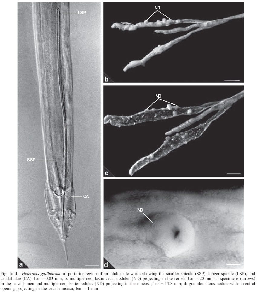
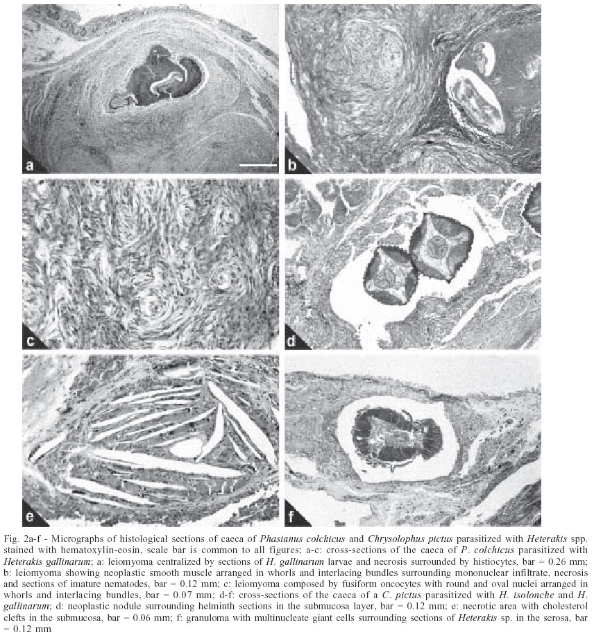
2003 Code Number: oc03197 An investigation related to the frequency and pathology of Heterakis gallinarum and pathology of Heterakis isolonche in pheasants from Rio de Janeiro, Brazil was conducted by means of clinical examinations, necropsies, and histopathological analysis in 50 ring-necked pheasants from backyard flocks of 11 localities; also, histological sections of caeca of golden pheasants deposited in the Helminthological Collection of the Oswaldo Cruz Institute (CHIOC) have been considered in the present study. During necropsies, only specimens of H. gallinarum were recovered with a prevalence of 90%, mean intensity of 81.9 and range of infection of 1-413. Gross lesions were characterized by congestion, thickening, petechial haemorrhages of the mucosa, intussusception, and nodules in the cecal wall. Under microscopy, chronic difuse typhlitis, haemosiderosis, granulomas with necrotic center in the submucosa and leiomyomas in the submucosa, muscular and serosa associated with immature H. gallinarum worms were observed. The examination of histological sections previously deposited in the CHIOC, revealed more severe alterations associated with concomitant infections with H. gallinarum and H. isolonche in golden pheasants, and were characterized by several necrotic areas with cholesterol clefts in the submucosa, giant cell granulomas in the submucosa, and serosa centralized by necrosis and worm sections and neoplastic nodules in the muscular and submucosa. Key words: nodular typhlitis - Heterakis gallinarum - Heterakis isolonche - pheasants - Phasianus colchicus - Brazil The nodular or verrucous typhlitis is a fatal parasitary disease, occurring worldwide and mostly affecting pheasants of different species and is characterized by the formation of inflammatory or granulomatous and even neoplastic nodules located in the cecal wall, mainly in the submucosa due, principally, to the infection with the nematode Heterakis isolonche Linstow, 1906 (Schwartz 1924, Beaudette 1942, Julini & Valenza 1975, Griner et al. 1977, Callinan 1987, Balaguer et al. 1992, Bhaskara Rao 1994). The nematode Heterakis gallinarum (Schrank, 1788) can frequently be found in the caeca of pheasants (Madsen 1941, Gilbertson & Huggins 1964, Greiner 1972, Draycott et al. 2000) and although considered of low pathogenicity in single infections (Lund & Chute 1974), previous studies report to a more severe pathology induced by this nematode in domestic Galliformes causing nodular typhlitis, with the formation of inflammatory or granulomatous cecal nodules (Meads & Taylor 1963, Kau-shik & Sharma Deorani 1969, Riddel & Gajadhar 1988, Khan et al. 1994). In Brazil, there is a single report of the occurrence of coinfection with H. isolonche and H. gallinarum and the appearance of verrucous typhlitis in ring-necked (Phasianus colchicus torquatus) and in golden (Chrysolophus pictus) pheasants from the Zoological Garden of Rio de Janeiro, with high mortality levels (Mendonça 1953). Nevertheless, there were no data on the pathology and frequency of these nematodes in the hosts, confirming the lack of information about this disease in our country. The main target of this investigation was to evaluate the prevalence, mean intensity, and range of infection of H. gallinarum and the associated gross and microscopic lesions in ring-necked pheasants (Phasianus colchicus) from backyard flocks in Rio de Janeiro, Brazil, comparing the microscopic findings to those previously reported for H. isolonche. MATERIALS AND METHODS Fifty specimens of ring-necked pheasants (Phasianus colchicus L., 1758), 25 males, 25 females, weigh 200-750 g from backyard flocks of 11 localities of the state of Rio de Janeiro, Brazil, were investigated. The localities and number of examined hosts are, respectively: Niterói (22º53'S-43º06'W): 9; Rio de Janeiro (22º54'S-43º12'W): 8; Tanguá (22º73'S-42º71'W): 9; São Francisco do Itabapoana (21º28'S-41º08'W): 8; Santo Antônio de Pádua (21º54'S-42º18'W): 2; Areal (22º14'S-43º65'W): 3; Petrópolis (22º30'S-43º10'W): 2; São José do Vale do Rio Preto (22º09'S-42º55'W): 2; Rio Bonito (22º43'S-42º37'W): 4; Engenheiro Paulo de Frontin (22º32'S-43º40'W): 2; Laje do Muriaé (21º12'S-42º07'W): 1. After individual clinical evaluation birds were killed and submitted to necropsy, according to the technique of Zander et al. (1997). Caeca were opened, with the aid of scissors, in Petri dishes containing 0.85% NaCl solution. Nematodes were collected, rinsed in the same solution, fixed with hot AFA (alcohol 70º GL, 93 ml; formaldehyde, 5 ml; acetic acid, 2 ml) and counted under a stereoscope microscope. Nematodes were clarified in acetic acid and phenol and identified in accordance with Vicente et al. (1995). Some specimens were preserved in Canada balsam and deposited in the Helminthological Collection of the Oswaldo Cruz Institute (CHIOC) 36213, 36.214 a-c, 36215 a-c, 36216, 36217, 36218, 36219, and 36220 (whole mounts); slides 36214 d-f, 36221 and 36222 refer to histological sections. Fragments of the parasitized caeca were removed and immediately fixed in 10% formalin. The material was then routinely processed (Behmer et al. 1976) for parafin embedding. Five micrometers thick sections were stained with hematoxylin and eosin (HE) and Van Gieson. Micrographs were obtained in a Zeiss Axiophot brightfield microscope. For the comparative study of the microscopic alterations observed in the case of coinfection of H. isolonche and H. gallinarum in C. pictus, original slides CHIOC 19502 a-g, were examined. The development of this research has been authorized by the Committee of Ethics for the Use of Animals (CEUA-Fiocruz), P0095-01. RESULTS The prevalence of H. gallinarum (Fig. 1a) was 90%, with a mean intensity of 81.9 and range of infection of 1-413. The nematodes were observed along the whole length of the caeca, mainly in the tips of these organs, in all the investigated localities. The nematode H. isolonche was not found and the parasitized birds with H. gallinarum did not present clinical signs. The gross lesions in the caeca, present in 37 (82%) of parasitized pheasants, were characterized by congestion, thickening, and petechial haemorrhages in the mucosa, intussusception in one of the birds, abscesses in the cecal tips of two birds, and nodules projecting in the mucosa and serosa. These birds presented a mean infection of 74.0 and a range of infection of 1-413. In 18 (40%) of the parasitized birds, 1-10/cecum small pink, dark-brown or reddish nodules, 1-3 mm in diameter projecting in the mucosa, with or without central openings were observed (Fig. 1d). The mean infection and range of infection in these animals were 41.8 and 1-193, respectively. In another 8 (17%) infected pheasants, for which these values were 111.4 and 5-310, light-pink, dark-brown or orange-reddish nodules also were present. Nevertheless, these lesions 1-25/cecum were bigger, 5-8 mm in diameter, without central opening, mainly projecting in the mucosa but also appearing in the serosa (Fig. 1b, c); when sectioned the nodules were white-grayish in color. The microscopic lesions in the parasitized ring-necked pheasants were diffuse chronic typhlitis with mononuclear cells infiltrate, haemosiderosis in the lamina propria and nodules in the mucosa, submucosa, and serosa. The 1-3 mm diameter nodules were localized in the submucosa and were represented by granulomas centralized with necrotic material suggesting the passage of helminth larvae and, in one out of two pheasants, were associated with lymphoid leucosis. The 5-8 mm diameter nodules appeared under several aspects; the smallest, compact and of fibrovascular nature, were located in the submucosa and muscular layer, projecting into the serosa. The biggest, of similar aspect, were present in the submucosa, and presented areas of myxoid characteristics with linear lymphocyte infiltrates. Multinuclear giant cells and histiocytes surrounded fragments of immature worms and cellular debris. Some of these nodules, covered with fibrotic tissue, were mainly located in the submucosa but also in the muscular layer and seldom in the serosa, showing characteristics of neoplasia, with fusiform oncocytes, with rounded to ovaled nuclei, forming bundles and whorls, without signs of atypical cells or invasion of adjacent tissues (Fig. 2a, b, c). Frequently, immature worms, some in a process of degeneration, surrounded by necrosis, were observed in cross-sections of these nodules (Fig. 2a, b). The staining by the Van Gieson's technique revealed, in despite of the discreet affinity to the stain, the presence of a leiomyoma. The mucosa in the site of the lesion showed the presence of a rich mononuclear infiltrate, erosion areas and marked atrophy (Fig. 2a). In the case of the intussusception, erosion of the caecal epithelial lining and predominant mononuclear infiltrates with few granulocytes in the mucosa, haemorrhagic areas in the lamina propria, a large edema in the submucosa and serositis were observed. Examination of slides CHIOC 19502 a-g containing the cross-sections of the caeca of C. pictus parasitized with H. gallinarum and H. isolonche in accordance with Mendonça (1953) revealed a great amount of viable worms, almost adults, mainly localized in the submucosa (Fig. 2d). The inflammatory reaction was absent around some of the parasites. Nevertheless, necrotic areas with cholesterol clefts in the submucosa (Fig. 2e) and giant cell granulomas in the submucosa and serosa, centralized by necrosis and sections of nematodes could be observed (Fig. 2f). Also, neoplastic nodules in the submucosa and muscular layer identical to those observed in our material could be seen. However, a more specific classification of the neoplasia could not be achieved by means of special staining methods, since the slides were prepared with hematoxylin and eosin and paraffin blocks were not available. DISCUSSION The high prevalence, mean intensity, and range of infection of H. gallinarum observed indicate that this is a very common nematode species infecting backyard flocks of ring-necked pheasants in the state of Rio de Janeiro. In this same locality this parasite was also the most frequent and highly prevalent (100%) in guinea fowls (Menezes et al. 2001) and (60%) in domestic chickens (Grisi & Carvalho 1974). H. gallinarum was also the most prevalent parasite recorded from ring-necked pheasants in England (Draycott et al. 2000), US (Olsen 1938, Gil-bertson & Huggins 1964, Greiner 1972, Pence et al. 1980), and Denmark (Madsen 1941). The absence of H. isolonche, reported only once in Brazil by Mendonça (1953) suggests that this may be an exotic parasite in our country. That case report referred above was on the basis of nematodes recovered from presumably imported birds, and historical data, shows that the birds were already parasitized on arrival in Rio de Janeiro, (departing from the state of São Paulo), reinforces this argument. In the scarce papers that refer to the frequency of H. isolonche in pheasants, Clapham (1961) reported a low prevalence (7.9%) in the United Kingdom, and Dowel et al. (1983) found this nematode in only 1.2% of the pheasants investigated. However, higher prevalences of H. isolonche have been reported in other birds, such as the bowhite quail (Colinus virginianus), and the lesser prairie chicken (Tympanucus pallidicinctus) in the US (Pence et al. 1983, Moore et al. 1986), and scavenging chickens in Africa (Permin et al. 1997, Poulsen et al. 2000), but with rather lower values than those observed for H. gallinarum when present. The cecal gross lesions associated with H. gallinarum in the present study were not always related to heavy worm burdens. These were similar burdens to those described for guinea fowls by Menezes et al. (2001) and in domestic chickens by León and Soldevila (1978). However, these authors did not refer to the occurrence of cecal nodules. Such nodules were noted by Khan et al. (1994), in guinea fowls, Kaushik and Sharma Deorani (1969), Mutalib and Riddell (1982), Riddell and Gajadhar (1988) in domestic chickens, and Meads and Taylor (1963) in pheasants, microscopically classified as inflammatory (Meads & Taylor 1963, Kaushik & Sharma Deorani 1969) or granulomas (Mutalib & Riddell 1982, Riddell & Gajadhar 1988, Khan et al. 1994), associated with the presence of H. gallinarum, as observed in this study. Helmboldt and Wyand (1972) reported the occurrence of neoplastic nodules in the golden pheasant, identified to leiomyomas caused by immature specimens of Heterakis sp., in despite of finding only H. gallinarum in the cecal lumen. Taking into account that the results of these authors were similar those obtained here, it can be affirmed that the leiomyomas were due to H. gallinarum infection alone, because if specimens of H. isolonche were present, then adult nematodes of this species would have been observed in the lumen or burrowed in the cecal submucosa of the parasitized birds, their principal site of infection (Schwartz 1924, Griner et al. 1977). The cecal nodules associated with H. isolonche, mainly in the submucosa, have already been micros-copically classified as granulomas and fibrous hyperplastic tissue (Griner at al. 1977, Callinan 1987), of fibrohistiocytic nature (AFIP 1998) or fibrovascular (Julini & Valenza 1975) and leiomyoma (Krahnert 1952, Balaguer et al. 1992), demonstrating controversial diagnoses. These descriptions are similar to the nodules observed in this study having, as a common characteristic, formation by fusiform mesenchymal cells with elongate nuclei arranged in dense whorls and interlacing bundles, sarcomatous in aspect, but without metastasis, mitotic figures or in-filtrations. On the basis of these characteristics, showing a situation of uncontrolled cell proliferation and in accordance with the definition of neoplasia (Jones et al. 1997), the nodules observed herein can be considered benign neoplasms, in disagreement with Julini and Valenza (1975), Griner et al. (1977), Callinan (1987), and AFIP (1998). A question to be answered is why leiomyomas were found in only eight of the pheasants parasitized with H. gallinarum, some with small worm burdens. In line with the present results, and those previously reported, the development of this neoplasia could be related to the immune response of pheasants to parasitism by Heterakis sp. The species H. isolonche, due to its higher pathogenicity related to its histotropic cycle (Levine 1980), would be the more frequent etiological agent of the neoplasia; nevertheless, the presence of immature H. gallinarum worms would be also responsible for the formation of tumors. The microscopic lesions present in the slides of Mendonça (1953) were more severe than those observed here, possibly due to the larger amount of more developed worms perforating the cecal wall and provoking peritonitis, the probable cause of death of the pheasants. It was impossible to relate the nodular lesions to specimens of H. isolonche alone, in despite of the stage of development of these helminths, since their association with H. gallinarum worms was observed. We agree with Riddell and Gajadhar (1988) in con-sidering that the probable causes for the appearance of granulomatous nodules, that in our opinion could evolve to neoplasias, would be continuous reinfections with H. gallinarum provoking a tissular phase for the parasite as observed by Kaushik and Sharma Deorani (1969), mainly by infections with different strains of nematode harbored by other host species. These strains are likely not adapted to the ring-necked pheasant, and thus induce a higher pathogenicity, as shown in previous results. Tompkins et al. (2001) in experimental infections of ring-necked pheasants and grey partridge (Perdix perdix), using H. gallinarum recovered from pheasants demonstrated that the latter hosts were more severely affected, in terms of weight, reduction of food intake and decrease of cecal activity. Perhaps related to this, strains of H. gallinarum from chickens were not so successful (in terms of the number of adult worms recovered, and the fecundity of females) when inoculated into turkeys (Lund et al. 1970). In addition, in young chuckar partridges, larvae strains of H. gallinarum recovered from other domestic Galliformes, and thus not physiologically adapted to this host, frequently perforate the cecal wall causing peritonitis (Lund & Chute 1974). Hence, the obtained results suggest a general pattern whereby both parasite fitness and host pathology can be observed. ACKNOWLEDGEMENTS To Jayade Machado de Mendonça, retired researcher of the Departamento de Helmintologia, Instituto Oswaldo Cruz, who pioneered the study of the nodular typhlitis of pheasants in Brazil 50 years ago, and to Bruno Eschenazi Vieira, Laboratório de Imagens, Instituto Oswaldo Cruz, for technical assistance with the figures. REFERENCES
Copyright 2003 Instituto Oswaldo Cruz - Fiocruz. Free, full-text also available from http://www.memorias.ioc.fiocruz.br The following images related to this document are available:Photo images[oc03197f2.jpg] [oc03197f1.jpg] |
| |||||||||
