 |
| About Bioline | All Journals | Testimonials | Membership | News |
|
||||||
|
||||||
Mem Inst Oswaldo Cruz, Rio de Janeiro, Vol. 98, No. 8, Nov, 2003, pp. 1115-1120 Antibacterial Activity of Extracts and Neolignans from Piper regnellii (Miq.) C. DC. var. pallescens (C. DC.) Yunck Greisiele Lorena Pessini++, Benedito Prado Dias Filho, Celso Vataru Nakamura, Diógenes Aparício Garcia Cortez*/+ Departamento de
Análises Clínicas *Departamento de Farmácia e Farmacologia,
Universidade Estadual de Maringá,
Av. Colombo 5790, 87020-900 Maringá, PR, Brasil Financial support: Conselho Nacional de Desenvolvimento Científico e Tecnológico, Capacitação de Aperfeiçoamento de Pessoal de Nível Superior, and Programa de Pós-graduação em Ciências Farmacêuticas, Universidade Estadual de Maringá Received 27 May
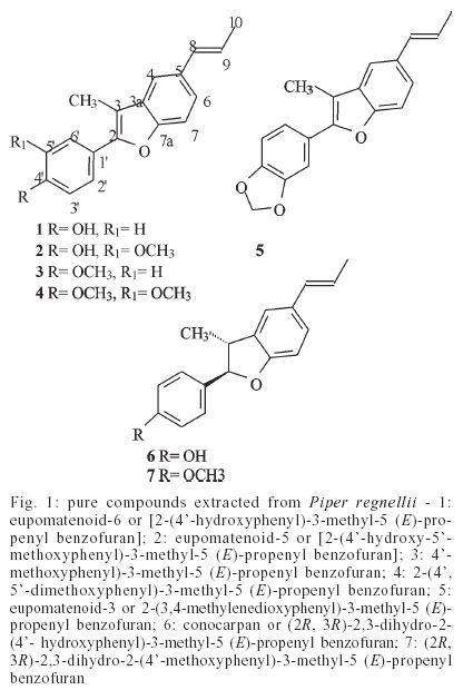
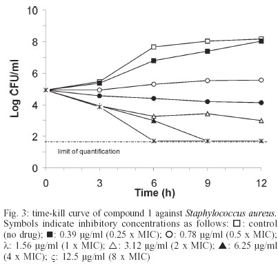
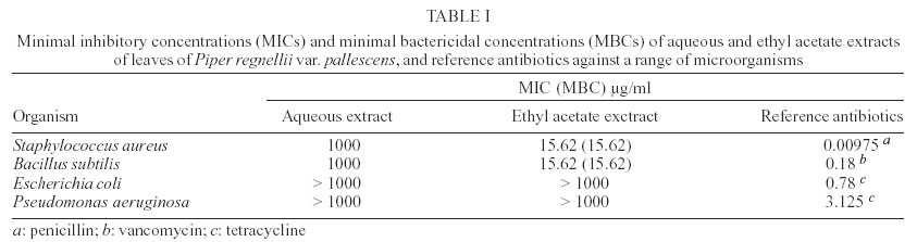
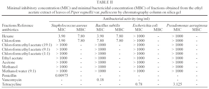
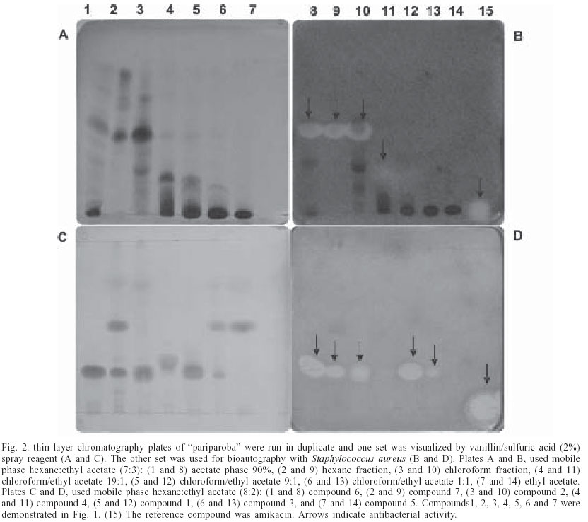
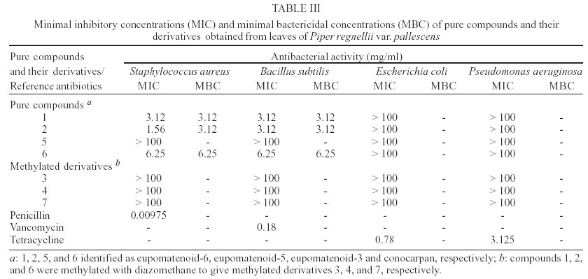
2003 Code Number: oc03217 The evaluation of the activity of the aqueous and ethyl acetate extracts of the leaves of Piper regnellii was tested against gram-positive and gram-negative bacteria. The aqueous extract displayed a weak activity against Staphylococcus aureus and Bacillus subtilis with minimal inhibitory concentration (MIC) and minimal bactericidal concentration (MBC) of 1000 µg/ml. The ethyl acetate extract presented a good activity against S. aureus and B. subtilis with MIC and MBC at 15.62 µg/ml. In contrast to the relative low MICs for gram-positive bacteria, gram-negative bacteria were not inhibited by the extracts at concentrations £ 1000 mg/ml. The ethyl acetate extract was fractionated on silica gel into nine fractions. The hexane and chloroform fractions were active against S. aureus (MIC at 3.9 µg/ml) and B. subtilis (MIC at 3.9 and 7.8 µg/ml, respectively). Using bioactivity-directed fractionation, the hexane fraction was rechromatographed to yield the antimicrobial compounds 1, 2, 5, and 6 identified as eupomatenoid-6, eupomatenoid-5, eupomatenoid-3, and conocarpan, respectively. The pure compounds 1 and 2 showed a good activity against S. aureus with MIC of 1.56 µg/ml and 3.12 µg/ml, respectively. Both compounds presented MIC of 3.12 µg/ml against B. subtilis. The pure compound 6 named as conocarpan was quite active against S. aureus and B. subtilis with MIC of 6.25 µg/ml. The antibacterial properties of P. regnellii justify its use in traditional medicine for the treatment of wounds, contaminated through bacteria infections. Key words: Piper regnellii - neolignans - antibacterial activity - bioautography Piper regnellii (Miq) C. DC. var. pallescens (C. DC.) Yunck popularly known as "pariparoba" are distributed in tropical and subtropical regions of the world (Cronquist 1981). Leaf and root are used in form of crude extracts, infusions or poultices to treat wounds, swellings, and skin irritations (Yuncker 1972, 1973, Corrêa 1984). In a screening of Brazilian medicinal plants, we reported the antimicrobial activity of the aqueous-ethanolic extract of the leaves of P. regnellii (Miq) C. DC. against the bacterium Sta-phylococcus aureus and Bacillus subtilis and against the yeasts Candida krusei and Candida tropicalis (Holetz et al. 2002). Chauret et al. (1996) have reported for the first time in a species of the Piperaceae three known neolignans (conocarpan, eupomatenoid-5, and eupomatenoid-6) isolated from Piper decurrens via insecticidal bioassay-guided fractionation. In another phytochemical study accomplished with P. regnellii leaves, the biosynthetic pathway was established which leads to the dihy-drobenzofuran neolignan (+)-conocarpan by means of in vivo administration of labeled l-phenylalanine [U-14C] and enantioselective conversion of p-hydroxypropenyl-benzene to (+)-conocarpan (85%) by an enzyme fraction from the leaves (Sartorelli et al. 2001). The purpose of this study was to analyse the chemical composition and the antibacterial activity of separated fractions of ethanolic extracts from the leaves of P. regnellii as well as of the bioactivity-directed isolates: eupomatenoid-6, eupomatenoid-5, eupomatenoid-3, and conocarpan. In addition, time-kill studies were performed to determine if eupomatenoid-6 had bactericidal activity. MATERIALS AND METHODS Instruments - NMR spectra were obtained in a BRU-KER DRX400 (9.4 T) and VARIAN GEMINI300 (7.05T), using deuterated solvent for field homogeneity, TMS as internal standard and a constant temperature of 298K. For gradient Heteronuclear Multiple Bond Coherence the coupling constants were optimized for 4, 6, 8, and 12 Hz. IR: film NaCl plates; Electrospray-Mass Spectrometry were recorded on a Micromass Quattro LC, High Resolution Mass Spectroscopy: Autospec-Micromass EBE, and Electronic Impact-Mass Spectroscopy on a Gas Chromatography/Mass Spectrometry-SHIMADZU QP 2000 A. CC: silica gel 60 (70-230 and 230-400 mesh); gel chromatography: Sephadex LH-20; TLC: silica gel plates F254 (0.25 mm in thickness). Plant material - The leaves of P. regnellii (Miq.) C. CD. var. pallescens (C. DC.) Yunck were collected in August of 2001 in the Medicinal Plants Garden "Profª. Irenice Silva" of the Universidade Estadual de Maringá campus, Maringá, PR. The plant material was identified by Marilia Borgo of the Botanical Department of the Universidade Federal do Paraná, and a voucher specimen (number HUM 8392) is deposited at the Herbarium of the Universidade Estadual de Maringá. Plant extract and fractionation - Dried leaves (200 g) of P. regnellii were extracted with ethanol:water (9:1) at room temperature. The solvent was removed under vacuum at 40°C to give an aqueous extract and a dark green residue. This aqueous extract was lyophilized (13.9 g) the dark green residue mentioned above was washed with ethyl acetate and the organic solvent removed to give the ethyl acetate extract (15.3 g). The aqueous and ethyl acetate extracts were assayed against S. aureus by bioautography and broth microdilution assay to determine the MICs described below. The active ethyl acetate extract (10.7 g) was submitted to vacuum column chromatography (silica gel 150 g) and eluted with hexane (1000 ml), chloroform (1400 ml), chloroform/ethyl acetate 19:1 (1000 ml), chloroform/ethyl acetate 9:1 (700 ml), chloroform/ethyl acetate 1:1 (500 ml), ethyl acetate (500 ml), acetone (700 ml), methanol (1400 ml) and methanol/water 9:1 (1800 ml). Further separation of the active hexane fraction (2.4 g) by column chromatography on silica gel 60 (230-400 mesh) eluted with hexane, hexane/chloroform (49:1, 19:1, 9:1 and 1:1), chloroform, ethyl acetate, acetone, and methanol showed 72 fractions, which were assayed against S. aureus as described below. Thin layer chromatography comparison and biograms of the fractions lead to their combination into four larger fractions (F10, F31, F38, F44) containing antimicrobial active compounds. The active fraction F44 was further separated on a ready made Silica gel 60 (230-400 mesh) column (1 x 20 cm) with the mobile phase hexane:dichlomethane:ethyl acetate to demonstrate 26 fractions. (F44-1 to F44-26). The most active fraction F44-5 was selected by means of biograms and further separated by means of column chromatography, using the above mentioned column in combination with the mobile phases. This procedure allowed the isolation of pure compounds 1 (98.5 mg), 2 (82.9 mg), 5 (55.1 mg), and 6 (181.3 mg) identified as eupomatenoid-6, eupomatenoid-5, eupo-matenoid-3, and conocarpan, respectively (Fig. 1), by spectroscopic analysis and by comparison with literature data (Achenbach et al. 1987, Chauret et al. 1996, Snider et al. 1997). Compounds 1, 2, and 6 were methylated with diazomethane to give methyl derivatives 3, 4, and 7, respectively (Fig. 1). Microorganisms used and growth conditions - The following strains were used as test organisms: Escherichia coli ATCC 25922, Pseudomonas aeruginosa ATCC 15442, Bacillus subtilis ATCC 6623, and Staphylococcus aureus ATCC 25923. The bacteria were grown in nutrient broth (Difco Laboratories, Detroit, MI) at 37°C and maintained on nutrient agar slants at 4°C. Antibacterial
susceptibility testing - The minimal inhibitory concentrations (MICs)
of the all extracts and reference antibiotics (tetracycline, vancomycin,
and penicillin - Sigma Chemical Co., St. Louis, MO, US) were determined
by microdilution techniques in Mueller-Hinton broth (Merck) for bacteria
(NCCLS 2000). Inoculates were prepared in the same medium at a density
adjusted to a 0.5 McFarland turbidity standard [108 colony-forming
units (CFU)/ml] and diluted 1:10 for the broth microdilution procedure.
Microtiter plates were incubated at 37ºC and the MICs were
recorded after 24 h of incubation. Two susceptibility endpoints were recorded
for each isolated. The MIC was defined as the lowest concentration of compounds
at which the microorganism tested did not demonstrate visible growth. Minimum
bacteriostatic concentration (MBC) was defined as the lowest concentration
yielding negative subcultures or only Thin layer chromatography - Kieselgel GF254 plates, 20 x 20 cm, 1 mm thick, was used. Plant extracts (1 mg/ml) were applied (50 µl) and the chromatogram developed using hexane:ethyl acetate (70:30) as solvent. TLC plates were run in duplicate and one set was used as the reference chromatogram. Spots and bands were visualized by UV irradiation (254 and 366 nm) and vanillin/sulphuric acid (2%) spray reagent. The other set was used for bio-autography. Amikacin (12.8 µg) (Bristol Myers Squibb) was used as reference antibiotic. Bioautography - Chromatograms developed as described above were placed in a square plate with cover and an inoculum of S. aureus containing 106 CFU/ml in molten Mueller-Hinton agar was distributed over the plate. After solidification of the medium, the TLC plate was incubated overnight at 37°C. Subsequently the bio-autogram was sprayed with an aqueous solution of 2,3,5-triphenyltetrazolium chloride (TTC) and incubated at 37°C for 4 h. Inhibition zones indicated the presence of active compounds. Time-kill curve methodology - Prior to experi-mentation, the lower limit of bacterial quantification and the potential of compound carryover during the plating process were determined as previously described by the NCCLS (2000). According to these sampling procedures antibacterial carryover was not observed. Additionally, the lower limit of bacterial quantification was 50 CFU/ml. The effects of compound 1 (Fig. 3) on the growth of S. aureus were determined by preparing a standardized suspension as described above. Dilutions yielded a starting inoculum of approximately 1x10⁶. The antibacterial activity of compound 1 was studied over a range of multiples of MIC encompassing 0.25 to 8 X MIC. Tests were performed in triplicate and incubated at 37°C. At predetermined time points (0, 3, 6, 9, 12 h) a 100 µl sample was removed from each test suspension, serially diluted in sterile saline, and plated on Mueller-Hinton Agar plates for colony count determination. Plates were incubated at 37°C for 24 h before colony count determination. Data from triplicate runs were averaged and plotted as log CFU/ml versus time (h) for each time point. RESULTS AND DISCUSSION The evaluation of the activity of the aqueous and ethyl acetate extracts of the leaves of P. regnellii against both gram-positive and gram-negative bacteria by using the microdilution technique is given in Table I. The in vitro results were classified as follows: if the extracts displayed a MIC less than 100 µg/ml, the antibacterial activity was considered good; from 100 to 500 µg/ml the antibacterial activity was considered moderate; from 500 to 1000 µg/ml the antibacterial activity was considered weak; over 1000 µg/ml the extracts were considered inactive. The aqueous extract displayed weak activity against S. aureus and B. subtilis with both MIC and MBC of 1000 µg/ml. The ethyl acetate extract presented good activity against S. aureus and B. subtilis with MIC and MBC at 15.62 µg/ml. In contrast to the relative low MICs for gram-positive bacteria, gram-negative bacteria were not inhibited by the extracts at concentrations ≤ 1000 µg/ml. As a result of this finding, the ethyl acetate extract was fractionated on silica gel into nine fractions. The hexane and chloroform fractions were active against S. aureus (MIC at 3.9 µg/ml) and B. subtilis (MIC at 3.9 and 7.8 µg/ml, respectively) (Table II). The chloroform/ethyl acetate, ethyl acetate, acetone, methanol, and methanol/water fractions showed no activity against the organisms tested. The minimal bactericidal concentrations were within two-twofold dilutions of the MIC for these organisms. To obtain some information on the active components, plant fractions were analyzed by TLC on silica gel. TLC plates were run in duplicate and one set was used as the reference chromatogram (Fig. 2 A, C) and the other set was assayed for bioautography (Fig. 2 B, D). Panels A and C show the chromatogram of plant extracts sprayed with vanillin/sulphuric acid (2%). Panels B and D show the appearance of same chromatogram after treatment with bacterial inoculum, indicating the location of bacterial inhibition zone. Antimicrobial components were present in, the ethyl acetate phase and hexane, chloroform, and chloroform/ethyl acetate (19:1) fractions (Fig. 2 B lines 8, 9, 10, 11) with the highest antimicrobial activities in the hexane and chloroform fractions and some components at a low concentration in the chloroform/ethyl acetate (19:1) fraction. The pure isolated compounds and their derivatives (1-7) were assayed against S. aureus for bioautography (Fig. 2 C, D). As shown in panel D the pure compound isolated from the hexane fraction shows an inhibition zone with Rf 0.25 for compound 6 (line 8), compound 2 (line 10) and compound 1 (line 12). The compound responsible for the inhibition zone seen in panel D, line 9, is due to the part of compound 6 that was not methylated. Compound 5 (line 14) did not present an inhibition zone. The results of tests with the pure compounds and their derivatives are shown in Table III. Compound 6, conocarpan, showed good activity against S. aureus and B. subtilis both with MIC of 6.25 µg/ml. Compounds 1 and 2 presented a good activity against S. aureus with MIC of 1.56 µg/ml and 3.12 µg/ml, respectively. Both compounds had MICs of 3.12 µg/ml against B. subtilis. Compound 5 and the methylated derivatives (3, 4, 7) were inactive against all the bacteria tested (Table III). Compound 5 possesses no phenolic hydroxyl group. The phenolic hydroxyl present in the active structures is apparently able through hydrogen bond links with the specific molecular structures on the bacterial surface, provoke the inactivation of essential bacterial metabolism (Graham 1995). The standard antibiotics used as control in the determination of MIC behaved in conformity with patterns determinede by the literature (Lorian 1996, NCCLS 2000). The kinetics of the bactericid action against S. aureus was performed with compound 1. As shown in Fig. 3, viable cells of S. aureus were reduced by a 3 log UFC/ml within 9 h after exposure to four times the MIC of compound 1. At the highest concentration studied (8 x MIC) more than 104 organisms/ml were eradicated (reductions to < 50 CFU/ml) within 6 h indicative of the potent bactericidal activity of compound 1. The control (no-drug) exhibited a 3-log-unit increase in CFU/ml in 6 h. Bacterial cultures were monitored for up to 24 h, and no regrowth was observed. The minimal concentration of an antimicrobial agent necessary to kill an organism, MBC, should be equal to or greater than the MIC. In this study, concentrations equal to 2x the MIC have an inhibitory effect on the growth of S. aureus. A time-kill result that corresponds to the MIC, plus or minus one dilution, shows a transition from minimal activity (subinhibitory con-centrations) to maximal antibacterial effect (suprainhibitory concentrations) and encompass the concentration that produces 50% of the maximal effect. In previous studies, the biosynthetic pathway of the (+)-conocarpan P. decurrens was determined by using insecticide bioassay-guided fractionation (Chauret et al.1996, Sartorelli et al. 2001). Conocarpan represents a class of the benzofuran neolignans with a variety of biological activities, including anti-PAF, antifungical, and insecticide activity. Several compounds of this class have been isolated from Piperaceae species. Antibacterial activity has also been found in P. nigrum (Pradhan et al. 1999) and related to the presence of 3,4-dihyroxyphenyl ethanol glucoside, effective at 2.25 mmol/l against bacteria E. coli, S. aureus and B. cereus, but inactive against Salmonella typhimurium. 3,4-dihydroxy-6-(N-ethylamino) benzamide in the same plant was also found to inhibit the growth of bacteria however, the minimal inhibitory concentration was 7.6 mmol/l. In a screening of Colombian medicinal plants anti-microbial activity has been demonstrated for methanol extract of leaves from P. lanceafolium against C. albicans, Klebsiella pneumoniae, Enterococus faecalis, Myco-bacterium phlei, B. subtilis, and S. aureus with significant inhibition zones (Lopez et al. 2001). Locher et al. (1995) studied extracts obtained from selected Hawaiian medicinal plants. They did not verify zones of growth inhibition with the leaves of P. me-thysticum against S. pyogenes, S. aureus, P. aeruginosa, and E. coli. On the other hand, when the stem of P. methysticum was tested against fungi, growth inhibition was observed at 1000 µg/ml to Microsporum canis, 125 µg/ml to Epidermophyton floccosum, and 1000 µg/ml to Trichophyton rubrum. Lentz et al. (1998) had screened Honduran medicinal plant species for antimicrobial activity. The extract from P. aduncum showed inhibition zones measuring 7 mm against both S. aureus and B. subtilis, and measuring 22 mm against Mycobacterium intracellulare. In the present work, the antibacterial properties of P. regnellii suggest its potential usefulness in traditional medicine for the treatment of wounds, contaminated whit gram-positive bacteria. The isolated compounds that showed antibacterial activity were identified as eupomatenoid-6, eupomatenoid-5, and conocarpan. These compounds were active against S. aureus and B. subtilis, and virtually inactive against E. coli and P. aeruginosa. Differences in the antimicrobial effect of the isolated compounds against gram-positive and gram-negative bacteria may be due to differences in permeability barriers. In gram-negative species, an outer membrane is a fairly effective barrier for amphipathic compounds. Further studies are needed in order to elucidate the mechanism(s) of action of these compounds and their derivatives, as well as the antimicrobial activity against other microorganisms. ACKNOWLEDGMENTS To Marinete Martinez Vicentim for help in the experiments. REFERENCES
Copyright 2003 Instituto Oswaldo Cruz - Fiocruz. Free, full-text also available from http://www.memorias.ioc.fiocruz.br The following images related to this document are available:Photo images[oc03217t2.jpg] [oc03217f2.jpg] [oc03217f1.jpg] [oc03217t3.jpg] [oc03217t1.jpg] [oc03217f3.jpg] |
| |||||||||
