 |
| About Bioline | All Journals | Testimonials | Membership | News |
|
||||||
|
||||||
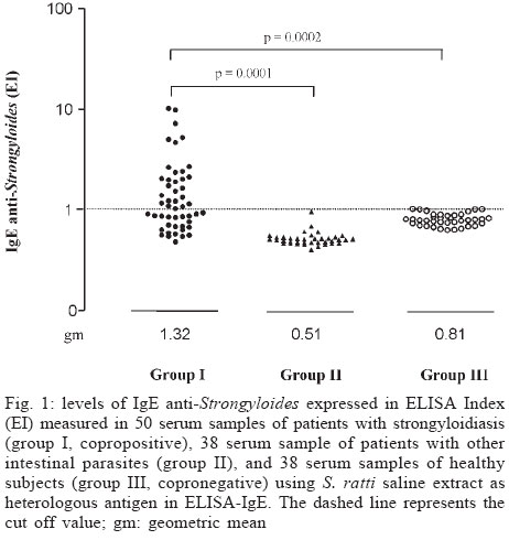
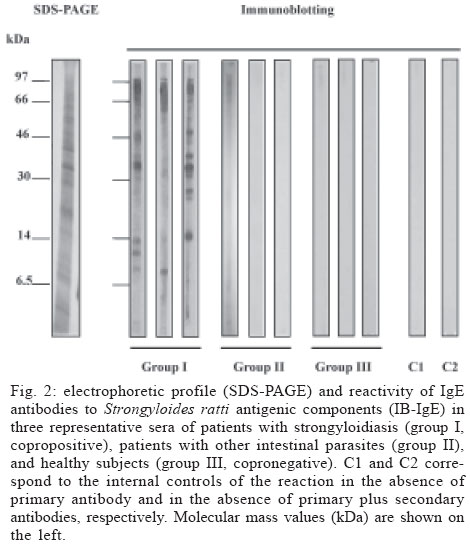
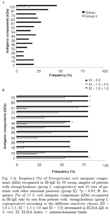
Mem Inst Oswaldo Cruz, Rio de Janeiro, Vol. 99, No. 1, Feb, 2004, pp. 89-93 Strongyloides ratti Antigenic Components Recognized by IgE Antibodies in Immunoblotting as an Additional Tool for Improving the Immunodiagnosis in Human Strongyloidiasis Rosângela Maria Rodrigues++, Mônica Camargo Sopelete, Deise Aparecida de Oliveira Silva, Jair Pereira Cunha-Júnior, Ernesto Akio Taketomi, Julia Maria Costa-Cruz+ Departamento de Imunologia, Microbiologia e Parasitologia, Instituto de Ciências Biomédicas, Universidade Federal de Uberlândia, Av. Pará 1720, 38400-902 Uberlândia, MG, Brasil Financial support: Conselho Nacional de Desenvolvimento Científico e Tecnológico, Coordenação de Aperfeiçoamento de Pessoal de Nível Superior, and Universidade Federal de Uberlândia, MG, Brasil Received 14 July 2003 Code Number: oc04016 ABSTRACT IgE antibody response in human strongyloidiasis was evaluated by enzyme-linked immunosorbent assay (ELISA) and immunoblotting (IB) using Strongyloides ratti saline extract as heterologous antigen. A total of 50 serum samples of patients who were shedding S. stercoralis larvae in feces (group I, copropositive), 38 of patients with other intestinal parasites (group II), and 38 of subjects with negative results in three parasitologic assays (group III, copronegative) were analyzed. Levels of IgE anti-Strongyloides expressed in ELISA Index (EI) were significantly higher in patients of group I (1.32) than in group II (0.51) and group III (0.81), with positivity rates of 54%, 0%, and 10.5%, respectively. Fifteen S. ratti antigenic components were recognized in IB-IgE by sera of group I, with frequency ranging from 8% to 46%. In group II, only two antigenic bands (101, 81 kDa) were detected in a frequency of 10% and no reactivity was found in group III. Sera with EI values > 1.5 recognized five from 13 specific antigenic bands (70, 63, 61, 44, 7 kDa). It can be concluded that these five antigenic components recognized by IB-IgE using S. ratti antigen might be employed as an additional tool for improving the immunodiagnosis in human strongyloidiasis. Key words: strongyloidiasis - Strongyloides ratti - IgE - immunoblotting - ELISA Strongyloides stercoralis is an intestinal nematode with worldwide distribution, predominantly in tropical and subtropical countries, and sporadically in temperate regions. Strongyloidiasis is a severe disease, but it can be clinically inapparent in the majority of patients with infection restricted to the gastrointestinal tract. Nevertheless, the systemic invasion of the parasite by its larval stage leads to a fatal hyperinfection syndrome or disseminated strongyloidiasis, particularly in immunocompromised subjects like patients with cancer, organ transplant recipients, acquired immunodeficiency syndrome (AIDS), and patients upon corticoids and other immunosuppressive therapy (Ferreira et al. 1999, Siddiqui & Berk 2001, Oliveira et al. 2002). Previous studies have shown that circulating IgE antibodies and eosinophils play an important role in the immune response to helminths (Meeusen & Balic 2000). High levels of specific IgE are usually demonstrated in immunocompetent patients with strongyloidiasis whereas lower levels are found in the disseminated disease and immunosuppressive conditions (Atkins et al. 1997, Vercelli et al. 1998). IgE anti-S. stercoralis antibodies have been detected by radioallergosorbent test (RAST), enzyme-linked immu-nosorbent assay (ELISA), and immunoblotting (IB) (McRury et al. 1986, Conway et al. 1993, Sato et al. 1995, Atkins et al. 1997). However, one of the most important limitations for immunodiagnosis in strongyloidiasis is the difficulty for obtaining S. stercoralis filariform larvae. Thus, studies have been conduced using heterologous antigen of filariform larvae from Strongyloides ratti and Strongyloides venezuelensis in the development of serological methods (Grove & Blair 1981, Costa-Cruz et al. 1997, Machado et al. 2001). The aim of this study was to evaluate IgE antibody response in human strongyloidiasis by immunoblotting using S. ratti saline extract as heterologous antigen and to compare it with ELISA-IgE results. MATERIALS AND METHODS Serum samples - A total of 126 serum samples of individuals divided into three groups according to the co-proparasitologic results obtained from the Ferrioli (1961), Lutz (1919), and Ritchie (1948) methods was analyzed. Group I consisted of 50 serum samples of patients from the Clinical Hospital, Federal University of Uberlândia and the Municipal Health Service, Uberlândia, state of Minas Gerais, Brazil, who were shedding S. stercoralis larvae in the feces (copropositive); group II consisted of 38 sera of patients with other intestinal parasites, such as Ascaris lumbricoides (10 cases); Enterobius vermicularis (4 cases); hookworm (5 cases); Taenia sp. (3 cases); Trichuris trichiura (2 cases); Giardia lamblia (12 cases); Entamoeba histolytica (2 cases); and group III comprised 38 serum samples of healthy individuals with negative results in three fecal samples (copronegative) and with no history of Strongyloides infection. In addition, subjects of groups II and III were non-reactive to Strongyloides as determined by ELISA-IgG. The Ethics Committee in Research of the Federal University of Uberlândia approved this study. Parasites - S. ratti larvae were obtained from the feces of experimentally infected rats (Rattus rattus). The fecal samples were mixed with an equal part of finely ground wood charcoal, moistened with water, spread in an uniform layer on Petri dishes and incubated at 25°C for 5 days. Filariform larvae were then harvested according to the Ferrioli (1961) method, concentrated by centrifugation for 5 min at 1000 g and stored at 20°C until being used in the heterologous antigen preparation. Heterologous antigen - S. ratti filariform larvae (500,000) were resuspended in 1 ml of 0.01 M phosphate buffered saline (PBS) pH 7.2 containing 2 mM EGTA, 2 mM EDTA and 0.3 mg/ml of protease inhibitors (Boehering Mannheim, Germany). For antigenic extraction, larvae on ice bath were disrupted using tissue homogenizer (Omnith International, US) with 5 cycles for 5 min and then submitted to 8 ultra-sound cycles for 20 s at 40 kHz (Thorton, Inspec Eletrônica, São Paulo, Brazil). After overnight incubation at 4°C under continuous agitation, the suspension was centrifuged at 3000 g for 30 min at 4°C and the supernatant (saline extract) was analyzed for protein content by the Lowry et al. (1951) method and stored at -20oC until being used in ELISA and IB. ELISA-IgE - Strongyloides-specific IgE antibodies were measured in serum samples of groups I, II, and III by indirect ELISA using heterologous antigen as described by Costa-Cruz et al. (2003). Preliminary experiments were carried out in order to determine the optimal conditions for ELISA-IgE, through block titration of the reagents (antigen, sera, and conjugate). High-binding microtiter plates (Costar, Sigma, US) were coated with 10 µg/ml of S. ratti saline extract in 0.06 M carbonate-bicarbonate buffer, pH 9.6 and incubated overnight at 4°C. Plates were washed three times for 5 min with PBS containing 0.05% Tween 20 (PBS-T) and blocked with PBS-T plus 1% bovine serum albumin (PBS-T-BSA) for 1 h at room temperature (RT). Subsequent steps were carried out using PBS-T-BSA as diluent and washings in PBS-T were done between the steps of the reaction. The plates were incubated with serum samples diluted at 1:2 in duplicate for 2 h at 37°C. Subsequently, biotinylated goat anti-human IgE (Kirkegaard & Perry Laboratories Inc, Baltimore, MD, US) was added (1:500) and incubated for 1 h at 37°C, and then streptavidin-peroxidase conjugate (Sigma) diluted at 1:500 was incubated for 30 min at RT. The assay was developed by adding the enzyme substrate consisting of 0.01 M 2,2'- azino-bis-3 ethyl-benzthiazoline sulfonic acid (ABTS) (Sigma) and 0.03% H202, and the absorbance was read at 405 nm in a plate reader (Titertek Multiskan Plus, Flow Laboratories, US). Three positive control sera obtained from patients who were shedding S. stercoralis larvae in the feces and three negative control sera from healthy subjects were included in each assay. The results were arbitrarily expressed as ELISA Index (EI) and determined as follows: EI = absorbance of test sample/cut off, where cut off was calculated as the mean absorbance of negative control sera plus five standard deviations. Values of EI > 1.0 were considered as positive. Additionally, EI values were arbitrarily ranked into four reactivity classes for comparison: (class 0) £ 1.0; (class 1) > 1.0 £ 1.5; (class 2) > 1.5 £ 5.0 and (class 3) > 5.0. IB-IgE - S. ratti saline extract (100 µg of final protein content) was diluted (v/v) in sample buffer (31 mM Tris pH 8.8, 2% SDS, 11 mM EGTA, 20% sucrose, 0.25% bromophenol blue) and boiled for 3 min at 100oC. The antigen extract was then separated by sodium dodecyl sulfate-polyacrylamide gel electrophoresis (SDS-PAGE) using a 14% resolving gel under reducing and non-reducing conditions (Hoefer Pharmacia Biotech Inc, San Francisco, US) according to the method of Laemmli (1970) and subsequently silver-stained (Friedman 1982). After electrophoretic separation, protein fractions were transferred to nitrocellulose membranes (0.45 µm; Sigma) using a semidry transfer system (Multiphor II Electrophoresis Unit, Pharmacia LKB, Uppsala, Sweden) as described by Towbin et al. (1979). Nitrocellulose membranes were divided into 3 mm-strips and blocked with 5% skim milk (SM) in 0.02M Tris buffered saline plus 0.05% Tween 20 (TBS-T) for 6 h at RT, and subsequently incubated with serum samples diluted at 1:2 in TBS-T-1% SM for 24 h at 4oC under continuous agitation. The strips were washed six times with TBS-T and then incubated with biotinylated goat anti-human IgE (Kirkegaard & Perry Lab. Inc.) at 1:500 in TBS-T-1% SM for 20 h at 4oC. After washing procedures, an amplification system (streptavidin-biotinylated peroxidase complex; Dako, Denmark) diluted at 1:500 in TBS-T-1% SM was added and incubated for 1 h at RT. After final washings, the strips were developed using a chemiluminescence system (ECL, Amersham Pharmacia Biotech, Buckinhamshire, England), following the manufacturer instructions. For negative controls, reactions were carried out in the absence of primary or secondary antibodies, i.e., substituting test sera with TBS-T-1% SM. Statistical analysis - Statistical analysis consisted of determinations of geometric means for specific IgE to Strongyloides and the differences between the means obtained in the different groups were analyzed by the Student t test. Frequencies of positivity in ELISA-IgE and S. ratti antigenic components recognized in IB-IgE among the different groups were compared using the analysis between two proportions by Z statistics. Differences were considered as statistically significant when p < 0.05. RESULTS Levels of IgE anti-Strongyloides expressed in EI measured in serum samples of groups I, II, and III are shown in Fig. 1. The geometric mean of specific IgE levels in patients of group I (1.32) was significantly higher than in group II (0.51; p = 0.0001) and group III (0.81; p = 0.0002), with positivity rates of 54% (27/50), 0% (0/38) and 10.5% (4/38), respectively. The electrophoretic profile in SDS-PAGE and reactivity of IgE antibodies to S. ratti by IB in sera of groups I, II, and III are illustrated in Fig. 2. Protein components of S. ratti extract, with relative molecular masses ranging from 101 to 7 kDa, were visualized predominantly in group I. Fifteen antigenic components (101, 81, 70, 63, 61, 57, 54, 44, 39, 36, 33, 28, 19, 15, 7 kDa) with frequency ranging from 8 to 46% were recognized in IB-IgE by sera of group I, whereas only two antigenic bands (101, 81 kDa) were detected in a frequency of 10% in group II (Fig. 3A) and no reactivity was found in group III. The frequency of the antigenic band of 101 kDa detected in the group I (46%) was significantly higher than in groups II (10%) and III (0%) (p < 0.05). However, no difference was found when comparing the frequency of the 81 kDa band in the group I (36%) and group II (10%). These two antigenic components (101, 81 kDa) were non-specific, since they showed 10% cross-reactivity with samples from patients with other parasitic diseases and samples with an EI £ 1.5. Thus, these two bands were not considered as specific immuno-dominant bands. The reactivity classes of specific IgE to Strongyloides obtained in ELISA-IgE were analyzed with regard to the number of S. ratti antigenic components recognized in IB-IgE by 50 sera of patients of group I, 10 sera of group II and 10 sera of group III. In group I, 2 (8.7%) of 23 sera presenting class 0 (EI £ 1.0, negative specific IgE) recognized only one antigenic band (101 kDa); out of 9 sera with class 1 (EI > 1.0 £ 1.5), only one (11.1%) recognized one band (101 kDa) and 2 (22.2%) recognized from 2 to 5 bands; 8 (61.5%) of 13 sera presenting class 2 (EI > 1.5 £ 5.0) recognized from 6 to 9 bands; and all of 5 sera with class 3 (EI > 5.0) recognized more than 6 antigenic bands. In group II, only one (10%) of 10 sera with negative specific IgE (EI £ 1.0) recognized two bands (101, 81 kDa), although they were weakly stained when compared to their reactivity in group I. No reactivity was found in group III. Considering as immunodominant antigenic components those that were recognized by sera in a frequency equal or more than 50%, 13 antigenic bands (70, 63, 61, 57, 54, 44, 39, 36, 33, 28, 19, 15, 7 kDa) were detected by sera from class 3 (EI > 5.0) whereas five of these bands (70, 63, 61, 44, 7 kDa) were detected by sera from class 2 (EI > 1.5 £ 5.0) in the group I. No immunodominant antigenic component was noticed in negative IgE sera (EI £ 1.0) while two of these immunodominant bands (61, 44 kDa) were recognized by a single serum of class 1 (EI > 1.0 £ 1.5) (Fig. 3B). In addition, all sera from class 3 and 77% of sera from class 2 recognized two to five of the specific immunodominant bands. DISCUSSION Regarding the difficulty for obtaining sufficient amount of S. stercoralis filariform larvae, it has become necessary the standardization and utilization of heterologous antigens from S. ratti, which can be used as reliable source of antigens for immunodiagnosis in human strongyloidiasis, thus replacing S. stercoralis antigen (Grove & Blair 1981, Rossi et al. 1993). In this study, patients with strongyloidiasis (group I) had significantly higher levels and positivity rates of specific IgE to S. ratti as compared to the patients with other intestinal parasites (group II) and healthy subjects (group III). Despite the presence of only 4/38 seropositive samples in the group III, these were detected in borderline cut off values and this positivity was not statistically significant in relation to the group II. Immunoblotting has been largely utilized to characterize Strongyloides protein fractions recognized by different antibody classes (Conway et al. 1993, Uparanukraw et al. 1999). In the present study, out of 15 antigenic fractions recognized by IgE in sera of patients with strongyloidiasis, three (44, 36, 33 kDa) had already been described in the literature (Atkins et al. 1999). Considering the different extracts used and the intrinsic variability of the reactions, it can be presumed that the bands of 28 and 57 kDa found in this study might correspond to bands of 29 and 56 kDa, respectively, described by Atkins et al. (1999) when using S. stercoralis homologous antigen for the detection of specific IgE. Different reactivity classes determined in ELISA-IgE to S. ratti showed diverse antigenic profiles in IB-IgE. Accordingly, sera from patients of group I presenting IgE reactivity classes 2 or 3 (EI > 1.5) recognized a higher number of different S. ratti antigenic fractions in IB-IgE. Theses results are in agreement with the findings of Hagan (1993) and Atkins et al. (1997) who showed that patients with high levels of specific IgE might be presenting an initial phase of S. stercoralis infection. In contrast, patients presenting low levels of specific IgE associated with a low frequency of antigenic components detected in IB-IgE might reflect cases of asymptomatic chronic infection. Additionally, several studies have demonstrated a predominance of specific IgG4 antibodies in S. stercoralis chronic infections and the important role of such antibodies as modulators in the IgE-mediated immune response (Ishizaka 1982, Atkins et al. 1999). In group II, only two antigenic fractions (101, 81 kDa) were recognized by a single serum sample and such bands were weakly stained when compared to their reactivity in group I. Therefore, since this serum sample was negative IgE (EI £ 1.0), there is a real possibility of cross-reactivity with other intestinal parasites. When analyzing the frequency of S. ratti antigenic components in IB-IgE according to different reactivity classes in ELISA-IgE to S. ratti in sera of group I, a broad spectrum of antigenic fractions was visualized. Thus, sera with EI > 5.0 recognized 13 immunodominant bands (70, 63, 61, 57, 54, 44, 36, 39, 33, 28, 19, 15, 7 kDa) with frequency equal or more than 50%, while sera presenting EI >1.5 £ 5.0 recognized only five from these immunodominant fractions (70, 63, 61, 44, 7 kDa). In our recent report (Silva et al. 2003) we have shown that in IB-IgG, 11 antigenic immunodominant bands were recognized by at least 25% of sera from patients with strongyloidiasis. In this study (IB-IgE), 5 different and highly specific antigenic bands were recognized by equal or more than 50% of sera from patients with EI > 1.5. Taken together, it can be concluded that the five antigenic components recognized by IB-IgE using S. ratti antigen might be employed as an additional tool for improving the immunodiagnosis in human strongyloidiasis. ACKNOWLEDGMENTS To Dr Dulcinéa Maria Barbosa Campos for supplying Strongyloides ratti larvae; to Maria do Rosário de Fátima Gonçalves-Pires and Ivanildes Solange da Costa Barcelos for their suggestions and technical helpful. REFERENCES
Copyright 2004 Instituto Oswaldo Cruz - Fiocruz. Free, full-text also available from http://www.memorias.ioc.fiocruz.br The following images related to this document are available:Photo images[oc04016f3.jpg] [oc04016f2.jpg] [oc04016f1.jpg] |
| |||||||||
