 |
| About Bioline | All Journals | Testimonials | Membership | News |
|
||||||
|
||||||
Mem Inst Oswaldo Cruz, Rio de Janeiro, Vol. 99, No. 2, March, 2004, pp. 147-152 Genetic Variability Analysis among Clinical Candida spp. Isolates Using Random Amplified Polymorphic DNA Patrícia M Pinto/+, Maria A Resende, Cristiane Y Koga-Ito**, Miriam Tendler* Departamento de
Microbiologia, Instituto de Ciências Biológicas, Universidade
Federal de Minas Gerais, Av. Antonio Carlos 6627, 31270-901 Belo Horizonte,
MG, Brasil *Departamento de Helmintologia, Instituto Oswaldo Cruz-Fiocruz,
Rio de Janeiro, RJ, Brasil **Departamento de Biociências e Diagnóstico
Bucal, Faculdade de Odontologia de São José dos Campos, Unesp,
São José dos Campos, SP, Brasil This study was approved by the Ethic Commitee of the Federal University of Minas Gerais, process ETIC 061/02. Financial support: CNPq Received 23 September
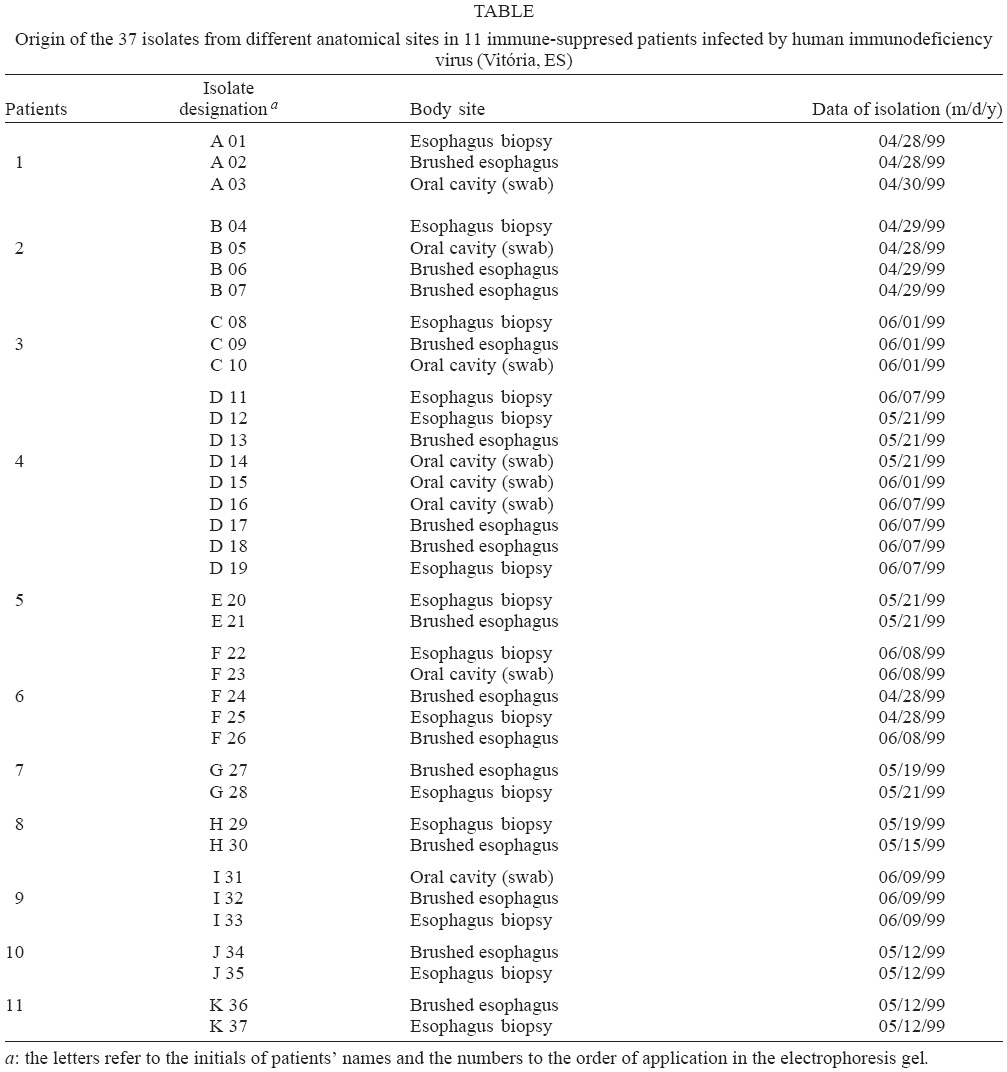
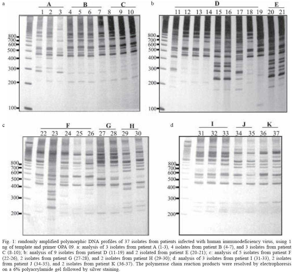
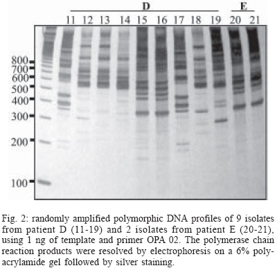
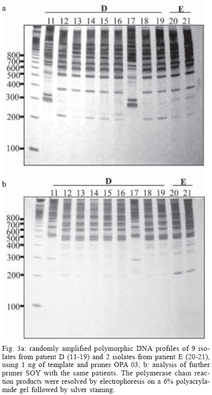
2003 Code Number: oc04027 The patterns of genetic variation of samples of Candida spp. isolated from patients infected with human immunodeficiency virus in Vitória, state of Espírito Santo, Brazil, were examined. Thirty-seven strains were isolated from different anatomical sites obtained from different infection episodes of 11 patients infected with the human immunodeficiency virus (HIV). These samples were subjected to randomly amplified polymorphic DNA (RAPD) analysis using 9 different primers. Reproducible and complex DNA banding patterns were obtained. The experiments indicated evidence of dynamic process of yeast colonization in HIV-infected patients, and also that certain primers are efficient in the identification of species of the Candida genus. Thus, we conclude that RAPD analysis may be useful in providing genotypic characters for Candida species typing in epidemiological investigations, and also for the rapid identification of pathogenic fungi. Key words: polymerase chain reaction - random amplified polymorphic DNA - candidosis - Candida spp. Several reports have described a significant increase in the incidence of systemic candidosis during the last decade (Hazen 1995, Voss et al. 1995, Coleman et al. 1997). The increase of the immune-suppressed population specially related to the human immunodeficiency virus (HIV), organ transplantation, and chemotherapy had dramatically raised the incidence of candidosis (Klein et al. 1984, Odds 1988, Samaranayake & Holmstrup 1989, Odds et al. 1990, Coleman et al. 1993, Pfaller 1995). Oropharingeal candidosis, caused mainly by the commensal yeast Candida albicans, is the most frequently occurring opportunistic infection, during acquired immunodeficiency syndrome (AIDS). Up to 90% of individuals infected with the HIV have presented at least one episode of oropharyngeal candidosis during the course of their disease (Klein et al. 1984, Barchiesi et al. 1997). C. albicans is the most pathogenic species of the Candida genus and the most frequently associated with candidosis (Odds et al. 1990). Earlier studies on the epidemiology of candidiasis used phenotypic properties to assess strain identity, but these methods lacked resolution power (Merz 1990, Pfaller et al. 1990). With the advent of molecular genetics more powerful DNA-based typing methods have emerged for clinical and epidemiological analysis (Scher & Stevens 1988, Schmid et al. 1990, Stevens et al. 1990, Clemons et al. 1997, Corlotti et al. 1997, Diaz-Guerra et al. 1997, Meyer et al. 1997, Xu et al. 1999b). These include fingerprinting methods such as karyotyping using pulse-field electrophoresis (PFGE), restriction fragment length polymorphism (RFLP), randomly amplified polymorphic DNA analysis (RAPD), and Southern hybridization with moderately repetitive DNA probes (Odds et al. 1992, Espinel-Ingroff et al. 1999). These methods have been used extensively for the detection and typing of Candida strains (Heimdahl & Nord 1990, Clemons et al. 1991, Bart-Delabesse et al. 1993, Miyakawa et al. 1993, Holmes et al. 1994) but have been used less frequently for differentiation of species. The most frequently used PCR-based technique currently in use is RAPD. In this technique, single or a pair wise combination of primers, typically 9 to 10 nucleotides in length, are used to amplify target genomic DNA by polymerase chain reaction (PCR). Fragments of DNA are generated by PCR amplification if the primer target sites for the primer happen to occur within approximately 5 kb of each other on opposite DNA strands. The amplified products form strain-specific fingerprints are then analyzed by separation through an agarose gel and ethidium bromide stained (Welsh & McClelland 1990). This procedure seemed to be efficient in distinguishing different isolates; it has a high discriminatory power, it is easy to perform, does not require radiolabelled probes, and it is applicable to several microorganisms (Robert et al. 1995). The aims of this work were to identify the species of the genus Candida and to analyze the genetic variability intra and inter-specific of 37 strains isolated from different anatomical sites of 11 immune-compromised patients infected with HIV, through the RAPD assay. Additionally, molecular biology techniques have been used to highlight information about genomic structure of these microorganisms and the epidemiology of the candidosis. MATERIALS AND METHODS Isolates - Thirty-seven strains were isolated from 11 immune-compromised patients infected by HIV, from Vitória, ES, Brazil. These samples were obtained through oropharingeal swabs, brushed from esophagus, and esophagus biopsy. The collection of the sample was performed with the aid of sterile swabs or citology brush during the endoscopic examinations (Table). All isolates were identified through tests of germ tube formation, fermentation, and assimilation of distinct carbohydrate and also through micro-morphological characterization (Kreger-Van Rij 1984, Kurtzman & Fell 1998). All the strains were maintained at 4°C on Sabouraud dextrose agar (SDA) until identification tests were carried out. Transfers were done at 3-month intervals. The isolate of C. albicans, ATCC 18804 was included in this study. DNA extraction - The DNA was extracted from all the isolates by the method described by Caligiorne et al. (1999) with modifications. The samples were grown on Sa-bouraud dextrose agar overnight at 37°C and a few colonies were transferred to a 1.5 ml Eppendorf tube containing 1 ml of 0.85% NaCl solution. Then, the pellets were recovered by centrifugation 10,000 X g for 15 min at 4°C and resuspended in 0.1 M Citrate, 1.1 M Sorbitol, and incubated with 3 mg/ml Glucanex (Glucanase/Novo Nordisk do Brasil, RS) for 3 h at 37°C so that the cellular walls were disrupted. After centrifugation, the precipitate was re-suspended in lysis buffer 0.04 M Tris (pH 8.5), 0.20 M NaCl, 1.5% sarcosil and 0.01 M EDTA 0.5 M (pH 8.0). Then, pellets were washed three times with phenol-chloroform and then precipitated with ethanol absolute and 0.3 M NaCl. After 15 min, the precipitate was centrifuged and washed twice with 70% ethanol dried and re-suspended in 50 µl of 10 mM Tris-HCl (pH 7.5). RAPD reactions - Each amplification was done in a final volume of 10 µl containing 1 X buffer (Promega), 0.2 mM each of dATP, dGTP, dCTP, and dTTP (Promega), 1 ng of genomic-DNA, 2 mM of MgCl2 (Promega), 0.8 µM of primer (Operon), and one unit of thermostable Taq DNA polymerase (Promega). Amplification parameters consisted of 35 cycles of denaturation at 95°C for 60 s, primer annealing at 36°C for 60 s and extension at 72°C for 60 s. In the first cycle, denaturation was done for 3 min and in the last cycle, final extension lasted for 5 min. Reactions were carried out using primers from Operon OPA 01 (CAGGCCCTTC), OPA 02 (TGCCGAGCTG), OPA 03 (AGTCAGCCAG), OPA 08 (GTGACGTAGG), OPA 09 (GGGTAACGCC) as well as SOY (AGGTCACTGA), RP1-4 (TAGGATCAGA), RP-2 (AAGGATCAGA) and RP4-2 (CACATGCTTC) primers (Lehmann et al. 1992). Polyacrylamide gel electrophoresis - Following the amplification, 3 µl of the reaction was mixed with DNA sample buffer (0.125% xylene cyanol, 0.125% bromophenol blue, 15% glycerol) and subjected to electrophoresis through a 6% polyacrylamide gel (acrylamide-bis-acrylamide 29/1) in TBE buffer (2 mM EDTA, 10 mM Tris-borate, pH 8.0). The gels were silver stained by the method described by Santos et al. (1993). For each experiment, the sizes of DNA fragments amplified by PCR were determined by direct comparison with the DNA marker (100 bp Ladder, Gibco, BRL). RESULTS All the 37 isolates studied were phenotypically identified (Kreger-Van Rij 1984, Kurtzman & Fell 1998). The identification of these samples was confirmed through the assimilation test of several carbohydrate sources. Among the 37 Candida isolates obtained from the 11 immune-compromised patients infected with HIV in the state of Espírito Santo, C. albicans was the most frequently isolated species (94.6%), followed by C. guilliermondii (5.4%). In order to examine genomic variability among the Candida isolates, molecular analysis was undertaken using RAPDs generated by 9 primers, OPA 01, OPA 02, OPA 03, OPA 08, OPA 09 as well as SOY, RP1-4, RP-2, and RP4-2. Fig. 1 a, b, c, d show the RAPD profiles of 37 isolates, amplified with primer OPA 09, from patients infected with HIV. Fig. 1a shows the 3 isolates of patient A, 4 isolates of the patient B, and 3 isolates of patient C, respectively. RAPD profiles of the same patient were similar, but not identical, whereas the profiles of different patients showed higher levels of polymorphisms. Fig. 1b shows the amplifications of 9 isolates from the patient D and 2 isolated of the patient E. Patient D presented 2 species, C. albicans and C. guilliermondii that were obtained from different infection episodes. During the first episode only C. albicans was isolated, but interestingly in a second episode an association between C. albicans and C. guilliermondii was observed (Table). The samples D 15 and D 16 were isolated from oral swabs, 10 and 16 days respectively after the first infection (Table). Isolate D 12 (biopsy), D 13 (brushed), and D 14 (swab) present a homogeneous profile, different from isolate D 19 collected 16 days after the first infection. The polymorphisms observed in isolate D 19 indicates that in the second episode the patient was infected with a genetically distinct isolate. Isolate D 18 also obtained from the second infection presented the same genotype of the first infection. In the second infection patient D was also infected with C. guiilliermondii, (D 11, D 17). Intra-specific polymorphisms were not observed between these isolates. Fig. 1c shows the profiles obtained for 5 isolates from patient F, 2 isolates from patient G and 2 isolates from patient H respectively. Isolates F 24 and F 25 were obtained from a first infection and isolates F 22, F 23, F 26 were obtained from a second infection (Table). In the second infection, 2 distinct genotypes were observed. One isolate was obtained through oropharingeal swab (F 23) and others from esophagus brushed and esophagus biopsy (F 22, F 26). RAPD profiles of isolates from the patients G and H were similar, but not identical. The profiles of the isolates from the same patient showed higher levels of similarity. Fig. 1 d shows the amplifications of 3 isolates from the patient I, 2 isolates from patient J, and 2 isolates from patient K. We can observe that profiles obtained for the isolates from the same patient were more homogeneous when compared with the inter-individual profiles. Fig. 2 shows the analysis of RAPDs derived from patients D and E samples obtained with primer OPA 02. The results obtained for primer OPA 09 were similar to those observed for primer OPA 02. The same was observed with the other samples (data not shown). Some primers were not capable to differentiate the genotypes of different isolates of the genus Candida. Fig. 3 a, b shows the profile of RAPD of the same samples of patients D and E obtained with the primers OPA 03 and SOY. We can observe the presence of a single genotype among all the isolates, obtained with both primers. However, we can observe that these primers were efficient in the identification of the species (D 11, D 17). The other primers generated results similar to the primers OPA 02 and OPA 09. All the C. albicans isolates studied showed an amplification profile similar to the strain ATCC 18804 (data not shown). We can conclude that some primers were efficient in the identification of species of the genus Candida and others were useful in the identification of inter-specific genotypes. DISCUSSION Oropharyngeal candidosis is the most common opportunistic infection during AIDS. It is associated with pain, limits food ingestion and can lead to cachexia (Cameron et al. 1993). With the increasing number of immune-compromised patients, such as infected-HIV patients, or those living together in the same environment in hospital wards, inter-human transmission of pathogenic fungi is likely to occur frequently. However, it is a rare event and has only recently been demonstrated by molecular typing methods for nosocomial Candida infections in patients at risk for candidosis (Romano et al. 1994, Bart-Delabesse et al. 1995, Voss et al. 1995). Molecular analysis was undertaken using RAPDs generated by 9 primers. This technique enables a large number of independent genetic loci to be examined that are not based on particular sequences or types of sequences and can thus be taken as representative of the genome (Welsh & McClelland 1990, Willians et al. 1990). RAPD was used to analyse the genomic variability of the 37 isolates obtained from 11 immune-compromised patients infected with HIV. We were particularly interested in determining whether isolates from a specific body site or isolated from the same patient might be genetically more similar to each other than among isolates from a different body sites or different patients. The yeast microflora of all patients had similar species, with exception of C. guilliermondii found just in the patient D, and genotypic diversities were found among the C. albicans isolates (Fig. 1a, b, c, d). Furthermore, a single patient can be colonized with multiple species or multiple genotypes of the same species at the same or different body sites, indicating that the yeast colonization is a dynamic process also in patients immune-suppressed with infection for HIV. This dynamic characteristic had been previously described (Xu et al. 1999a) for the yeast microflora of the vagina. The goal of this study was the contributions of two commonly recognized factors associated with candidiasis in women: HIV infection status and pregnancy. This technique produced a profile of bands that allowed the identification of intra- and inter-specific polymorphisms among isolates obtained from the same patient. Additionally, it differentiates C. albicans strains isolated from different patients (Fig. 1b). Fast and reliable identification of Candida genus species is important to define adequate therapeutic decisions, because the different species have highly variable susceptibilities to antifungal drugs. Accurate statistical records on case history and epidemiological studies also depend on effective identification. The traditional methods of identifying Candida species are often based on examination of phenotypic characteristics. This approach can be time-consuming, and the reliance on the variable expression of phenotypic characteristics can lead to inconsistent results. In the present study, we could observe that RAPD technique was able to point out clearly the genomic variability within the Candida genus. The identification of specific species markers and the definition of larger details on these yeasts were important aspects that can aid in controlling and best understanding the epidemiology of the candidiasis. The PCR genotyping has the advantage of being the least arduous procedure among the different typing methods, requiring only small amounts of DNA and being relatively easy to standardize among laboratories. It requires little knowledge of the molecular biology of the species being examined and no sequence information is necessary. In addition it is a quick, economical technique, with high reproducibility. It has been suggested that the use of a combination of different molecular typing procedures may help to distinguish certain isolates from one another (Pfaller et al. 1994). According to the results obtained, RAPD assay can be considered an important tool to identify as well as study the inter- and intra-specific genetic variability among Candida genus isolates. Moreover, this technique allowed the identification of distinct strains related to different episodes of candidosis in HIV patients, which can be of great importance in the control and therapeutic choice. ACKNOWLEDGEMENTS To Dr Reynaldo Dietze, Dr Elenice Moreira Lemos, and Maristela Vicente Araújo from Infectious Diseases Center of Federal University of Espírito Santo for the collection of the HIV strains. REFERENCES
Copyright 2004 Instituto Oswaldo Cruz - Fiocruz. The following images related to this document are available:Photo images[oc04027f3a-b.jpg] [oc04027f1a-d.jpg] [oc04027f2.jpg] [oc04027t1.jpg] |
| |||||||||
