 |
| About Bioline | All Journals | Testimonials | Membership | News |
|
||||||
|
||||||
Mem Inst Oswaldo Cruz, Vol. 99, Suppl. 1, August, 2004, pp. 67-71 Hypertensive Portal Colopathy in Schistosomiasis Mansoni - Proposal for a Classification Maria Angelina C Miranda+, Ana Lúcia C Domingues, Heloisa S Dias, Renata C Miranda, Norma T Jucá, Maria Fátima M Albuquerque, Fernando T Cordeiro Faculdade de Medicina,
Universidade Federal de Pernambuco, Av. Moraes Rego s/no, Cidade
Universitária, 50640-900 Recife, PE, Brasil Received 28 May
2004 Code Number: oc04089 Portal hypertension is a frequent complication of chronic liver disease, detected not only in schistosomiasis, but also in cirrhosis of any etiology. Vascular alterations in the colonic mucosa are a potential source for acute or chronic bleeding and have been observed in patients with portal hypertension. The purpose of this prospective study was to describe and propose a classification for the vascular alterations of portal hypertension in the colonic mucosa among patients with hepatosplenic schistosomiasis mansoni. One or more alterations of portal colopathy were observed in all patients and they were classified according to their intensity, obeying the classification proposed by the authors. Portal colopathy is an important finding in hepatosplenic schistosomiasis and might be the cause of lower gastrointestinal bleeding in patients with severe portal hypertension. Key words: Schistosoma
mansoni - portal colopathy - vascular alterations - telangiectasia
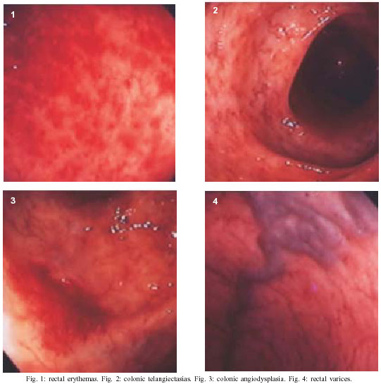
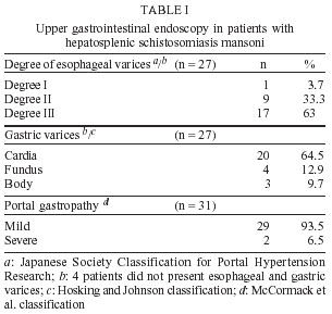
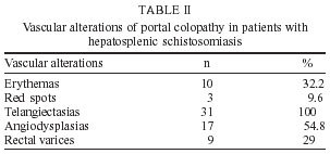
- angiodysplasia - erythema - Schistosomiasis mansoni is a public health problem of major importance in Brazil. The infection reaches extensive areas in the Northeast affecting a large number of people, particularly youngsters and adults in their most productive phases of life in which 4 to 10% present serious forms of the disease (Coutinho & Domingues 1993). Portal hypertension is a frequent complication of chronic liver disease, detected not only in schistosomiasis, but also in cirrhosis of any etiology. Endoscopic abnormalities in the colonic mucosa of patients with portal hypertension due to hepatic cirrhosis with risk of intermittent hemorrhage are well described (Kosareck et al. 1991, Viggiano & Gostout 1992, Rabinovitz et al. 1995, Ganguly et al. 1995, Chen et al. 1996, Misra et al. 1996, Bresci et al. 1998, Bini et al. 2000). The most frequent alterations are defined as telangiectasias or vascular ectasias, angiodysplasia-like lesions, red spots, and erythema. The initial histologic findings on these lesions showed dilated capillaries, submucosal edema, and non-specific inflammatory infiltrate (Kosareck et al. 1991). The term ectasia should be used for the combination of dilatation and structural alterations of the blood vessels (wall thickening and tortuousity), and not for dilatation only (Viggiano & Gostout 1992). Alterations in the colonic mucosa caused by Schistosoma mansoni eggs are described as increased vascular bed, edema, congestion, scattered petechial spots with the aspect of "fleabites", ulceration, and polyps (Pereira 1962, Mohamed et al. 1990, Sanguino et al. 1993). Considerable controversy still exists regarding the colonic abnormalities in patients with portal hypertension due to schistosomiasis mansoni (Mohamed et al. 1990, Sanguino et al. 1993, Geboes et al. 1995). The aim of this study was to better define the colonic alterations that are present in patients with portal hypertension (portal colopathy) due to schistosomiasis mansoni, to classify the severity of these abnormalities and to associate them with esophageal varices and portal gastropathy. MATERIALS AND METHODS After approval from the Research Ethics Committee of the Federal University of Pernambuco, 31 patients over 18 year of age, presenting the hepatosplenic form of schistosomiasis mansoni participated in this study. All patients had a history of specific treatment for S. mansoni and antecedents of upper or lower gastrointestinal bleeding. Other parasites eventually identified were treated before the inclusion of the patients in the study, in order to prevent possible lesions in the colon that might alter the results (Pereira 1962). Patients with alcohol ingestion up to 60 g/day were excluded, as were those with other hepatic disease, positive HbsAg and anti-HCV, or other disease that could cause vascular alterations, such as chronic nephropathy, stenosis of the abdominal aorta, abdominal surgery (ileostomy and colostomy), and inflammatory bowel disease. After the feces, hematologic and biochemistry exams, an abdominal ultrasonography was performed to confirm the diagnosis of hepatosplenic schistosomiasis mansoni, as well as to characterize the portal hypertension and to exclude other hepatopathies. The ultrasonography classifications of peripheral fibrosis used in this study were proposed by WHO (Niamey 2000). An upper gastrointestinal endoscopy was carried out to characterize esophageal (Japanese Research 1980), gastric varices (Hosking & Johnson 1988) and the portal hypertensive gastropathy. The entire colon was visualized using an Olympus CF-VI colonoscope with conventional resolution. The patients had their colon prepared by using manitol at 10% (Habar-Gama et al. 1981). During the examination, they received an intravenous sedation of meperidine associated with midazolan, monitored through pulse oximetry. In accordance to criteria described by Tam et al. (1995), the lesions were characterized as: erythemas (diffuse hyperemia in the mucosa), red spots (hyperemic focus in the mucosa), telangiectasia (dilated and tortuous small vessels), angiodysplasia (coiled vessels measuring about 1 cm), rectal varices (dilated, crooked vessel). The current authors elaborated a classification for the colopathy in patients with schistosomiasis and portal hypertension, based on both literature concerning cirrhotic patients and personal experience. It was classified into three levels: degree I (erythemas and telangiectasias); degree II (erythemas, telangiectasias, and angiodyspla-sias); and degree III (erythemas telangiectasias, angio-dysplasias, red spots, and rectal varices). During the colonoscopy, mucosal biopsy specimens were obtained, by way of the FB-24U-1 Olympus forceps, in normal appearing mucosa of the rectum, sigmoid and in areas with lesions, avoiding to perform biopsy in areas with angiodysplasia-like lesions. Histological sections were stained with hematoxilina and eosine (HE) (Figs 1-4). RESULTS The 31 hepatosplenic schistosomotic patients studied were between 20 to 73 years old. Fifteen patients were male and 16 female. All were from endemic schistosomiasis region. Twenty-six patients (83%) reported having one or more episodes of upper gastrointestinal bleeding and 8 (25.8%) had had lower gastroentestinal bleeding. The stool examinations were negative for S. mansoni eggs in 30 patients (96.7%). The HbsAg and anti-HCV serology were negative in all cases. The upper gastrointestinal endoscopy findings are displayed in Table I. All patients presented portal hypertensive colopathy. One or more alterations were identified simultaneously, in the same patient. Erythemas were observed in 10 (32.2%), telangiectasias in 31 (100%), angiodysplasia-like lesions in 17 (54.8%), and red spots in 3 patients (9.6%). Rectal varices were identified in 9 cases (29%). Erythemas predominated in the distal segments (70%). Telangiectasias were distributed throughout the entire colon in 93.5% of the patients. Angiodysplasia-like lesions also had a pan-colonic presentation in 58.8% of the cases. The frequencies of the vascular alterations in portal colopathy are displayed in Table II. After grouping the cases according to the classifications proposed by the authors, mild colopathy was found in 10 patients (32.2%), moderate colopathy was found in 10 patients (32.2%), and severe colopathy was found in 11 (35.5%). The histological examination identified mononuclear inflammatory infiltrated (lymphocytes and plasma cells) of mild intensity in 28 cases (90.3%), moderate intensity in 3 (9.7%); mild hyperemia in 12 cases (38.7%), and moderate in 1 (3.3%); moderate edema in 13 cases (41.9%) and severe in 3 (9.7%), moderate capillary ectasia in 12 cases (38.6%) and severe in 2 (6.5%). S. mansoni eggs were observed in three cases: viable eggs in the sigmoid with and without schistosomotic granuloma in one; viable egg without granuloma in the distal ileo in another; and calcified eggs in the sigmoid in the last one. While vascular lesions in the colonic mucosa of cirrhotic patients with portal hypertension have been reported (Kosareck et al. 1991, Viggiano & Gostout 1992, Rabinovitz et al. 1995, Chen et al. 1996, Ganguly et al. 1995, Misra et al. 1996, Bresci et al. 1998, Bini et al. 2000), studies on hepatosplenic schistosomiasis mansoni are scarce (Mohamed et al. 1990, Sanguino et al. 1993, Geboes et al. 1995). For ethical reasons, we did not study patients without prior episode of upper or lower gastrointestinal bleeding and the patients selected had already been treated for schistosomiasis to avoid the alterations due to the parasitose per si (Pereira 1962, Mohamed et al. 1990, Sanguino et al. 1993, Geboes et al. 1995). The characteristic alterations of the portal colopathy in schistosomiasis are similar to those of portal colopathy in cirrhotics (erythemas, telangiectasias, angiodysplasias, red spots, and rectal varices), but they tend to be more frequent and accentuated. It has been suggested that the schistosomotic portal colopathy happens due to the intense and permanent rise of the portal venous pressure (Lamps et al. 1998). This is associated to the neovascu-larization process stimulated during the active phase of the disease, through the blockage of the vessels caused by the schistosome eggs, as well as the secretions released by them (Wyler et al. 1987). Telangiectasias are the most frequent finding in this series and in Justo's work (2003). These vascular changes, which are similar to the injuries of the upper gastrointestinal tract, can be the cause of lower gastrointestinal bleeding, a process that is often obscure and co-responsible for the chronic anemia found in these patients. This may have occurred in 8 (25.8%) cases of this present study. The patients in this series had advanced portal hypertension, as determined by endoscopy and ultrasonography. All of them had vascular alterations of portal colopathy, 32.2% mild, 32.2% moderate, and 35.5% severe. A retrospective study by Bini et al. (2000) on cirrhotic patients with portal hypertension identified both medium and large esophageal varices in 51% of the cases, gastropathy in 47%, and at least one characteristic alteration of the colopathy in just 57%. However, in a study by Eleftheriadis et al. (1993) on cirrhotic patients with accentuated portal hypertension who had undergone sclerotherapy for the erradication of esophageal varices and had no prior history of lower gastrointestinal bleeding, a high frequency of portal colopathy (93%) was observed, with lesion-like telangiectasias predominating (70%). In the present series, colitis-like (ery-themas) alterations in the mucosa were seen in 32.2% of the cases in relation to what was observed in cirrhotic patients, 27% (Misra et al. 1996), 32% (Rabinovitz et al. 1995) and 38% (Bini et al. 2000). Rectal varices are collateral portosystemic dilated veins in patients with portal hypertension and communicate the venous flow of the superior hemorrhoid veins to the medium and inferior hemorrhoid veins (Jacobs et al. 1989, Hosking et al. 1989). Therefore, they may occasionally be subjected to massive bleeding (Weinshall & Cheer 1986). The prevalence of the rectal varices has varied from 0% to 92% in cirrhotic patients ( Hosking et al. 1989, Rabinovitz et al. 1995, Misra et al. 1996). Our findings are in agreement with Hosking et al. (1989), who concluded that rectal varices reflect a later stage in the development of the portal hypertension, being observed more frequently in patients with large esophageal and gastric varices. This fact was demonstrated in this series, in which 6 of the 9 cases with rectal varices (66.6%) had large esophageal varices. In the histological studies, a mild mononuclear inflammatory infiltrate was evidenced, particularly lymphocytes and plasma cells in 90.3% of the cases, excluding inflammatory bowel disease. Edema and the loss of the villous architecture of the lamina (propria?) of the colonic mucosa are secondary to the vessel wall thickness present in this study with moderate to accentuated intensity in 51.6% of the cases. This can be explained by the chronic increase of the intraluminal pressure (Misra et al. 1997). Endoscopic biopsies of the vascular lesions in the colonic mucosa were avoided due to the high risk of bleeding (Tam et al. 1995). However, when it is obtained, the descriptions include dilatation and tortuosity of the mucosa vessels, as well as an increase in the number of small vessels and the wall thickness in the lamina propria (Viggiano & Gostout 1992). In this study, we followed the criteria established by Viggiano and Gostout (1992) and observed moderate to severe capillary ectasia in only 45.1% of the cases. In these two cases, who had viable eggs in the biopsia, probability due to re-infection, an increase in the degree of colopathy was not verified when compared to the other patients. There was no correlation between the severity of portal gastropathy and the presence of hypertensive colo-pathy. Meanwhile, it was observed a predominance of severe portal colopathy (87.5%) in patients with large esophageal varices. We conclude that in a well-characterized portal hypertension in hepatosplenic schistosomiasis mansoni, hypertensive colopathy is an important entity, as it was present in all cases. The identification and classification of these vascular lesions in the colonic mucosa is of great significance for the diagnosis and control of the evolution of the disease, as these alterations can be the cause of anemia, hypoproteinemia, and obscure lower bleeding, contributing to a higher morbidity from the disease. We suggest that colonoscopy should be done in every patient with hepatosplenic schistosomiasis who has anemia without a prior episode of upper gastrointestinal hemorrhage. The classification proposed in this work would be useful to standardize the categorized alterations according to intensity.
Copyright 2004 Instituto Oswaldo Cruz - Fiocruz. The following images related to this document are available:Photo images[oc04089t1.jpg] [oc04089t2.jpg] [oc04089f1-4.jpg] |
| |||||||||
