 |
| About Bioline | All Journals | Testimonials | Membership | News |
|
||||||
|
||||||
Mem Inst Oswaldo Cruz, Rio de Janeiro, Vol. 99, No. 5, August, 2004, pp. 493-498 Histopathological and
Ultrastructural Effects of CFG Cavados/+, S Majerowicz*, JQ Chaves*, CJPC Araújo-Coutinho**, L Rabinovitch Departamento de
Bacteriologia *Departamento de Virologia, Instituto Oswaldo Cruz- Fiocruz,
Av. Brasil 4365, 21045-900 Rio de Janeiro, RJ, Brasil **Superintendência
de Controle de Endemias, Secretaria de Saúde do Estado de São
Paulo, São Paulo, SP, Brasil Received 26 February
2004 Code Number: oc04106 The bacterium Bacillus
thuringiensis (Bt) produces parasporal crystals containing Key words: Bacillus thuringiensis serovar israelensis - Simulium pertinax - midgut - histopathology - ultrastructure Bacillus thuringiensis (Bt)
is a Gram-positive, aerobic bacterium, producing parasporal crystals containing
Most of the simuliids are hematophagus insects; they are involved in the transmission of human onchocerciasis, mainly in the North of Brazil. Simulium pertinax is the most important species in Southeast region inducing a negative socio-economic impact. The frequent and intensive attacks by S. pertinax on the transitory populations in this area helps to reduce tourism during summer months (Gerais & Ribeiro 1986, Araújo-Coutinho 1995, Maia-Herzog et al. 1999, Cavados et al. 2001, Araújo-Coutinho et al. 2003). After isolation of the B. thuringiensis serovar israelensis (Bti) by Goldberg and Margalit (1977) and its characterization by De Barjac (1978), several studies were realized confirming the action of Bti as a larvicide mainly for controlling Simulium and Aedes species (De Barjac 1978, Lacey et al. 1982, Charles & De Barjac 1983, Becker 1990, Araújo-Coutinho 1995, Mardini et al. 1999, Rabinovitch et al. 1999, Cavados et al. 2001, Regis et al. 2001). Histophatological investigations in larvae of S. vittatum infected with B. thuringiensis serovar kurstaki HD 255 (Lacey & Federici 1979) and S. variegatum infected with Bti (Rey et al. 1998) demonstrated morphological lesions in the intestinal epithelium which exhibited swollen cells, degenerated brush borders, disorganized nuclei, enlargement of intercellular spaces and cell lysis. The present report
describes sequential changes in the midgut of S. pertinax larvae infected
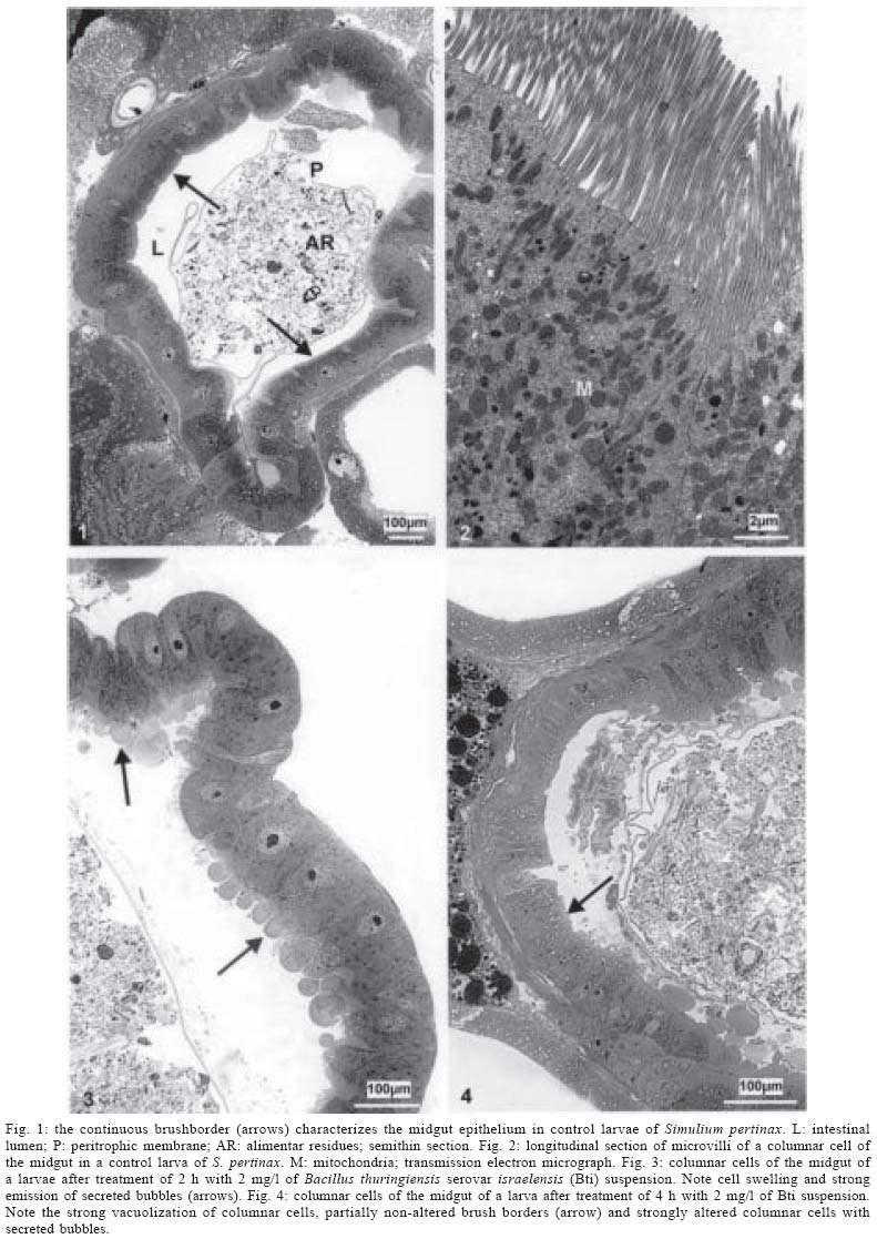
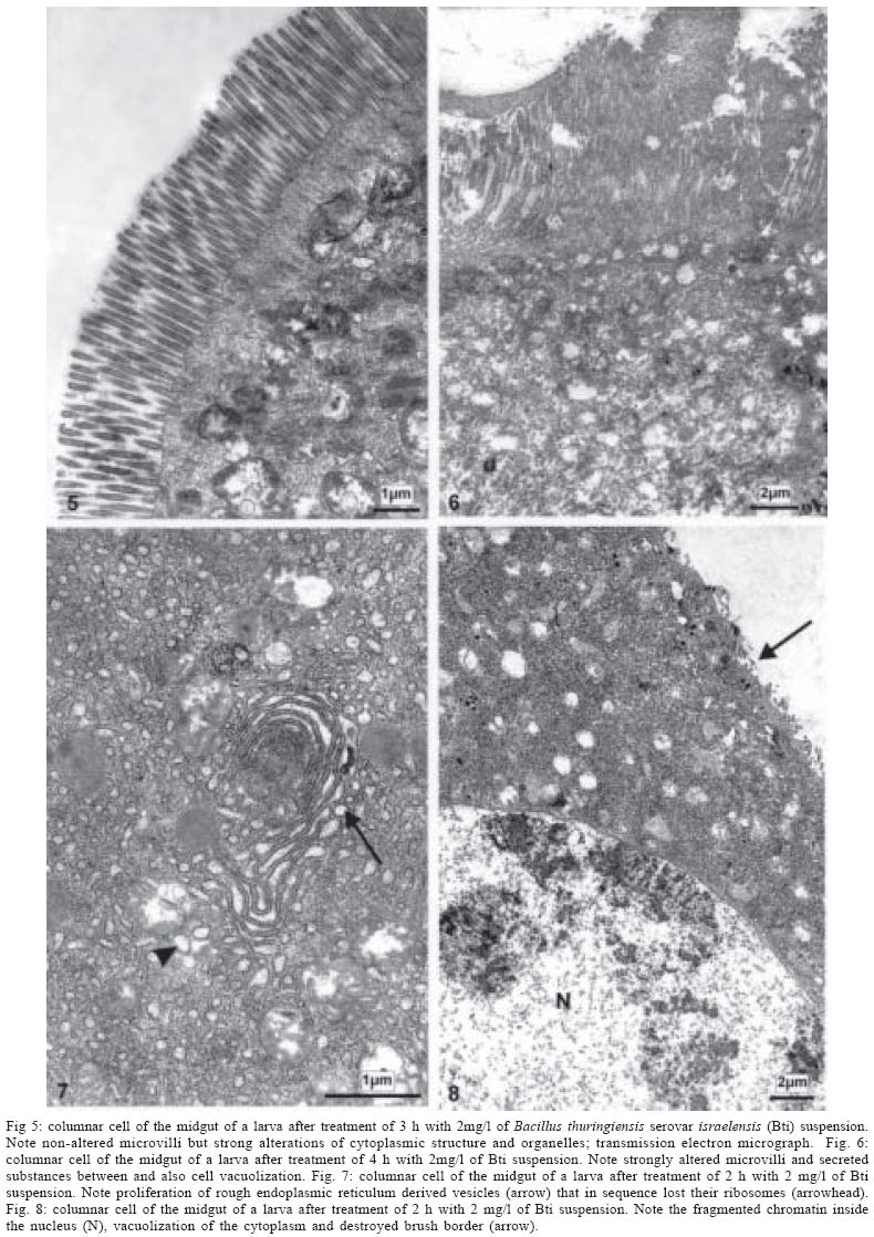
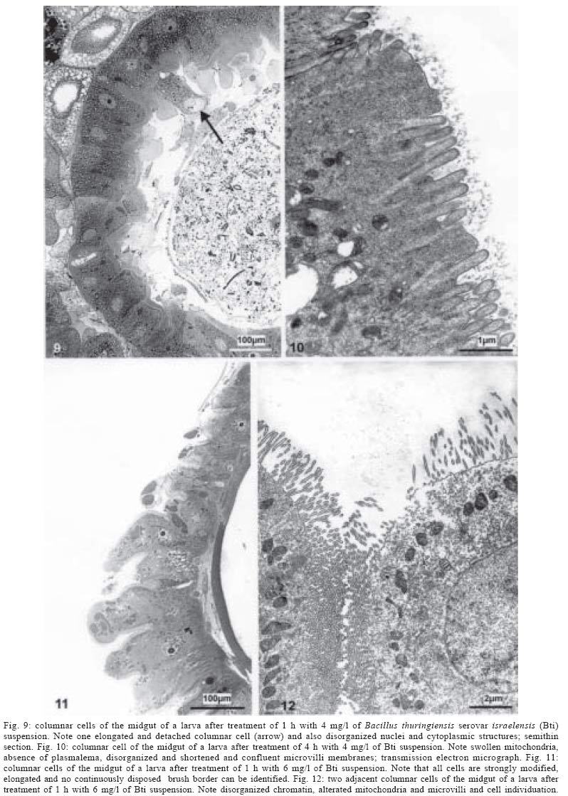
with increasing concentrations of MATERIALS AND METHODS Bacterial strain - Bti strain IPS-82 (LFB-FIOCRUZ 584), serotype H-14 type strain of the Institute Pasteur, Paris. It was maintained in agar medium with metals-ANM at room temperature (Rabinovitch et al. 1975). Culture medium - The bacterial biomass was prepared using a fermentation medium based on soya flour and metals (such as Mg2+, Mn2+, Zn2+, Fe2+ and Ca2+) developed in the Laboratório de Fisiologia Bacteriana, Departamento de Bacteriologia, IOC-Fiocruz (Cavados et al.1998, Rabinovitch et al.1998). Inoculum and biomass production - Growth started with a pre-inoculum to reduce the duration of the lag-phase of bacterial growth. After inoculation in 125 ml Erlenmeyer flasks containing 50 ml of the medium, the flasks were incubated in a New Brunswick Scientific agitator series 25 D, at 175 opm and 30oC for 6 h. Subsequently, 3 ml were transferred to 500 ml Erlenmeyer flasks containing 150 ml of the soya flour and metals medium and incubated as previously described for a further 72 h period. Once sporulation had reached a level of 95% of free spores and crystals, each culture was centrifuged (6000 g, 10oC), the biomasses were kept in an amber container with the pH adjusted to 5.0 with propionic acid and then formulated (Rabinovitch et al. 1998). Bioassay with S. pertinax larvae - S. pertinax larvae were collected in the Soberbo river in the municipality of Guapimirim, state of Rio de Janeiro. Field-collected larvae were maintained in chambers where the water was aerated by a continuous stream of air bubbles. Biological insecticide doses equivalent to 2, 4, 6 mg/l were applied to the different groups of larvae. The exposure times employed ranged from 1 to 4 h. Only live larvae were examined. At the end of each time period the larvae were observed under a stereoscopic microscope and the head and anal region were dissected and discarded (Cavados 2000). The remainder of the larval body was fixed and processed for observation using light and electron microscopy. Light microscopy (LM) - Semi-thin sections were made from intestine samples previously embedded in Epon, stained with a methilene blue-azure II solution in phosphate buffer 0.2 M, pH 6.9 (Richardson et al. 1960, Humprey & Pittman 1974) and observed in a Zeiss Axiophot microscope. Electron microscopy (TEM) - Samples of the intestine were fixed in 2.5% glutaraldehyde in 0.2M cacodylate buffer, pH 7.2, then washed in cacodylate buffer containing 7.2% sucrose, post-fixed in 1% osmium tetroxide for 1:45 h, dehydrated in graded acetone and embedded in Epon. Ultrathin sections were stained with uranyl acetate and lead citrate (Reynolds 1963) and examined with a Zeiss EM-900 electron microscope. RESULTS The non-infected Simulium control midgut shows a well-preserved layer of epithelial cells. The ovoid shaped nuclei are located in the center of the cell (Fig. 1). Long and regularly placed microvilli border the midgut lumen (Fig. 2). The midgut of the larvae exposed to Bti using 2 mg/l shows some cells presenting an irregularly structured brush border within 1 to 2 h (Fig. 3). The cells begin to be swollen by a slight vacuolization and increasing of secretion vesicles. (Fig. 4). This feature is confirmed when observed by ultrastructure (Fig. 5). From 3 to 4 h after applying the endotoxin, structural changes occur in some of the epithelial cells (Fig. 8), whereas other ones maintain its morphology (Fig. 7). In addition, cell groups observed at the basis of the epithelium suggest a beginning of tissue recovery (Fig. 6). When the midgut of larvae exposed to 4 mg/l of the endotoxin is analyzed after 1 to 2 h, it shows increased morphological changes of the epithelium with most of the cells swollen, vacuolated, with an increased number of secretion vesicles and an irregularly disposed brush border (Fig. 9). After 3 to 4 h, the pathological effects are observed in nearly all of the intestinal cells. The epithelium presents detached cells also with bubble shape tips (Fig. 10) and cells with short and thick (Fig. 11), irregularly and modified microvilli (Fig. 12). When 6 mg/l of the bioinsecticide are employed, the midgut can only be analyzed during the first hour of exposure to the endotoxin, since after this time all the larvae are dead. The structural disorganization of the intestinal epithelium is evident, showing cells without the characteristic morphology (Fig. 13), becoming elongated, presenting destroyed tips and sometimes budding into the intestine lumen (Figs 14, 15). DISCUSSION In our experiments, which were stopped after 4 h of exposure to the Bti endotoxin, S. pertinax larvae could survive when a low concentration of Bti (2 mg/l) was applied. Histological alterations of some columnar cells of the midgut epithelium during the exposure to the endotoxin were observed. Nevertheless, after 4 h of toxin action well preserved groups of cells located at the base of the epithelium next to the basal membrane (Fig. 6) indicated that cell recovery was in progress. Increasing the endotoxin concentration (4 mg/l) nearly all columnar-cells were affected after 3 h of exposure to Bti endotoxin and no preserved cell groups next to the basal membrane of the midgut could be detected, though the larvae had not yet died. When 6 mg/l of the endotoxin were applied, all the larvae died after 1 h. Using Bti at a very low concentration (0.4 mg/l) during routine field application against S. variegatum, Rey et al. (1998) observed that 72 h after the beginning of the treatment all black flies died, but only 15.7% after 24 h of treatment. Lacey and Federici (1979) using a concentration of 10 mg/l of B. thuringiensis serovar kurstaki against S. vittatum larvae noted that mortality was increased by temperature elevation. In another experiment, Charles and de Barjac (1983) using 0.08 mg/l of purified Bti crystals against the larvae of Aedes aegypti, reported that all were dead after 10 h. Lahkim-Tsor et al. (1983) used 10 mg/l of Bti for Ae. aegypti larvae feading and found that the larvae died between 37 and 120 min after the beginning of exposure to the endotoxin. Regarding these experiments, the cytopathic effects observed in larvae midguts were proportional to Bti endotoxin concentrations applied and inversely proportional to the time of exposure. The endotoxin concentrations used in our experiments gave a clear idea of S. pertinax larvae resistance. Ultrastructural observations showed that the first cell damages due to the Bti endotoxin in the S. pertinax larvae midgut were related to brush border microvilli degeneration. As illustrated in Percy and Fast (1983) using purified Bt crystal toxin (1 g/l) against silkworm larvae, the dissolution of cytoskeleton structures inside and at the basis of the microvilli were responsible for its decrease in size and further disappearance, when bubbles of cytoplasmic substances protrude into the midgut lumen as in S. pertinax (Figs 4, 7, 14). At this stage, before the cell death, the columnar cells appeared more elongated in light microscope observations (Fig. 9). The use of B. thuringiensis endotoxins originated vacuolization of the midgut epithelial cells in the different experimental models (Percy & Fast 1983, Charles & Barjac 1983, Rey et al. 1998), as also in S. pertinax (Figs 5, 8). These vacuoles proceeded from enlarged rough endoplasmic cisterns that had lost their ribosomes (Percy & Fast 1983). This article is the first report of the histopathological effects of the Bti endotoxins in the midgut of S. pertinax larvae and the data obtained may contribute for better understanding the mode of action of this bacterial strain used as bioinsecticide against black fly larvae. ACKNOWLEDGEMENTS To Dr OM Barth, head of the Laboratory Electron Microscopy, Department of Virology, Instituto Oswaldo Cruz, for helpfull electron microscope utilities and critical review of the manuscript. REFERENCES
Copyright 2004 Instituto Oswaldo Cruz - Fiocruz. The following images related to this document are available:Photo images[oc04106f5-8.jpg] [oc04106f1-4.jpg] [oc04106f9-12.jpg] |
| |||||||||
