 |
| About Bioline | All Journals | Testimonials | Membership | News |
|
||||||
|
||||||
Mem Inst Oswaldo Cruz, Rio de Janeiro, Vol. 99, No. 5, August, 2004, pp. 499-502 Diagnostic of Biomphalaria Snails and Schistosoma mansoni: DNA Obtained from Traces of Shell Organic Materials Roberta L Caldeira, Liana K Jannotti-Passos, Pollanah M Lira, Omar S Carvalho+ +Corresponding
author. Fax: +55-31-3295.3115. E-mail: omar@cpqrr.fiocruz.br Received 8 September
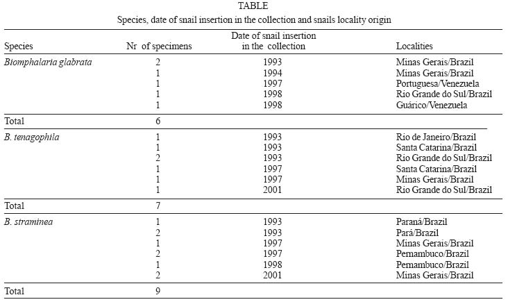
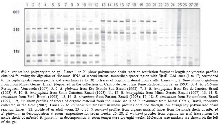
2003 Code Number: oc04107 Freshwater snails belonging to the genus Biomphalaria act as intermediate hosts for the parasite trematode Schistosoma mansoni in Africa and in the neotropical region. Identification of such molluscs is carried out based on morphological characters and the presence of cercariae is verified through squeezing snails between two glass slides or by exposing them to artificial light. However, sometimes, the material collected includes molluscs with decomposed bodies or, yet, only empty shells, which precludes their identification and S. mansoni detection. Due to these difficulties, we have developed a methodology in which DNA may be extracted from traces of organic material from inside shells in order to identify molluscs through polymerase chain reaction and restriction fragment length polymorphism and to detect S. mansoni into these snails, by using low stringency polymerase chain reaction. Species-specific profiles obtained from B. glabrata, B. straminea, and B. tenagophila snails and their shells, maintained in laboratory for ten years, showed the same profiles. S. mansoni profiles showed to be present in shell specimens as far as the eighth week after being removed from aquarium. Key words: Biomphalaria - Schistosoma mansoni - molluscs - shell - DNA - polymerase chain reaction There are 34 identified species of the genus Biom-phalaria (Mollusca: Planorbidae) in Africa and in the Neotropic region, out of which B. glabrata, B. tena-gophila, B. straminea, B. prona, B. pfeifferi, B. sudanica, B. alexandrina B. choanomphala, B. camerunensis, and B. stanley are regarded as intermediate hosts of the trematode Schistosoma mansoni, the aethiological agent of human intestinal schistosomiasis (Malek 1985, Brown 1994, Noya et al. 1999). The identification of such molluscs is normally carried out based on morphological characters of the shell, renal, and reproductive systems (Paraense 1975). However, the identification of some species may become complicated due to the similarity among these characters (Paraense 1988). Recently, molecular tools based on polymerase chain reaction and restriction fragment length polymorphism (PCR-RFLP) of the ribosomal RNA intergenic spacer regions (ITS) have been used in order to overcome this problem (Vidigal et al. 1998, Caldeira et al. 1998, 2000). The detection of Biomphalaria snails infected with S. mansoni is usually performed by cercariae shedding induced by artificial light exposure or by squeezing snails between two glass slides. However, these methods are not able to detect the parasite neither in dead snails nor in the pre-patent period. In the latter, infection diagnosis is only possible after the parasite has completed its life cycle (3 to 4 weeks after infection), when cercariae release is started. Thus, molecular methods have been used to detect S. mansoni infection for both the situations (Hanelt et al. 1997, Jannotti-Passos et al. 1997, Hamburger et al. 1998). Sometimes, the material collected and sent to laboratory for identification purposes includes molluscs with decomposed bodies or, yet, only empty shells, which precludes their identification and S. mansoni detection. Due to these difficulties, we have developed a methodology in which DNA may be extracted from traces of organic material from inside the shells in order to identify molluscs using PCR-RFLP of the ITS2 region and to detect possible infections by S. mansoni, through low stringency (LS-PCR) using tandem repeated region of mtDNA from S. mansoni. MATERIALS AND METHODS Snails population - Biomphalaria shells and their respective cephalopodal regions from the following species: B. glabrata (8-19 mm diameter), B. tenagophila (5-13 mm), and B. straminea (5-7 mm) were used. This material had been maintained in a Malacological Collection at the Laboratory of Intestinal Helminthiasis of Centro de Pes-quisas René Rachou-Fiocruz (Table). In addition, three empty shells, randomly collected in the field, were used (Jaboticatubas, Minas Gerais, Brazil). Artificially dried B. glabrata snails infected with S. mansoni - Fifty specimens of B. glabrata (10-12 mm), shedding S. mansoni cercariae (experimental infection with LE strain: 10 miracidia/mollusc), were recovered from aquarium and kept in platters at room temperature for decomposition. DNA was weekly extracted, during eight weeks, from three shells that had already had their soft part decomposed. DNA extraction - The shells were washed with distilled water and then had their central whorl perforated in a single side. Following, they were immersed into 50 mM Tris HCL pH 8.0, 100 mM NaCl, 50 mM EDTA, 0.5% SDS and incubated with 25 µg/ml proteinase K, at 37o C for five days. Afterwards, the shells were removed from this solution, washed with distilled water, and returned to the collection. Then, phenol/chloroform extraction and ethanol precipitation were carried out. DNA was resuspended in 10 mM Tris-HCl, 1 mM EDTA pH 8.0. As control group, DNA from the cephalopodal region of S. mansoni infection-free snails (without infection) and S. mansoni (LE) adult worms were used. PCR-RFLP analysis - The entire ITS2 region from snails was amplified using the primers ITS2F (5'-CGTCCGT CTGAGGGTCGGTTTGC-3') and ETTS1 (5-TGCTTAA GTTCAGCGGGT-3), anchored, respectively, in the conserved extremities of the 5.8S and 28S ribosomal genes (Kane & Rollinson 1994, Vidigal et al. 2000). PCR amplification and RFLP conditions, using HpaII enzyme were the same as used by Vidigal et al. (2004). Products were visualised on 6% silver stained polyacrylamide gels and the results were recorded with the camera Mavica (Sony). LS-PCR - DNA extracted from artificially dried snails was amplified through LS-PCR, using the primers ER 5'ACCTACCGTACTATGACG 3' and EF 5'GGT TTCTT AGTGTTATAGCC 3' (Jannotti Passos et al. 1997), as well as adult worm DNA. These primers amplified the tandem repeated region of mitochondrial DNA from S. mansoni. The reaction conditions were the same as those used by Jannotti-Passos et al. (1997). Products were visualised on 6% silver stained polyacrylamide gels and the results were recorded with the camera Mavica (Sony). RESULTS PCR-RFLP analysis - DNA amplification with the ITS2F and ETTS1 primers generated one fragment of approximately 460 bp for all specimens (data not shown). All samples had their profiles reproducible, but only three specimens of each species are demonstrated here. The Figure shows RFLP profiles obtained after digestion of rRNA ITS2 with HpaII enzyme. From traces of shell organic material distinct profiles were obtained for B. glabrata (four fragments), B. straminea (two), and B. tenagophila (four), which showed to be identical to those obtained from their cephalopodal region. LS-PCR - Profiles obtained through LS-PCR, for artificially dried snails infected with S. mansoni (lanes 23-28), were similar to the adult worm profile (lane 22), which corresponds to the amplification of the tandem repeated region of the 62 bp fragment from S. mansoni. The current study reports on DNA obtained from shells of Biomphalaria snails, which have been deposited in a malacological collection for 10 years. From such DNA, it was possible to perform a specific identification of Brazilian molluscs, S. mansoni intermediate hosts, through PCR-RFLP. Further, the detection of S. mansoni from artificially infected snails, dried throughout eight weeks, was also carried out. The methodology based on PCR-RFLP, using the ITS region of rDNA, was successfully used in order to define species-specific patterns of Neotropical Biomphalaria snails and in study of species usually difficult to be identified by morphological techniques (Vidigal et al. 1998, 2000, 2001, Caldeira et al. 1998, 2000, Spatz et al. 1999, 2000). It is known that the amplification of such region generates a fragment of 1300 bp, however, in the present study, a shorter region was used, once target DNA could be degraded. Aimed at confirming the profiles obtained by PCR-RFLP of shells, we used, as a comparison parameter, DNA extracted from the cephalopodal region from the snails under study. This strategy turned our results more reliable, precluding the possibility of amplifying other organisms DNA, which would be contaminating our material. Molecular techniques for S. mansoni detection in snails have been used as a complementary tool when the conventional techniques are not efficient to do so. Indeed, Hamburger et al. (1992) diagnosed S. mansoni in Biomphalaria sp., through a DNA probe marked with 32P directed to a repeated genome region of the parasite. However, such method offers the inconvenient use of a radioactive substance. Hanelt et al. (1997) were able to detect the presence of S. mansoni in B. glabrata snails, during the pre-patent period, and distinguished S. mansoni between two other trematode by amplifying its 18S region from rDNA through "nested" PCR. This methodology involves two PCR reactions, which is long lasting and laborious. Besides, the lack of an intern control turns a possible negative result, corresponding to the absence of infection, to be undetectable. By amplifying a 121 bp repeated region from S. mansoni, Hamburger et al. (1998) detected its presence in B. glabrata snails during the pre-patent period through one PCR reaction. In further studies, Jannotti-Passos et al. (1997) were able to detect the presence of S. mansoni in B. glabrata snails until 72 h after death, and distinguished S. mansoni among other trematode through mitochondrial DNA repeated region amplification using LS-PCR. This reaction is rapidly executed with a high sensitivity. In the present study, this technique was carried out in order to detect S. mansoni by obtaining traces of organic material from inside the shells, artificially dried up to eight weeks, enabling its possible use for epidemiological studies. The results from the current work are an important breakthrough once it shows to be possible to recover sufficient DNA from traces of organic material into the shells for molecular studies. Furthermore, they represent promising possibilities for retrospective studies on geographical distribution of snail species or shells with a questionable classification in malacological collections or empty shells in collection sites. ACKNOWLEDGEMENTS To José Geraldo Amorim for technical assistance and Dr Naftale Katz for suggestions. REFERENCES
Copyright 2004 Instituto Oswaldo Cruz - Fiocruz. The following images related to this document are available:Photo images[oc04107f1.jpg] [oc04107t1.jpg] |
| |||||||||
