 |
| About Bioline | All Journals | Testimonials | Membership | News |
|
||||||
|
||||||
Mem Inst Oswaldo Cruz, Rio de Janeiro, Vol. 99, No. 8, December, 2004, pp. 845-854 Identification of Casein Kinase 1, Casein Kinase 2, and cAMP-dependent Protein Kinase-like Activities in Trypanosoma evansi José Manuel Galán-Caridad, Maritza Calabokis, Graciela Uzcanga++, Frank Aponte, José Bubis+ Departamento de
Biología Celular, Universidad Simón Bolívar, Apartado
89.000, Valle de Sartenejas, Baruta, Received 3 August
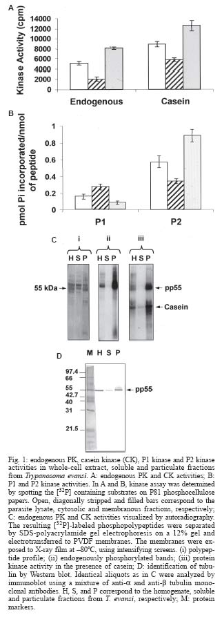
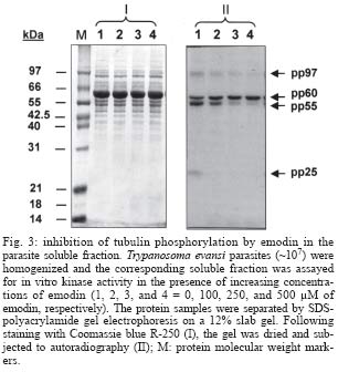
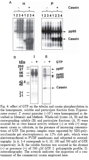
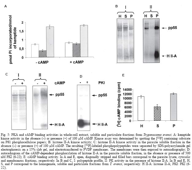
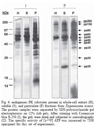
2004 Code number: oc04167 Trypanosoma evansi contains protein kinases capable of phosphorylating endogenous substrates with apparent molecular masses in the range between 20 and 205 kDa. The major phosphopolypeptide band, pp55, was predominantly localized in the particulate fraction. Anti-a and anti-b tubulin monoclonal antibodies recognized pp55 by Western blot analyses, suggesting that this band corresponds to phosphorylated tubulin. Inhibition experiments in the presence of emodin, heparin, and 2,3-bisphosphoglycerate indicated that the parasite tubulin kinase was a casein kinase 2 (CK2)-like activity. GTP, which can be utilized instead of ATP by CK2, stimulated rather than inactivated the phosphorylation of tubulin in the parasite homogenate and particulate fraction. However, GTP inhibited the cytosolic CK2 responsible for phosphorylating soluble tubulin and other soluble substrates. Casein and two selective peptide substrates, P1 (RRKDLHDDEEDEAMSITA) for casein kinase (CK1) and P2 (RRRADDSDDDDD) for CK2, were recognized as substrates in T. evansi. While the enzymes present in the soluble fraction predominantly phosphorylated P1, P2 was preferentially labeled in the particulate fractions. These results demonstrated the existence of CK1-like and CK2-like activities primarily located in the parasite cytosolic and membranous fractions, respectively. Histone II-A and kemptide (LRRASVA) also behaved as suitable substrates, implying the existence of other Ser/Thr kinases in T. evansi. Cyclic AMP only increased the phosphorylation of histone II-A and kemptide in the cytosol, demonstrating the existence of soluble cAMP-dependent protein kinase-like activities in T. evansi. However, no endogenous substrates for this enzyme were identified in this fraction. Further evidences were obtained by using PKI (6-22), a reported inhibitor of the catalytic subunit of mammalian cAMP-dependent protein kinases, which specifically hindered the cAMP-dependent phosphorylation of histone II-A and kemptide in the parasite soluble fraction. Since the sum of the values obtained in the parasite cytosolic and particulate fractions were always higher than the values observed in the total T. evansi lysate, the kinase activities examined here appeared to be inhibited in the original extract. Key words: Trypanosoma evansi - protein kinases - casein kinases - cAMP-dependent protein kinases - signal transduction Protein phosphorylation and dephosphorylation constitute a cardinal mechanism for the regulation of many physiological processes such as metabolic pathways, gene transcription, membrane transport of ions and metabolites, cell division, and synaptic transmission. Processes modulated by reversible protein phosphorylation require not only a protein kinase (PK), but also a protein phosphatase (PP). Target proteins are phosphorylated at specific sites by one or more PK, and these phosphates are removed by specific PP. The activation of the correct pool of PK or PP by individual hormones, to trigger specific intracellular events, is controlled in part by where PK and PP are located in the cell. PK can be classified according to the amino acid species they covalently phosphorylate: namely, protein serine/threonine kinases (PSK); protein tyrosine kinases (PTK); protein histidine kinases (PHK); and also, a small subfamily of PSK can phosphorylate both serine/threonine and tyrosine residues. PSK and PTK are structurally related to each other and are most likely derived from a common ancestral enzyme, whereas PHK are a distinct family of enzymes. In most cases, PHK are part of the so-called two-component signal transduction systems, which are characterized by a histidine-to-aspartate phospho-transfer mechanism. Correspondingly, there are protein serine/threonine phosphatases (PSP), protein tyrosine phosphatases (PTP), protein aspartate phosphatases (PAP), and dual specificity phosphatases capable of dephosphorylating both phosphoserine/phosphothreonine and phosphotyrosine residues. Based on the similarity in the primary structure of their catalytic domain, the eukaryotic PSK and PTK have been organized into five distinct classes that share basic structural and functional properties (Hanks & Quinn 1991). These are: (1) the AGC group, which includes the cAMP- and cGMP-dependent protein kinase family (PKA and PKG), the diacylglycerol-activated/phospholipid-dependent protein kinase C family (PKC), the family of kinases related to PKA and PKC (PKB), and the family of protein kinases that phosphorylate G protein-coupled receptors; (2) the CaMK group, which includes the Ca+2/CaM-dependent protein kinase families; (3) the CMGC group, which includes the cyclin-dependent kinase family (CDK), the ERK kinase family (MAPK), the glycogen synthase kinase 3 family (GSK3), and casein kinase 2 family (CK2); (4) the PTK group, which includes the conventional non-membrane spanning and membrane spanning protein tyrosine kinases; and (5) the OPK group, which contains other protein kinases not falling in any of the groups just mentioned, for example the casein kinase 1 family (CK1). The hemoparasitic protozoa Trypanosoma evansi is the causative agent of the disease known as "derrengadera" or "surra", which affects equines and other domestic animals. T. evansi is widely spread since it occurs in tropical Africa, Southeastern Asia, and South America. The syndromes associated with the infection of T. evansi vary from chronic to acute and fatal, being the major clinical symptoms progressing weakness, emaciation, fever, anemia, and death. At the present time, the tools available to control T. evansi proliferation are inadequate, and an immediate prospect for a vaccine is uncertain. Even accurate diagnostic kits are not available yet. Thus, there is an urgent need to obtain fundamental knowledge about the biology of T. evansi at a molecular level, in order to identify susceptible targets. Since reversible protein phosphorylation is one of the most widely used mechanisms of signal integration in eukaryotic cells, and no information about signaling cascades in T. evansi have been reported, one of our immediate goals has been to identify and characterize the various protein kinases present in this parasite. In this manuscript we initiated the study of protein phosphorylation events in T. evansi. MATERIALS AND METHODS Materials - Reagents were purchased from the following sources: [g-32P] ATP (3000 Ci/mmol), New England Nuclear or Amersham; [8-3H] cAMP (23 Ci/mmol), Amersham; monoclonal anti-a tubulin (clone DM 1A), anti-b tubulin (clone TUB 2.1), dephosphorylated casein, emodin, GTP, heparin, 2, 3-bisphosphoglycerate (BPG), benzamidine, kemptide, L-trans-epoxysuccinyl-leucylamido(4-guanidino)butane (E-64), phenylmethyl-sulfonylfluoride (PMSF), fibrous DEAE-cellulose, PKI (6-22), Sigma; anti-mouse IgG alkaline phosphatase conjugate, 5-bromo-4-chloro-3-indolyl phosphate, nitro blue tetrazolium, Promega; P81 phosphocellulose chromatography paper, Whatman; polyvinylidene difluoride (PVDF) membranes, filters type HA (0.45-µm), Millipore Corporation. Dr Susan S Taylor (University of California, San Diego, US) generously donated synthetic P1 (RRKDLHDDEEDEAMSITA) and P2 (RRRADDS DDDDD) peptides. All other chemicals were of the highest quality grade available. Parasites - Albino rats (Sprague Dawley) were infected with cryopreserved T. evansi from the Venezuelan TEVA1 isolate (Desquesnes & Tresse 1996). When the number of parasites reached ~107 trypanosomes/ml of blood, the rats were bled by cardiac puncture using 2% EDTA as anticoagulant. T. evansi was purified according to Lanham and Godfrey (1970), with slight modifications. The blood was centrifuged (3000 g), for 10 min, at 4ºC. The supernatant containing the plasma was discarded, and the pellet was diluted 1:3 in PBSG (60 mM NaH2PO4/Na2HPO 4, 150 mM NaCl, 1% glucose, pH 8.0) and centrifuged under identical conditions. This procedure was repeated and the resulting pellet was finally diluted 1:3 in PBSG and loaded onto a fibrous DEAE-cellulose column, previously reconstituted and equilibrated in PBSG. We used a proportion of approximately 20 ml of packed resin per 5 ml of diluted blood. The column was thoroughly washed with PBSG, and the parasites remaining in the non-absorbing fraction were collected into tubes maintained on ice. The elution profile of T. evansi was followed by light microscopy. Parasites were collected by centrifugation (3000 g), for 10 min, at 4ºC, and washed twice with PBSG. The concentration of parasites was determined using a Neubauer chamber. The final parasite pellet was kept frozen at -80ºC until further use. Homogenization of T. evansi, and preparation of parasite soluble and particulate fractions - All the operations were performed at 4ºC. Approximately 107 T. evansi parasites were suspended in 4 ml of Buffer A (50 mM Tris, 1 mM CaCl2, 1 mM MgCl2, 50 µM PMSF, 1 mM benzamidine, 10 µM E-64, pH 8.0) and homogenized by sonication on ice (4 cycles, 30 s each, with a resting period of 2 min between cycles, 70% power output). A 1-ml aliquot of the crude extract was separated and kept frozen at -80ºC for later use. The remaining homogenate was ultracentrifuged (Beckman L8-70, SW 50.1 rotor) at 100,000 g, for 1 h, to yield the clarified soluble and the particulate fractions. The parasite soluble fraction was collected and stored at -80ºC. The pellet was reextracted twice in Buffer A to eliminate contaminating soluble proteins. The final pellet, corresponding to the parasite particulate fraction, was resuspended in 3 ml of Buffer A and was kept frozen at -80ºC until further use. Protein kinase assays - Protein kinase activity was determined using [g-32P] ATP, either in the absence or presence of exogenous substrates. The incorporation of 32P was quantitatively measured by spotting an aliquot of the reaction mixture on Whatman P81 phosphocellulose papers. Endogenous kinase activity was assayed in a 35 µl-reaction mixture containing 20 µl of the sample and 15 µl of kinase Buffer {50 mM Tris, 12 mM MgCl2, 20 mM KF, 30 mM [g-32P] ATP (specific activity 1800 cpm/pmol), pH 8.0}. Exogenous substrates were also added to the mixtures to determine specific kinase activities. The substrates employed were: dephosphorylated casein (1 mg/ml), histone II-A (0.5 mg/ml), P1 (400 µM), P2 (80 µM), and kemptide (100 µM). Some assays were performed in the presence of cAMP (100 µM). Reactions were incubated for 15 min, at room temperature. Then, 30 µl of the re-actions were spotted onto 2 x 2 cm squares of P81 filter papers. The papers were subsequently washed three times (15 min per wash) in 50 mM phosphoric acid, dried, and analyzed for radioactivity by liquid-scintillation counting (Rackbeta 1209, LKB-Walac). Protein kinase activities were also qualitatively determined by autoradiography after SDS-polyacrylamide gel electrophoresis separation of the radioactively labeled substrates. In these cases, the specific activity of [g-32P] ATP was increased to 3600 or 7200 cpm/pmol. The reactions were terminated by addition of 4X SDS-polyacrylamide gel electrophoresis sample buffer (Laemmli 1970), followed by a 5-min incubation at 100ºC. Labeled gels were either dried or electroblotted onto PVDF membranes (250 mA, for 1 h, at 4ºC), according to Towbin et al. (1979). Then, the dried gels or the PVDF membranes were exposed to Kodak X-Omat x ray film, at -80ºC, with intensifying screens. Phosphorylation assays in the presence of protein kinases inhibitors - Aliquots of the parasite homogenate, clarified supernatant fraction, and particulate fraction, containing 30 µg of total protein, were assayed for kinase activity, in the presence or in the absence of various concentrations of GTP (0-500 µM), BPG (0-10 mM), emodin (0-500 µM), or heparin (0-500 µg/ml). Assays were performed with and without dephosphorylated casein (1 mg/ml). Additionally, inhibition assays were also performed by incubating aliquots of the T. evansi lysate, cytosolic and membranous fractions with 500 nM of the cAMP-dependent protein kinase inhibitor PKI (6-22), in the absence or presence of 100 µM cAMP, and using histone II-A as exogenous substrate. The phosphorylation reactions were incubated for 15 min at room temperature and then stopped with 4X SDS-polyacrylamide gel electrophoresis sample buffer (Laemmli 1970). The protein samples were separated by SDS-polyacrylamide gel electrophoresis. The gels were either dried or electroblotted onto PVDF membranes and subjected to autoradiography as described above. Measurement of cAMP binding - Cyclic AMP binding was measured by Millipore filtration as described by Bubis and Taylor (1985). The assay was carried out in binding buffer (50 mM Tris, 1 mM EDTA, 5 mM b-mercaptoethanol, 200 nM [3H] cAMP, pH 8.0), final volume = 100 µl. After 1 h incubation at 4ºC, an 80-µl aliquot of the reaction mixture was transferred to a filter reservoir (0.45-µm Millipore HA filter) containing 5 ml of ice-cold binding buffer. The filters were washed under vacuum with 25 ml of additional binding buffer, placed in scintillation vials, dried, and counted in 3 ml of scintillation cocktail. Miscellaneous procedures - Protein concentration was determined using bovine serum albumin as protein standard (Bradford 1976). Protein gel electrophoresis was carried out in the presence of SDS on 1.5-mm thick slab gels containing 12 or 15% polyacrylamide (Laemmli 1970). Coomassie blue R-250 or silver staining was used for protein visualization on gels. For Western blot analyses, proteins separated by SDS-polyacrylamide gel electrophoresis were electrotransferred from the gels to nitrocellulose sheets as described (Towbin et al. 1979). The filters were blocked with 3% bovine serum albumin in TBST [20 mM Tris-HCl (pH 8.0), 150 mM NaCl, 0.05% Tween 20] for 1 h, at room temperature, and then incubated with a mixture of anti-a and anti-b tubulin monoclonal antibodies (1:1000) for 1 h. After three washes with TBST, the filters were incubated with anti-mouse IgG coupled to alkaline phosphatase (1:7500) and washed with 100 mM Tris-HCl (pH 9.5). The substrate and the chromophore, 5-bromo-4-chloro-3-indolyl phosphate and nitro blue tetrazolium, were then added in alkaline phosphatase buffer, containing 100 mM Tris-HCl (pH 9.5), 100 mM NaCl and 5 mM MgCl2. Reactions were stopped with 20 mM Tris-HCl (pH 8.0) and 5 mM EDTA. RESULTS To determine the presence of protein kinase activities in T. evansi, kinase assays were performed using aliquots from the complete parasite lysate, and from the corresponding soluble and particulate fractions. The reactions were carried out using equal volumes of the fractions in order to equivalently maintain the protein relation in the three different samples. As illustrated in Fig. 1A (left), T. evansi contains kinases capable of phosphorylating endogenous substrates. However, the kinase activity present in the whole-cell extract was less than the sum of the activities present in the soluble and particulate fractions, which indicated the existence of kinase inhibitors in the homogenate. All three samples were also capable of phosphorylating exogenously added casein (Fig. 1A, right), which demonstrated the presence of CK-like activities in both, the parasite soluble and particulate fractions. As previously seen with the phosphorylation of endogenous substrates, the addition of the CK activities from the soluble and particulate fractions was higher than the total CK activity observed in the original lysate, suggesting that these kinases were inhibited in the parasite homogenate. To further characterize the CK activities evidenced in T. evansi, we carried out assays in the presence of P1 and P2, peptide substrates for mammalian protein kinases CK1 and CK2, respectively (Marin et al. 1994). In both cases the endogenous phosphorylation obtained in the absence of P1 and P2 was subtracted. We detected kinase activities capable of phosphorylating P1 in whole-cell extracts and in the soluble and particulate fractions (Fig. 1B, left). However, P1 phosphorylation was higher in the soluble than in the membranous fraction, indicating that the parasite CK1-like activities were preferentially located in the cytosol. As shown in Fig. 1B (right), kinase activities capable of phosphorylating P2 were also determined in the parasite lysate, suggesting that T. evansi contains CK2-like enzymatic activities. The parasite CK2-like activities seemed to be distributed in both, the soluble and the particulate components, although they were preferentially localized in the membranous fraction (Fig. 1B, right). Additionally, more stoichiometric incorporation of [32P] inorganic phosphate into P2 than into P1 was obtained, which demonstrated a higher expression of CK2-like than CK1-like enzymes in T. evansi. In agreement with the endogenous phosphorylation of T. evansi substrates, and with the total CK activities, the sum of the individual phosphorylating activities for both P1 and P2, in the parasite soluble and particulate fractions, was always higher than the corresponding activities obtained in the original lysate. As illustrated in Fig. 1C (panel ii), autoradiographic analysis showed that the major phosphorylated polypeptide in whole-cell lysates of T. evansi corresponded to a 55-kDa band (pp55). Under the conditions used here, this polypeptide band and the kinase(s) that phosphorylate(s) it predominantly remained in the parasite membranous fraction. In agreement with the quantitative P81 filter measurement shown above, the kinase assay followed by autoradiography also indicated that the parasite extract, the soluble and the particulate fraction, contained protein kinases capable of phosphorylating exogenously added casein (Fig. 1C, panel iii). The corresponding polypeptide profiles for the homogenate, soluble and particulate fractions are shown in Fig. 1C (panel i). The relative abundance of pp55 and its molecular weight, which is consistent with tubulin, prompted us to test monoclonal antibodies against this cytoskeletal component in a Western blot. Identical samples from the complete parasite lysate, and from the corresponding cytosolic and membranous fractions were analyzed. Specific anti-a and anti-b tubulin monoclonal antibodies recognized the 55-kDa phosphorylated polypeptide band (Fig. 1D), indicating that pp55 corresponds to phosphorylated tubulin. Additionally, tubulin primarily compartmentalized with the parasite particulate fraction (Fig. 1D), similar to pp55 (Fig. 1C). Thus, under the conditions employed here, the protein kinase responsible for phosphorylating tubulin, as well as the fraction of tubulin that became phosphorylated remained tightly associated with the T. evansi particulate fraction. Kinase assays were carried out in the presence of several well-known mammalian CK2 inhibitors, in order to examine whether this protein kinase was responsible for the phosphorylation of tubulin in T. evansi. Specifically, we tested the effect of emodin, heparin and BPG on the endogenous phosphorylation of total homogenates, and parasite soluble and particulate fractions. Additionally, kinase assays were also carried out in the presence of exogenously added casein for comparison. A prominent dose-dependent inhibition on the phosphorylation of tubulin and casein was observed in both the whole-cell lysate and parasite membranous fractions, following addition of increasing concentrations of emodin (Fig. 2B), heparin (Fig. 2C) or BPG (data not shown). As illustrated with emodin in Fig. 3, similar results were obtained for the phosphorylation of the minor amount of tubulin that compartmentalized with the parasite cytosolic fraction. The phosphorylation of two additional soluble polypeptides, pp97 and pp25, were also inhibited by emodin in a concentration dependent manner (Fig. 3). Interestingly, a 60-kDa endogenously phosphorylated substrate, which was enriched in the parasite cytosol, was not affected by emodin (Fig. 3) or by any other mammalian CK2 inhibitor (data not included). These results demonstrated that other protein kinases, different than CK2, were responsible for the phosphorylation of the 60-kDa-phosphopolypeptide band. Accordingly, GTP, which is known as a good phosphate donor for CK2 (KM for GTP = 5-100 µM vs. 5-30 µM for ATP), inhibited both the phosphorylation of tubulin and exogenously added casein in the parasite soluble fraction (Fig. 4B). GTP also inhibited the phosphorylation of other phosphopolypeptide bands in this fraction, including pp97. However, the phosphorylation of pp60 was not affected by GTP (Fig. 4B). The effect of GTP on the protein kinases present in the original lysate and parasite membranous component was also determined. Interestingly, a slight activation on the phosphorylation of tubulin and casein was observed in the whole-cell homogenate and particulate fraction, when increasing amounts of GTP were added (Fig. 4A). Kemptide (LRRASVA), a peptide that serves as substrate for other PSK including cyclic nucleotide-dependent protein kinases, was also monitored as exogenous substrate for the protein kinases of T. evansi. Specific kemptide phosphorylation was determined by subtracting the endogenous incorporation of [32P] inorganic phosphate. In the absence of cAMP, we detected kinase activities capable of phosphorylating kemptide in the parasite homogenate, and in both, the cytosolic and membranous components (Fig. 5A, left), yet the kemptide phosphorylation was higher in the particulate than in the soluble fraction. We also measured the phosphorylation of kemptide in the presence of cAMP to examine for PKA-like activities in T. evansi. The kemptide kinase activities seen in both, the whole lysate and the particulate fraction remained approximately the same in the absence or presence of cAMP (Fig. 5A). However, the kinase activity observed in the parasite soluble fraction was approximately doubled when cAMP was added, indicating the presence of PKA-like activities in the soluble fraction of T. evansi (Fig. 5A, right). The kemptide kinase activity observed in the whole-cell extract was lower than the sum of the kemptide kinase activities obtained in the soluble and particulate fractions, evidencing again the presence of kinase inhibitors in the parasite homogenate. We also detected kinase activities capable of phosphorylating histone II-A in T. evansi extracts, and in the parasite cytosolic and membranous components (Fig. 5B). Similar to the results with kemptide, the histone II-A phosphorylation was higher in the particulate fraction than in the soluble fraction. These results evidence that other PSK exist in T. evansi, which are preferentially located in the parasite membranous fraction. Moreover, compatible with the cAMP-dependent kemptide phosphorylation data shown previously in Fig. 5A, the phosphorylation of histone II-A increased in the parasite soluble fraction when cAMP was added, which again suggested the existence of cytosolic PKA-like activities in the parasite (Fig. 5C). However, no endogenous substrates for this enzyme were identified in this fraction. As no PKA-like activities were observed in the original parasite lysate, when kemptide or histone II-A was used, these activities must be unavailable or somewhat inhibited in the cell homogenate. Fig. 5D shows the cAMP-dependent phosphorylation of exogenously added histone II-A by the parasite cytosolic fraction, in the absence or presence of PKI (6-22), a specific inhibitor of the catalytic subunit of mammalian cAMP-dependent protein kinases. The autoradiogram revealed that the labeling of histone II-A, in the presence of cAMP, was 90-95% inhibited when the parasite soluble fraction was incubated with 500 nM PKI (6-22). Although the endogenous phosphorylation of various polypeptide bands was inhibited by PKI (6-22), the phosphorylation of tubulin was not affected by this inhibitor peptide (Fig. 5D). These results indicated that PKA was not involved in phosphorylating this cytoskeletal component. PKI (6-22) also inhibited the cAMP-dependent phosphorylation of exogenously added kemptide by the T. evansi soluble fraction (data not shown). Interestingly, the original homogenate, the parasite soluble fraction and the parasite particulate fraction, were capable of binding [3H] cAMP (Fig. 5E), indicating the presence of cytosolic and membranous cAMP-binding proteins in T. evansi. Besides pp55, other endogenous phosphorylated polypeptide bands with apparent molecular masses in the range between 20 and 205 kDa, were also detected in the parasite soluble and particulate fraction (Fig. 6). Some of these bands were absent in the original lysate. DISCUSSION Phosphorylation by protein kinases has been implicated as an important control mechanism in signal transduction, growth and differentiation in parasitic protozoa. However, very little is known about the mechanisms of signal transduction in salivarian American trypanosomes. In this paper, we have initiated the identification of protein kinase activities from T. evansi, an important animal hemoparasite that infects horses, donkeys, and capybaras in South America. A 55-kDa protein, pp55, which was identified as phosphorylated tubulin by Western blot analysis, represented the major phosphoprotein in T. evansi total extracts. Under the conditions used here, the fraction of tubulin that became phosphorylated and its corresponding kinase, were primarily located in the parasite particulate fraction. The kinase activity responsible for phosphorylating tubulin was inhibited by emodin, heparin and BPG, characteristics that are shared by protein kinases CK2 from higher eukaryotes. These results established that the parasite tubulin kinase appeared to be a CK2-like enzyme. In addition, P2, which is a specific substrate for CK2 enzymes, was predominantly phosphorylated by the parasite membranous portion. All these results strongly indicated the presence of CK2-like activities in T. evansi. Emodin and heparin have been reported to inhibit mammalian CK2s with IC50 values of 1 µM (Yim et al. 1999) and 150 ng/ml (Hathaway & Traugh 1983), respectively. The effective inhibitor concentrations used for the putative CK2 enzyme from T. evansi were slightly higher, showing about 95% inhibition in its ATP-phosphotransferase activity with 100 µM emodin and 10 µg/ml heparin. Since the enzyme investigated here belongs to a hemoprotozoan parasite, it is expected to diverge from its homologous counterparts in higher eukaryotes. In addition, the parasite CK2 was not purified to homogeneity, and inhibitors' IC50 values may change according to the purity of the material. Consistently, tubulin has been reported to be the major phosphoprotein in another trypanosome, T. cruzi (Casas et al. 2002), and likewise, T. cruzi tubulin has also been shown to be phosphorylated by a CK2-like activity (Casas et al. 2002, Uzcanga et al. 2003). In T. brucei, an evolutionarily more related parasite, two CK2 genes have been identified: CK2a1 and CK2a2 (Park et al. 2002). Although, the protein encoded by CK2a1 was capable of associating with Nopp44/46, an abundant nucleolar phosphoprotein of T. brucei (Park et al. 2002), no substrates for the kinase encoded by CK2a2 have been described yet. On the basis of the results shown here, it is plausible that T. brucei tubulin could also serve as a substrate for the CK2a2-encoded protein kinase. Interestingly, CK2 is also responsible for b-tubulin phosphorylation in neuroblastoma cells (Diaz-Nido et al. 1990). Microtubules are polymers of globular tubulin subunits, which are organized in a cylindrical tube, composed of protomers of a and b tubulin heterodimers. A number of posttranslational modifications of tubulin have been reported, which include acetylation, tyrosination, phosphorylation, polyglutamilation, and polyglycylation, and these posttranslationally modified tubulin appeared to be associated with specific populations of microtubules (Scherwin et al. 1987, Gallo et al. 1988). Two new members of the tubulin superfamily, g and d, have been described in T. brucei using a genetic approach (Gull 2001). Additionally, several microtubule-associated proteins (MAPs) seem to be involved in interconnecting microtubules to each other, as well as to the plasma membrane (Gull 1999). Previous reports have suggested that CK2 plays an important role in the maintenance of cell morphology and polarity (Ulloa et al. 1994, Rethinaswamy et al. 1998). Also, coimmunoprecipitation and in vitro binding studies have evidenced that CK2 is strongly associated to tubulin in mammalian cell extracts (Faust et al. 1999). More recently, chicken CK2 has been identified as a structural MAP that stabilizes microtubules and mediates microtubule dynamics (Lim et al. 2004). However, the CK2 microtubule assembling and stabilizing function was independent of its kinase activity, since a kinase-inactive mutant of CK2 displayed the same microtubule assembling activity as the wild-type protein (Lim et al. 2004). Interestingly, tubulin and the CK2 responsible for its phosphorylation have also been shown to be associated in T. cruzi (Casas et al. 2002, Uzcanga et al. 2003). It is plausible that the parasite CK2 also acts as a MAP regulating microtubule dynamics in trypanosomes through its direct physical association with tubulin, similar to what has been shown in higher eukaryotes. Since GTP can be used instead of ATP as the phosphoryl donor by mammalian CK2s, GTP frequently has been employed as a competitor for these kinases in radioactive labeling assays. In T. evansi, GTP did not inhibit the incorporation of [32P] inorganic phosphate from radioactive ATP onto the fraction of tubulin present either in whole-cell lysates or in the parasite particulate fraction. In contrast, a slight increase in the phosphorylation of tubulin was observed in the presence of GTP, in both samples. These results indicate that either a GTP-dependent activation of the protein kinase in charge of phosphorylating tubulin or a GTP-dependent inactivation of a potent protein phosphatase responsible for desphos-phorylating tubulin occurs in the T. evansi homogenate and particulate fractions. On the contrary, a clear competition between GTP and radioactive ATP was only assessed with the tubulin kinase that compartmentalized together with the parasite soluble fraction. Similar results were observed when exogenously added casein was used as a substrate for the kinases present in the parasite soluble fraction. GTP enhancement on the phosphorylation of proteins by [g-32P] ATP is not a new phenomenon. Van Dijk et al. (1981) showed that the presence of non-radioactive GTP could modulate [g-32P] ATP-mediated protein phosphorylation using rat brainstem. Similarly, Kondratev et al. (1986) reported the modulation of [g-32P] GTP-driven phosphorylation by unlabeled ATP of synaptic membrane proteins. Lelong-Rebel et al. (2003) have also reported that the ratio of ATP/GTP was capable of modulating the phosphorylation level of P-glycoprotein and of various plasma membrane proteins of KB-V1 multidrug resistant cells. Several possible explanations could be proposed for the GTP stimulatory effect shown here: 1) the existence of two CK2 isoenzymes in T. evansi, one compartmentalizing with the parasite particulate component and the other one with the soluble fraction. The soluble CK2 is inhibited by GTP, while the membranous form is more active than the soluble form, and is stimulated by GTP; 2) the existence of a parasite protein phosphatase inactivated by GTP, which uses phosphorylated tubulin as a substrate, and is compartmentalized with the membranous fraction. Alternatively, 3) the presence of GTP might be linked to the phosphorylation of an intermediate "regulatory" element present in the parasite membranes, which would in turn be involved in the activation of the [g-32P] ATP-driven phosphorylation of tubulin by CK2. Interestingly, competition assays have revealed that, unlike most CK2s, the protein encoded by CK2a1 from T. brucei was capable of discriminating between ATP and GTP (Park et al. 2002). These evidences imply that in comparison to mammalian CK2s, CK2s from salivarian trypanosomes have different affinities toward intracellular triphosphate nucleotides. P1, which is a specific substrate for CK1 protein kinases, was predominantly phosphorylated by the parasite cytosolic fraction, suggesting the presence of soluble CK1-like enzymes in T. evansi. Calabokis et al. (2002) have reported a soluble CK1-like activity in T. cruzi, which showed similar biochemical properties to other CK1s from higher eukaryotes. The importance of CK1 for the control of some essential cellular functions was demonstrated by genetic studies in yeast (Robinson et al. 1992). However, the precise physiological function and possible mode of regulation of this multipotential enzyme have remained poorly characterized. The CK1 isoforms from yeast have been linked genetically to vesicular trafficking, DNA repair, cell cycle progression and citokinesis (Gross & Anderson 1998). The CK1-like activities from T. evansi may have similar functions. Histone II-A and kemptide were also recognized as exogenous substrates by the various parasite fractions, implying the existence of other Ser/Thr kinases in T. evansi. Moreover, the addition of cAMP increased the phosphorylation of histone II-A and kemptide in the parasite soluble fraction, which supported that T. evansi contains cytosolic PKA-like activities. Yet, no endogenous substrates were identified for the parasite cAMP-dependent protein kinase. The specific inhibition of the cAMP-dependent phosphorylation of histone II-A and kemptide in the presence of 500 nM PKI (6-22), a potent inhibitor of the mammalian PKA catalytic subunit (Ki = 1.7 nM), provided further evidences for the existence of PKA-like activities in the parasite soluble fraction. The cAMP-PKA pathway is one of the most common and versatile signal pathways in eukaryotic cells, and is involved in the modulation of cellular functions in almost all tissues in mammals (Taskén & Aandahl 2004), including: (1) the regulation of ion channels in the nervous system; (2) the regulation of the cell cycle which involves microtubule dynamics, chromatin condensation and decondensation, nuclear envelope disassembly and reassembly, and numerous intracellular transport mechanisms; (3) control of exocytotic events in polarized epithelial cells; (4) regulation of cardiovascular function; (5) modulation of metabolism in adipose tissue; (6) regulation of steroidogenesis, reproductive function and immune responses etc. Thus, further studies should be carried out in order to reveal the physiological role of these PKA-like enzymes in the T. evansi parasite. T. evansi also contained cAMP binding proteins in the homogenate, and in both, the soluble and particulate fractions. Since adenylyl cyclase genes have been reported in various trypanosome species (Sanchez et al. 1995, Taylor et al. 1999), cAMP must be of importance in the intracellular signaling of these parasites. The current view in higher eukaryotic cells is that the majority of the cAMP-mediated effects are due to the activation of PKAs and the subsequent phosphorylation of metabolic enzymes and transcriptional factors. However, today there is evidence that the cAMP-dependent kinases are not the only intracellular receptors involved in intracellular cAMP signaling in eukaryotes. Other cAMP-binding proteins have been identified, including some cyclic nucleotide-gated channels and Epac (exchange protein directly activated by cAMP) proteins (Dremier et al. 2003). Cyclic AMP-binding proteins that do not correspond to the PKA regulatory subunits have been described in T. cruzi (Rangel-Aldao et al. 1988) and in Leishmania (Banerjee & Sarkar 2001). Several biochemical approaches have failed to detect the presence of the regulatory subunits of PKA in Leishmania donovani, although catalytic subunits of PKA have been identified and cloned from Leishmania spp. (Siman-Tov et al. 2002). Similarly, Walter and Opperdoes (1982) have attempted to detect PKA activities in crude extracts of T. brucei, with no avail. Interestingly, a cGMP-dependent PKA has recently been identified in T. brucei (Shalaby et al. 2001). Other endogenously phosphorylated polypeptides with apparent molecular weights between 20 and 205 kDa were also detected in T. evansi. The phosphorylation of a 97-kDa and a 25-kDa-polypeptide band was inhibited in the presence of various CK2 inhibitors, suggesting that these polypeptides are endogenous substrates for a parasite CK2. In contrast, a 60-kDa-polypeptide band was phosphorylated even in the presence of these inhibitors. A 50-kDa phosphoprotein was also detected in T. evansi. Interestingly, Garcia-Salcedo et al. (2002) reported a 50-kDa cytoplasmatic protein kinase from T. brucei, TBPK50, which belongs to a family of kinases involved in the regulation of cell cycle, cell shape and proliferation. TBPK50 was able to autophosphorylate but was unable to transphosphorylate a range of classical acceptor substrates in vitro (Garcia-Salcedo et al. 2002). It is plausible that the pp50 detected in the T. evansi soluble fraction corresponds to the homologous TBPK50 from T. evansi. The presence of these kinase activities in T. evansi suggest that signal transduction cascades similar to the ones found in mammalian cells may exist in trypanosomes. Moreover, growth in these parasites may be controlled by some of the same components that have been reported in mammalian cells. In addition to potential roles in the regulation of parasite functions, protein kinases could also play an important role in the modulation of some host functions. For example, Leishmania major possesses an extracellular protein kinase capable of phosphorylating the C3 and 3Cb components of the human complement system (Hermoso et al. 1991). We have yet to further characterize these proteins; their functional roles as their precise relevance in T. evansi development and physiology remain to be determined. Interestingly, the phylogenetic isolation of parasitic protozoa has reflected atypical structural and functional properties of many of their protein kinase homologues (Doerig 2004). Likewise, evidence is emerging, which suggests that the organization of some otherwise well-conserved signal transduction pathways is divergent in some parasitic species (Doerig 2004). The differences between protein kinases of a parasite and their homologues in its host cell imply that specific inhibitors of the former can be designed as anti-parasitic drugs. The work reported here represents only the beginning of the task of defining, purifying and functionally characterizing the various protein kinases from T. evansi. ACKNOWLEDGEMENTS To Dr Susan S Taylor (University of California, San Diego, US) for donating the synthetic P1 and P2 peptides. REFERENCES
Copyright 2004 Instituto Oswaldo Cruz - Fiocruz The following images related to this document are available:Photo images[oc04167f2.jpg] [oc04167f1.jpg] [oc04167f4.jpg] [oc04167f3.jpg] [oc04167f6.jpg] [oc04167f5.jpg] |
| |||||||||
