 |
| About Bioline | All Journals | Testimonials | Membership | News |
|
||||||
|
||||||
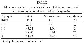
Memórias do Instituto Oswaldo Cruz, Vol. 100, No. 03, May 2005, pp. 237-239 SHORT COMMUNICATION DNA evidence of Trypanosoma cruzi in the Chilean wild vector Mepraia spinolai (Hemiptera: Reduviidae) Carezza Botto-Mahan/+, Sylvia Ortiz*, Marlene Rozas*, Pedro E Cattan**, Aldo Solari* Departamento
de Ciencias Ecológicas, Facultad de Ciencias *Programa de
Biología Celular y Molecular, Facultad de Medicina **Departamento
de Ciencias Biológicas Animales, Facultad de Ciencias Veterinarias
y Pecuarias Universidad de Chile, Santiago, Chile +Corresponding author. E-mail: cbotto@uchile.cl Received
15 December 2004 Code number: oc05075 Molecular evidence showed 46.2% of Trypanosoma cruzi infection in Mepraia spinolai insects from North-Central Chile, which is significantly higher than previous reports of up to 26% by microscopic observation. Our results show similar infection levels among nymphal stages, ranging from 38.3 to 54.1%, indicating that younger nymphs could be as important as older ones in parasite transmission. A cautionary note must be stressed to indicate the potential role of M. spinolai in transmitting T. cruzi in country areas due to the high infection level detected by molecular analysis. Key words: Triatominae - Chagas disease - Kissingbug Chagas disease is a serious human parasitic disease in America that is caused by the flagellate protozoan Trypanosoma cruzi, and transmitted by blood-sucking insects of the subfamily Triatominae (Hemiptera: Reduviidae) (Panzera et al. 2004). Detection of T. cruzi can be carried out through different methodologies such as direct microscopic observation, hemoculture, xenodiagnosis, and in the last decade the polymerase chain reaction (PCR). It is well known that PCR-based detection from feces or urine of reduviid bugs, and blood samples from mammals is more efficient than the other techniques (Moser et al. 1989, Breniere et al. 1992, Russomando et al. 1992, 1996). However, scarce information has been reported about infection levels of wild triatomine populations using molecular techniques. Mepraia spinolai is one of the two triatomine species responsible of T. cruzi transmission in arid and semiarid Chile (Lent et al. 1994). This strictly hematophagous and diurnal species distributes between 18º and 34ºS, and its main habitat includes stay grounds, bird nests, rock crevices, and caves although it has been also found in rustic and abandoned houses (Lent & Wygodzinsky 1979, Schofield et al. 1982, Canals et al. 1997). Even though, human blood index for M. spinolai indicates that this species is not an important vector of T. cruzi, the insect reaches high population densities in quarries near human dwellings suggesting an increasing risk of Chagas disease transmission in these zones (Cattan et al. 2002). Studies using microscopic methods have reported high variability in T. cruzi infection levels of wild insect populations, ranging 0-26%, depending on the locality (Apt & Reyes 1986, Frias et al. 1995, Ordenes et al. 1996, Canals et al. 2001). In this paper we document the level of T. cruzi infection in a wild population of M. spinolai using PCR and direct microscopic techniques. Individuals of M. spinolai were collected from April to August 2002 at Las Chinchillas National Reserve (31°30'S, 71°06'W), located approximately 300 km north from Santiago (Chile). In this area, the climate corresponds to a semiarid Mediterranean type with most rainfall concentrated in the winter season (di Castri & Hajek 1976). First to fifth instar nymphs were collected from the same ecotope, characterized by stony slopes with low to moderate human activity and traffic of cattle yard animals. In the collecting site, M. spinolai individuals feed on free ranging introduced rabbits (Oryctolagus cuniculus), and small native mammals (Phyllotis darwini, Octodon degus, Abrothrix olivaceus, Oligoryzomys longicaudatus, and Thylamys elegans) inhabiting the area (Rengifo 2000). Captured nymphs were kept separately inside a climate chamber at 27ºC, 70% RH and 14:10 h L:D cycle. The intestinal content of 182 M. spinolai individuals were removed through abdominal extrusion, and inspected by light microscopy (Nikon Diaphot-FXA) for the presence of T. cruzi (Schenone et al. 1980). For microscopic observation, 5 µl of fresh intestinal content was compressed between a slide and an 18 ´ 18 mm cover slip. The presence of motile parasites in 50 microscopic fields was registered using ´ 400 magnification. The remaining intestinal sample was mixed with 200 µl of PBS buffer, centrifuged at 10,000 xg, and frozen at -20º C for PCR assay. Most wild insects were naturally fasted implying that intestinal contents were free of fresh blood. Therefore, intestinal samples did not require DNA extractions as previously described for other insect vectors (Russomando et al. 1996). The amplification reactions were performed with oligonucleotides 121 and 122, which anneal to the four constant regions present in the minicircles of T. cruzi (Wincker et al. 1994). This PCR test is directed to minicircle DNA that is present in more than 10,000 copies per parasite, therefore, the assay is highly sensitive and appropriate for diagnosis. The intestinal sample was boiled for 10 min, centrifuged at 10,000 ´ g, and 5 µl of the supernatant was used as DNA template. Each experiment included a negative control where the DNA sample was changed by H2O, and a positive control with a purified DNA of T. cruzi. PCR products of 330 bp were analyzed by electrophoresis in a 2% agarose gel and visualized by ethidium bromide staining. For each sample, PCR assay was performed thrice. As confirmatory assay, amplified DNAs were transferred to nylon membranes, denatured, crosslinked with UV irradiation, hybridized using a total kinetoplast DNA from T. cruzi as a universal probe, and labeled by a random priming method with [a32P]dCTP (Solari et al. 1991). Direct microscopic observation indicated that 9.89% of the nymphs were infected with T. cruzi. Molecular results showed 100% correspondence between the ethidium bromide and the autoradiography intensity bands (Southern analysis). PCR assays indicated that 46.15% of the nymphs were infected with T. cruzi, indicating as expected that previous microscopic observations as well as the ones reported here provide underestimated infection levels (Schenone et al. 1980, Ordenes et al. 1996). Interestingly, PCR assays revealed a similar level of infection among the different nymphal stages, ranging 38.3-54.1% (Table). Microscopic observation does not only underestimate the level of infection of this wild triatomine but also the importance of younger nymphs in parasite transmission. Assessment of T. cruzi infection levels in wild vectors may have important consequences for the disease control. Relatively low infection levels in M. spinolai have been described in different endemic areas of Chile (Schenone et al. 1980, Ordenes et al. 1996), and therefore, almost no attention has been paid on this wild species. Even though the main vector of T. cruzi in Chile (Triatoma infestans) has been virtually eliminated, the potential importance of M. spinolai in transmitting T. cruzi cannot be overlooked. Consistent PCR measurements of infection levels in the wild vector and mammal populations are needed for disease epidemiology assessments in the wild cycle of T. cruzi. ACKNOWLEDGMENTS Rodrigo Medel and Mauricio Canals made important suggestions that improved the clarity of this manuscript. REFERENCES
Copyright 2005 Instituto Oswaldo Cruz - Fiocruz The following images related to this document are available:Photo images[oc05075t1.jpg] |
| |||||||||
