 |
| About Bioline | All Journals | Testimonials | Membership | News |
|
||||||
|
||||||
Memórias do Instituto Oswaldo Cruz, Vol. 100, No. 03, May 2005, pp. 245-247 Myxobolus insignis sp. n. (Myxozoa, Myxosporea, Myxobolidae), a parasite of the Amazonian teleost fish Semaprochilodus insignis (Osteichthyes, Prochilodontidae) JC Eiras/+, JCO Malta*, AMB Varella*, GC Pavanelli** Departamento de Zoologia e Antropologia, and CIIMAR, Faculdade de Ciências, Universidade do Porto, 4099-002 Porto, Portugal *Laboratório de Parasitologia e Patologia de Peixes, Inpa, Manaus, AM, Brasil **Departamento de Biologia, Universidade Estadual de Maringá, Maringá, PR, Brasil +Corresponding author. E-mail: jceiras@fc.up.pt Received 30 July
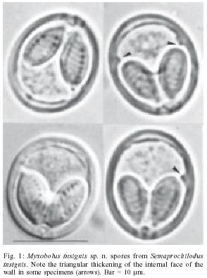
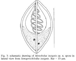
2004 Code number: oc05077 A new myxosporean species is described from the fish Semaprochilodus insignis captured from the Amazon River, near Manaus. Myxobolus insignis sp. n. was located in the gills of the host forming plasmodia inside the secondary gill lamellae. The spores had a thick wall (1.5-2 µm) all around their body, and the valves were symmetrical and smooth. The spores were a little longer than wide, with rounded extremities, in frontal view, and oval in lateral view. They were 14.5 (14-15) µm long by 11.3 (11-12) µm wide and 7.8 (7-8) µm thick. Some spores showed the presence of a triangular thickening of the internal face of the wall near the posterior end of the polar capsules. This thickening could occur in one of the sides of the spore or in both sides. The polar capsules were large and equal in size surpassing the mid-length of the spore. They were oval with the posterior extremity rounded, and converging anteriorly with tapered ends. They were 7.6 (7-8) µm long by 4.2 (3-5) µm wide, and the polar filament formed 6 coils slightly obliquely to the axis of the polar capsule. An intercapsular appendix was present. There was no mucous envelope or distinct iodinophilous vacuole. Key words: Myxozoa - Myxosporea - Myxobolus insignis sp. n. - Semaprochilodus insignis - Brazil Myxobolus spp. are the most common Myxosporea parasitizing fish. Landsberg and Lom (1991) listed 444 valid species, and Eiras et al. (in press) provided a synopsis of the species giving the full characterisation of 744 nominal species, including nearly all the species described so far in fish and amphibians. It is surprising that from such a high number of species only 21 were described for South America 2 from Argentina, 1 from Guiana, and 18 from Brazil. This is surprising because the fish diversity in Brazilian waters is very high, comprising about 8000 species representing nearly 24% of all the known fish species (Cellere et al. 2002). It is likely that the small number of Myxobolus species known so far for this region (as well as of other Myxosporea species) does not mean a scarcity of parasites in this part of the world, but a lack of research on Myxosporea (Cellere et al. 2002). In this paper we describe Myxobolus insignis sp. n. (Myxozoa, Myxosporea, Myxobolidae) from the "jaraqui", Semaprochilodus insignis Jardine & Schomburgk, 1841 (Osteichthyes, Prochilodontidae), a freshwater fish from the Amazon River, Brazil. This fish species, jointly with S. taeniurus, is most important for a large part of the Central Amazon human population once they represent its main food resource. MATERIALS AND METHODS Three specimens of S. insignis (total length: 22.3-24.5 cm) were net-fished from the Amazon river, at Manaus, and transported to the laboratory. The specimens were thoroughly dissected under a compound microscope and all the organs were inspected for the presence of parasites. Measurements were made from fresh spores (30 specimens), and spores were observed under Nomarski differential interference-contrast. For detecting the presence of an iodinophilous vacuole fresh spores were treated with Lugol's iodine solution. Spores were also stained with India ink for revealing any mucous envelope (Lom & Vávra 1961). RESULTS Two specimens of S. insignis exhibited the gills infected by Myxobolus. The parasites formed whitish, rounded or slightly irregular plasmodia (about 0.02-0.08 mm in diameter) located intra-lamellarly within the secondary gill lamellae. Most of the plasmodia were located at the base of the lamellae, but could be present also more or less far from the base. The spores (Figs 1-3) had a valvular wall 1.5-2 µm thick all around their body, and the valves were symmetrical and smooth. The spores were a little longer than wide, with rounded extremities, in frontal view, and oval in lateral view. They were 14.5 (14-15) µm long by 11.3 (11-12) µm wide and 7.8 (7-8) µm thick. Some spores showed the presence of a triangular thickening of the internal face of the wall near the posterior end of the polar capsules. This thickening could occur in one of the sides of the spore or in both sides. The polar capsules were large and equal in size surpassing the mid-length of the spore. They were oval with the posterior extremity rounded and converging anteriorly with tapered ends. They were 7.6 (7-8) µm long by 4.2 (3-5) µm wide, and the polar filament formed 6 coils slightly obliquely to the axis of the polar capsule. A small intercapsular appendix was present. There was no mucous envelope or distinct iodinophilous vacuole. Host: teleost fish Semaprochilodus insignis Jardine & Schomburgk, 1841 Locality: Amazon River, near Manaus, Brazil Site of infection: inside secondary gill lamellae Prevalence: two out of three fish were infected Etymology: the specific name derives from the name of the host species. Specimens deposition: the syntipes are deposited at the "Collection of Myxozoa from the National Institute for the Amazonian Research" (Inpa), Manaus, AM, Brasil, under the reference INPA-002, in the Section of Animal Pathology from the Department of Zoology and Anthropology from the Faculty of Sciences of Porto, Portugal, and in the Museum of Natural History from the Faculty of Sciences of Porto, Portugal. Taxonomic affinities Comparing our species with other Myxobolus described for the South American fishes it can be seen that they are different from all of them. The most similar, concerning the morphometric characteristics, are M. absonus Cellere, Cordeiro & Adriano, 2002, M. associatus Nemeczek, 1926, M. galaxii Szidat, 1953, M. paranensis Bonetto & Pignalberi, 1965 and M. pygocentris Penido, 1927. M. absonus despite some similarities in morphometric characters has unequally sized polar capsules. M. associatus has narrower spores and smaller polar capsules. M. galaxii has spores that are narrower, and infect all the organs of the hosts except the gills, exactly the contrary occurring with our specimens. M. paranensis has smaller spores and smaller polar capsules, and M. pygocentris has longer spores and longer polar capsules. Besides, the spores under examination have a very thick wall, a characteristic not reported for any of the South American Myxobolus species. On the other hand, the spores from our material are ovoid with rounded extremities. This is clearly a different shape of a number of species that have the spores pyriform (M. maculatus Casal, Matos & Azevedo, 2002, M. inaequus Kent & Hoffman, 1984, M. cunhai Penido, 1927, M. lutzi Aragão, 1919, M. paranensis, M. desaequalis Azevedo, Corral & Matos, 2002, M. braziliensis Casal, Matos & Azevedo, 1996, M. associatus, M. chon-drophilus Nemeczek, 1926 and M. inaequalis Gurley, 1893), or the anterior end slightly pointed (M. absonus and M. kudoi Guimarães & Bergamim, 1938). The species which have a similar shape of the spores can not be identified as our species because they are quite different in spore size, in size and position of the polar capsules, in the number of coils of the polar filament, or do not have intercapsular appendix. Our material was also compared with the characteristics of 744 species corresponding to nearly all the species described so far (Eiras et al. in press). Despite some similarities of some species, our specimens could not be identified as any of these species. M. alievi Gasi-magomedov (1970) described from Rutilus rutilus caspicus, and M. lampiformis Chen & Ma (1989) infecting Aristichthys nobilis have dimensions similar in some aspects, but have unequally sized polar capsules and do not have an intercapsular appendix. M. angustus Kudo (1934) from the host Cliola vigilax has spores with the same length, but they are not so wide (7-8 µm), the polar capsules are larger and thinner (8-9.5 and 2.5-3 µm, respectively), and do not have intercapsular appendix. M. baueri Chernova, 1970, infecting Tinca tinca, has similar spore dimensions, but the polar capsules are distinctly smaller (5.3-6 by 2.7-3.3 µm). M. bibullatus Grinham & Cone, 1990 infecting Catostomus commersonii has spores of identical dimensions and the polar capsules are a little smaller, but do not has intercapsular appendix. M. enoblei Lom & Cone (1996) parasitizing Ictiobus bubalus has spores not so wide (10.5-11.5 µm), and longer and larger polar capsules (7.9-8.5 µm and 4.5-5.5 µm, respectively). Finally, M. spalli Landsgerg & Lom, 1991, has spores of the same length but much more thin, smaller polar capsules, and do not has intercapsular appendix. Furthermore, none of the above mentioned species has a spore wall as thick as observed in our specimens. It is concluded that the present material represents a species not yet described, and the name Myxobolus insignis sp. n. is proposed. REFERENCES
The following images related to this document are available:Photo images[oc05077f1.jpg] [oc05077f3.jpg] [oc05077f2.jpg] |
| |||||||||
