 |
| About Bioline | All Journals | Testimonials | Membership | News |
|
||||||
|
||||||
Memórias do Instituto Oswaldo Cruz, Vol. 100, No. 3, May 2005, pp. 277-279 SHORT COMMUNICATION Detection of Brazilian spotted fever infection by polymerase chain reaction in a patient from the state of São Paulo Elvira Maria Mendes Nascimento, Flávia de Sousa Gehrke***, Rosa Amélia Maldonado**, Silvia Colombo, Luiz Jacintho da Silva*, Teresinha Tizu Sato Schumaker***/+ Laboratório de Rickettsias, Instituto Adolfo Lutz, São Paulo, SP, Brasil *Departamento de Clínica Médica, FCM, Unicamp e Superintendência do Controle de Endemias, Secretaria de Estado da Saúde de São Paulo, São Paulo, SP, Brasil **Dept. of Biological Sciences, University of Texas at EL Paso, TX-79968, US ***Departamento de Parasitologia, Instituto de Ciências Biomédicas, Universidade de São Paulo, Av. Professor Lineu Prestes 1374, 05508-900 São Paulo, SP, Brasil Financial support:
Fapesp Received 22 September
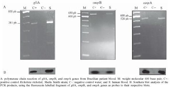
2004 Code number: oc05083 Brazilian spotted fever (BSF) cases have been increasing in the state of São Paulo but no genomic information about local rickettsia isolated from humans has been well documented. We recovered spotted-fever group rickettsiae from a sample of patient blood cultured in Vero cells using the shell vial technique. Rickettsial DNA fragments (gltA, ompA, and, ompB genes) were detected, and analysis of the ompB gene base sequences showed identity with the Rickettsia rickettsii ompB sequence available in the GenBank. Key words: Brazilian spotted fever - Rickettsia rickettsii - ompB gene Brazilian spotted fever (BSF) is a rickettsiosis very similar to the North American Rocky Mountain spotted fever (RMSF) caused by Rickettsia rickettsii, which is maintained in nature in a cycle involving ticks and mammals. The initial signs and symptoms of the disease include the sudden onset of fever, headache, and muscle pain, followed by the development of a rash (Lemos et al. 2001). This disease can be difficult to diagnose in its early stages, and without prompt and appropriate treatment it can be fatal. BSF cases are known to occur in the Southeastern Brazilian states of São Paulo, Minas Gerais, Rio de Janeiro, and Espírito Santo (Sexton et al. 1993, Lemos 2002, Galvão et al. 2003). Although several Ixodid tick species of the genus Amblyomma may be involved in the BSF cycle, A. cajennense is considered the main vector species (Magalhães 1952, Lemos 2002). Isolated reports from areas of the country that are far apart, with differing ecology, suggest that the disease may be more common than is shown by existing data. In recent years, an upsurge of cases has been recorded in the state of São Paulo, particularly in the Atibaia, Jaguari, and Camanducaia river basins, principally in the Pedreira municipality (Lemos et al. 2001). Increased pressure on the remaining forests and a population growth of the mammalian tick host, the capybara (Hydrocherus hydrocherus), could be causes for the apparent increase in human cases. The majority of studies of R. rickettsii characteristics rely on serology and immunostaining of tissues and culture, individually or in combination (Melles et al. 1999, Lemos et al. 2001), but no genomic information about local rickettsia isolated from humans has been well documented. From the ticks A. cajennense and A. cooperi collected in this focus, Spotted Fever Group (SFG) rickettsiae were isolated by culture in Vero cell and confirmed by immunofluorescent antibody assay (Lemos et al. 1996, Nascimento 2003), but none of these studies went as far as species identification. We report the use of molecular biology methods for the identification of BSF-causing-Rickettsia, in this important Brazilian endemic area. The protocol utilized was approved by the Ethical Committee on Human Experimentation of the Instituto de Ciências Biomédicas (Biomedical Sciences Institute/USP). The patient was a five-year old boy from Artur No-gueira, a municipality of the state of São Paulo, with history of recent tick exposure. During the course of the disease the patient presented fever, malaise, myalgia, headache, abdominal pain, and maculopapular rash. The blood sample was collected six days after the onset of symptoms in September 1998. The sample clot was diluted to a 10% suspension in brain heart infusion broth (BHI) and was stored at -70ºC in the Laboratory of the Adolfo Lutz Institute (IAL) until the use. For cultures, blood clot aliquots were centrifuged and the supernatant was inoculated in a confluent monolayer of Vero cells on circular slides adapted to the flat-bottomed tubes (shell vials) as previously described (Melles et al.1999). Infection of Vero cells was monitored by immunofluorescence reaction prepared with R. rickettsii-positive human serum, which permitted us to observe the presence of fluorescent microorganisms in form of intracellular bacteria (Marrero & Raoult 1989). Part of the remainder blood clot sample was used for rickettsial gene access. No patient serum was available for serology. Prior to DNA extraction through the use of phenol/phenol-chloroform, the frozen blood clot was incubated at 56ºC for 2 h for rickettsiae inactivation (Tzianabos et al. 1989). Rickettsial DNA was detected by polymerase chain reaction (PCR) using previously described conditions (Regnery et al. 1991) and the three sets of primers (GIBCO BRL), RpCs.877p (5'- GGGGGCCTGCTCACGGCGG) and RpCs.1258n (5'- ATTGCAAAAAGTACAGTGAACA) to amplify a 381-bp fragment of the citrate synthase gene (gltA) of Rickettsia species (Wood et al. 1987); Rr190.70p (5'-ATGGCGAATATTTCTCCAAAA) and Rr190.602n (5'- AGTGCAGCATTCGCTCCCCCT) for a 532-bp fragment of the 190-kDa surface protein gene (ompA) of SFG rickettsia (Regnery et al. 1991); and BG1-21 (5'- GGCAATTAATATCGCTGACGG) and BG2-20 (5'- GCA TCTGCACTAGCACTTTC) for a 650-bp fragment of the 120 kDa surface protein gene (ompB) of SFG and Typho Group rickettsiae (Eremeeva et al. 1994). PCR products to be sequenced were cloned into plasmid vector pGEMT-Easy (Promega). Escherichia coli strain DH5 a cells were transformed by the method described by Sambrook et al. (1989). The transformants carrying the vector with the DNA insert were screened by color on plates containing X-Gal, IPTG and ampicilin. The clones were digested with the restriction endonuclease Eco RI. The sequence analysis was carried out using the Thermo Sequenase Fluorescent Labelled Primer Cycle Sequencing kit (Amersham Pharmacia) and an ALF express automatic sequencer (Amersham Pharmacia) with ALFwin 2.1 software. The sequence of both DNA strands was determined twice. For Southern blot analysis, 10 µl of PCR product was separated by electrophoresis on a 2% agarose gel and transferred to a positively charged nylon membrane (Hybond N+, Amersham-Pharmacia Biotech) (Sambrook et al. 1989). Hybridization and signal detection were performed using the Gene Images labelling and detection kit (Amersham-Pharmacia-Biotech). The PCR amplification of 381 bp, 650 bp, and 532 bp fragments respectively for gltA, ompB, and ompA genes, and the positive hybridization shown in the Southern blot (Figure) confirms the SFG rickettsia propagation through cell Vero culture. The sequence analysis of ompB gene fragment showed 98% identity with R. rickettsii (GenBank access number Gi46939). The GenBank accession number for the partial ompB reported in this communication is AY751299 (R. rickettsii strain Brazil). Recently, molecular techniques were used to detect Rickettsia in blood sucking arthropods collected in the BSF transmission foci in the state of São Paulo. The rickettsial gltA gene fragments were detected in A. cooperi, by PCR, and the sequence analyzed showed homology with R. belli, a non-pathogenic rickettsia species (Horta 2002, Estrada 2003). The ompA gene of SFG rickettsia was accessed in A. cajennense collected from different localities (Nascimento 2003). R. felis, a pathogenic SFG rickettsia, was detected in Ctenocephalides felis collected from local domestic dogs (Horta 2002). In addition, R. felis was detected in Ctenocephalides fleas (Oliveira et al. 2002) in the state of Minas Gerais, Brazil, employing these same molecular techniques, collected in an area where patients with R. felis rickettiosis were reported (Raoult et al. 2001, Galvão et al. 2004). These findings suggest that, although R. rickettsii is accepted as a BSF causing agent other rickettsiae species or different R. rickettsii variants may be involved, playing roles as yet to be determined. Based on our present result, we can say that R. rickettsii can be one of the etiological agents of BSF in the state of São Paulo and the use of PCR amplification of the ompB gene coupled with partial DNA sequencing can be used as a valuable technique for establishing a definitive diagnosis of BSF during the critical stage. For a better characterization of the BSF agent sequence analysis of the amplified ompA and gltA fragments is in progress. ACKNOWLEDGEMENTS To HHB Melles (IAL/São Paulo) who provided the Rickettsia rickettsii (Sheilla Smith strain) to use as positive control for our experiments. REFERENCES
The following images related to this document are available:Photo images[oc05083f1.jpg] |
| |||||||||
