 |
| About Bioline | All Journals | Testimonials | Membership | News |
|
||||||
|
||||||
Memórias do Instituto
Oswaldo Cruz, Vol. 100, No. 4, July 2005, pp.
359-363
Marcia Regina Franzolin/+, Rosely Cabette Barbosa Alves, Rogéria Keller, Tânia Aparecida Tardelli Gomes*, Lothar Beutin**, Mauricio Lima Barreto***, Craig Milroy***, Agostino Strina***, Hugo Ribeiro****, Luiz Rachid Trabulsi Laboratório
Especial de Microbiologia, Instituto Butantan, Av. Vital Brazil 1500, 05503-900
São Paulo, SP, Brasil *Departamento de Microbiologia, Imunologia e
Parasitologia, Universidade Federal de São Paulo, São Paulo,
SP, Brasil Received 24 January
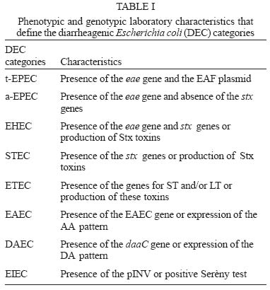
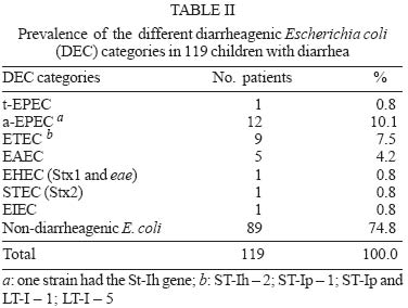
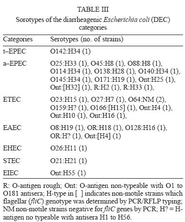
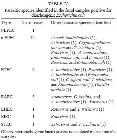
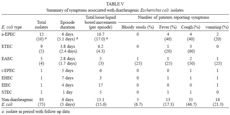
2005 Code Number: oc05097 We report the frequency of the different diarrheagenic Escherichia coli (DEC) categories isolated from children with acute endemic diarrhea in Salvador, Bahia. The E. coli isolates were investigated by colony blot hibridization whit the following genes probes: eae, EAF, bfpA, Stx1, Stx2, ST-Ih, ST-Ip, LT-I, LT-II, INV, and EAEC, as virulence markers to distinguish typical and atypical EPEC, EHEC/STEC, ETEC, EIEC, and EAEC. Seven of the eight categories of DEC were detected. The most frequently isolated was atypical EPEC (10.1%) followed by ETEC (7.5%), and EAEC (4.2%). EHEC, STEC, EIEC, and typical EPEC were each detected once. The strains of ETEC, EAEC, and atypical EPEC belonged to a wide variety of serotypes. The serotypes of the others categories were O26:H11 (EHEC), O21:H21 (STEC), O142:H34 (typical EPEC), and O?H55 (EIEC). We also present the clinical manifestations and other pathogenic species observed in children with DEC. This is the first report of EHEC and STEC in Salvador, and one of the first in Brazil. Key words: typical and atypical enteropathogenic Escherichia coli (EPEC) - Shiga toxin producing E. coli (STEC) - diarrhea - virulence factors Diarrheagenic Escherichia coli (DEC) is an important cause of endemic and epidemic diarrhea worldwide (Nataro & Kaper 1998). Currently these organisms are classified in six categories, but for practical purposes, we have divided them in eight categories: typical enteropathogenic E. coli (t-EPEC), atypical enteropathogenic E. coli (a-EPEC), enterotoxigenic E. coli (ETEC), enteroinvasive E. coli (EIEC), diffusely adhering E. coli (DAEC), enteroaggregative E. coli (EAEC), enterohemorrhagic E. coli (EHEC), and Shiga-toxin producing E. coli (STEC) (Nataro & Kaper 1998). Table I shows the laboratory characteristics that define the DEC categories. The aim of this study was to evaluate the contribution of the different DEC categories (and subcategories) to children's diarrhea in poor neighborhoods of Salvador, Bahia (Northeast Brazil). MATERIALS AND METHODS Study population - As part of an ongoing study to evaluate the health impacts of recently implemented basic sanitation (Bahia Azul Program), a sample of 1233 pre-school children from diverse areas of Salvador (Teixeira et al. 2002) were monitored for 66 weeks (October 30, 2000 to January 31, 2002). The overall study has been described elsewhere (Barreto, unpublished observations, Strina et al. 2003). In summary, children were visited twice weekly (72 to 96 h apart) and caretakers were questioned about the occurrence of diarrhea and other symptoms (number and consistency of stools, fever, vomiting, and blood in stool) since the preceding home visit. Any day in which the caretaker reported diarrhea and/or loose/liquid stools was classified as a "day with diarrhea". All "days with diarrhea" that were separated by fewer than three days of "normal" stools were considered to be part of the same diarrheal episode, while all days/episodes of diarrhea separated by at least three consecutive days of "normal" stools were considered distinct. Sample collection - Fecal samples were collected during a 6-month period (July 24, 2001 to January 31, 2002). Caretakers received stool-collection recipients (plastic collection pots, plastic sacks, and disposable diapers) for collecting their child's feces, and were instructed to do so on any day that they judged their child to have diarrhea (regardless of the number of stools passed), and request collection of material by: (1) calling collect to the study center (Monday-Thursday, Saturday and Sunday, 7 am- 4 pm), or (2) bringing their child directly to the study center. When requested by telephone, a technician was sent on motorcycle to the subject's house: two swabs of material were collected from the diaper/pot/sack, placed in Cary-Blair transport media and taken on ice (along with any remaining fecal material) for cataloguing and sample preparation. Whenever children were brought to the study center, two rectal swabs were taken, set in transport media, and refrigerated. Culture - Culture for E. coli was made in MacConkey agar (Merck) and incubated overnight. Colonies were confirmed as E. coli using biochemical tests. As a rule 2-3 lactose-fermenting colonies, presumed to be E. coli by colony morphology were examined for DEC. Bacterial testing for Salmonella, Shigella, Yersinia, Aeromonas, and Vibrio was conducted using standard procedures (Ko-neman et al. 1997). Campylobacter jejuni-coli was identified through a sample-filtration technique (Steele & McDermott 1984). Adenovirus and rotavirus (group A) were identified by enzyme immunoassay (Biomanguinhos, Oswaldo Cruz Foundation) and polyacrilamine gel electrophoresis (PAGE). Parasitological examinations for helminthes and protozoa were conducted using the Kato-Katz (Katz et al. 1972) and sedimentation (Koneman et al. 1997) methods. To test for Cryptosporidia sp., stool specimens were sieved and submitted to the formol-ether concentration method (Allen & Ridley 1970). DEC identification - Colony hybridization using specific radiolabelled DNA probes was used to identify the following DEC virulence factors: eae (E. coli attaching and effacing gene encoding intimin), EAF (E. coli adherence factor), bfpA (bundle forming pilus structural gene), daaC (accessory gene for F1845 fimbriae biogenesis), EAEC (enteroaggregative E. coli adherence plasmid), INV (E. coli invasiveness plasmid), LT-I and II (heat-labile enterotoxin type I and II), ST-Ih and ST-Ip (heat-stable enterotoxin type I of human and porcine origin respectively), Stx-1 and 2 (Shiga toxin 1 and 2 genes) (Ghilard et al. 2003). Serotyping and PCR typing of flagellar (fliC) genes of E. coli - Serotyping of E. coli O and H-antigens was performed as described previously (Orskov & Orskov 1984), at the National Reference Laboratory for Escherichia coli, Germany, by L Beutin. Determination of flagellar (fliC) genotypes was performed by PCR followed by analysis of HhaI digested fliC specific PCR products as described previously (Beutin et al. 2004). RESULTS A total of 175 samples were considered; of these, 169 samples were tested for Salmonella, Shigella, Yersinia, Aeromonas, Vibrio, and E. coli, 124 for Campylobacter, 139 for virus, 158 for Cryptosporidia, and 164 for hel-minthes/other protozoa. E. coli was suspected in 138 samples (i.e. 81.7% of all samples examined for bacteria); of these, 119 were confirmed positive (86.2%) and 30 were found to contain diarrheagenic strains (Table I) (i.e. 17.1% of all samples received, 21.7% of all samples tested for E. coli, and 25.2% of all confirmed samples). The prevalence of each category of DEC is shown in Table II. The most frequently identified DEC were a-EPEC (10.1%), followed by ETEC (7.5%), and EAEC (4.2%). Nine ETEC strains were identified: 5 reacted only with the probe for LT, 3 for ST and 1 with the probes for both enterotoxins. In addition, one of the a-EPEC strains reacted with the probe for ST. t-EPEC, STEC, EHEC, and EIEC were detected only once each and DAEC was not detected. The DEC serotypes were attributed to 18 O-groups and 17 different H-types (Table III). Seven strains were O-nontypeable (Ont) and four had a rough LPS (R). Five strains were non-motile and were investigated for their fliC genotype by PCR. The EHEC strain belonged to serotype O26:H11, and the t-EPEC strain belonged to serotype O142:H34. The serotype of the EIEC strain could not be determined because it was rough. This strain was submitted to test of keratoconjuntivitis in guinea pigs (Sèreny 1955), but presented negative result. All a-EPEC and EAEC strains belonged to different serotypes. Other enteric pathogens identified in samples containing DEC are shown in Table IV, and clinical manifestations are shown in Table V. Duration of the diarrheal episodes was similar for the more frequent DECs, and no blood was reported in the feces of the children with EHEC and with EIEC. DISCUSSION In this study, seven of the eight different DEC categories were detected in children with endemic diarrhea in Salvador, Bahia. Atypical EPEC was the most commonly isolated category. In addition, we identified two EHEC/STEC strains and observed a very low frequency of t-EPEC. The frequency of the remaining categories was similar to that reported in previous studies (Gomes et al. 1991, Rosa et al. 1998, Regua-Mangia et al. 2004). Tornieporth et al. (1995) conducted an epidemiological study with children in Salvador, Bahia. They evaluated 76 children with diarrhea and 16 children without diarrhea, using PCR technique and identified 16% of the children with enterotoxi-genic E. coli, 8% with enteropathogenic E. coli (BFP positive), and 1% with enteroinvasive E. coli infections, but nevertheless they did not investigate the genes for others categories. Atypical EPEC are emergent enteric pathogens that have only recently began to attract the attention of investigators (Trabulsi et al. 2002). The high frequency of these organisms in this study reinforces the need for further studies on their epidemiology, pathogenesis, and role in the different forms of diarrhea. In contrast to typical EPEC, they seem to be common in domestic animals (Trabulsi et al. 2002), have been implicated in large food poisoning outbreaks in adults (Viljanen et al. 1990, Hedberg et al. 1997) and are relatively unknown in regard to virulence determinants (Pelayo et al. 1999). The isolation of two EHEC/STEC strains was also interesting and a cause of concern, since this is the first time that these organisms have been registered in Salvador, and one of the first in Brazil. Although a few EHEC/STEC strains were isolated in the 80s (Giraldi et al. 1990, Gomes et al. 1996), they seem to be occurring more frequently in the last few years. In addition to the strains detected in this study, three O157:H7 strains (Irino et al. 2002) and one O26:H11 strain (strain isolated from a case of HUS Guth et al. 2002) have been reported in São Paulo. Cantarelli et al. (2000) also reported one instance of O91:H21 in Porto Alegre. EHEC/STEC are frequently isolated from cattle in Brazil (Cerqueira et al. 1999), and we recently isolated an O157:H7 strain from a child with diarrhea in Salvador (Trabulsi, unpublished observations). Taken together, these findings suggest that EHEC/STEC infections are on the rise in Brazil and require more attention from our public health services. The finding of only one typical EPEC strain was somewhat surprising, but appears to confirm a recent trend that has been observed in Brazil (Girão et al. 2001, Da Silva Duque et al. 2002, Rodrigues et al. 2002). Until the 90s, these organisms were the main cause of infantile diarrhea in Brazil (Gomes et al. 1991, Rosa et al. 1998, Regua-Mangia et al. 2004), but it seems that they are becoming more and more rare. The reason for this fall in the typical EPEC frequency has not been established but it may be a consequence of recent public health measures such as more efficient control of hospital infections and implementation of regular water supply in many slums. Another factor may have been the more frequent adoption of breast feeding, which is highly protective against intestinal infections since breast milk is extremely rich in antibodies against enteric pathogens (Loureiro et al. 1998). Regarding EHEC and both EPEC categories, the present situation in Brazil is becoming similar to the one that exists in developed countries were typical EPEC are very rare and atypical EPEC and EHEC are relatively frequent. It is interesting to mention that typical EPEC were rather frequent in these countries in the past (until the 60s). As t-EPEC have as host basically humans (Nataro & Kaper 1998) it is likely that their frequency has been more influenced by the public health measures recently adopted than the frequency of a-EPEC and EHEC which are hosted by humans and animals (Trabulsi et al. 2002). Several studies conducted in Brazil and in other countries have shown that ETEC and EAEC are frequently detected in children with diarrhea and that EIEC are much less frequent (Gomes et al. 1991, 1996, Nataro & Kaper 1998, Regua-Mangia et al. 2004). The results of these studies corroborates the one reported here. Serotyping of the DEC strains revealed some interesting aspects. For example, the single t-EPEC strain belonged to serotype O142:H34. This strain is not a "traditional" EPEC serotype, but it is the most frequently isolated serotype among serogroup O142 strains in Brazil (Ghilard et al. 2003). The EHEC strain belonged to serotype O26:H11, one of the most common EHEC serotypes (Nataro & Kaper 1998). The single EIEC strain was motile, a rare characteristic among EIEC strains (Toledo & Trabulsi 1983), and was Sereny negative, possibly due to lost of the INV plasmid after storage. Finally, serotyping revealed the diversity of serotypes characteristic of ETEC and EAEC (Gyles 1994). Approximately the half of the patients infected with DEC also carried parasites or viruses (Table IV) but as some of these are not associated with acute diarrhea and no other bacterial pathogens were isolated (the feces were also plated in selective media for Shigella, Salmonella, and Vibrio). Therefore, it is probable that at least some of the clinical manifestations presented by the patients (Table V) were to a major part due to the DEC infections. It is interesting to observe that the children carrying a-EPEC and EHEC had longer periods of diarrhea than those infected with other DEC. Further studies are needed to investigate the ecological, socio-economical, and epidemiological basis of atypical EPEC infections as an emerging pathotype in children in Brazil. ACKNOWLEDGMENTS To Monica Aparecida Midolli Vieira for her technical assistance in the colony hybridization assays. L Beutin was supported by funds from the European Commission project "Attaching and Effacing E. coli infections" (reference no. QLK2-CT-2000-00600). REFERENCES
The following images related to this document are available:Photo images[oc05097t3.jpg] [oc05097t2.jpg] [oc05097t1.jpg] [oc05097t5.jpg] [oc05097t4.jpg] |
| |||||||||
