 |
| About Bioline | All Journals | Testimonials | Membership | News |
|
||||||
|
||||||
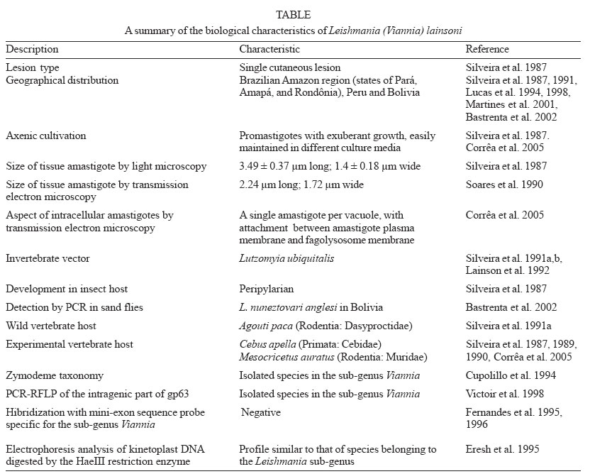
Memórias do Instituto Oswaldo Cruz, Vol. 100, No. 6, October 2005, pp. 687-692 Leishmania (Viannia) lainsoni (Kinetoplastida: Trypanosomatidae), a divergent Leishmania of the Viannia subgenus - A mini review José R Corrêa, Reginaldo P Brazil*, Maurilio J Soares/+ Laboratório
de Biologia Celular de Microrganismos, Departamento de Ultra-estrutura e
Biologia Celular *Laboratório de Bioquímica e Fisiologia de
Insetos, Departamento de Bioquímica e Biologia Molecular, Instituto
Oswaldo Cruz-Fiocruz, Financial support: CNPq, Fiocruz Received 29 March
2005 Code Number: oc05145 Leishmania (Viannia) lainsoni is the Leishmania species that presents the most distinct biological (morphology, growth in axenic culture medium), biochemical (enzymatic electrophoresis profile), and molecular biology characteristics, when compared to other species of the Viannia subgenus. Development of promastigote forms of this parasite attached to the wall of the pyloric and hind gut regions of sand fly vectors is a solid characteristic that allows its positioning in the Viannia subgenus. However, taxonomic data from biochemical and molecular techniques on this Leishmania species are still not conclusive. It is evident the difficulty in taxonomically positioning this borderline Leishmania species. In this review we present the data accumulated since L. (Viannia) lainsoni has been described and we discuss its position in the Viannia subgenus. Key words: Leishmania (Viannia) lainsoni - trypanosomatid - taxonomy - review Human leishmaniasis is a disease with a wide spectrum of clinical manifestations, caused by different species of flagellate protozoa belonging to the Leishmania genus (Grimaldi et al. 1991, Grimaldi & Tesh 1993). These heteroxenic flagellates (Protozoa: Kinetoplastida: Try-panosomatidae) are obligate intracellular parasites (under the amastigote form) of vertebrate hosts, being transmitted (as promastigote forms) by sand flies (Diptera: Psychodidae: Phlebotominae). There are two main clinical manifestations of leishmaniasis: cutaneous and visceral (WHO 2003). The cutaneous form is polymorphic, affects the skin and/or mucoses, and is characterized by the presence of either (a) single/multiple painless lesions (simple cutaneous form), (b) nodular lesions spread over the body (diffuse cutaneous form) or (c) muco-cutaneous lesions that affect the nasofaringeal region (muco-cutaneous form). Leishmania flagellates are grouped into two sub-genera, Leishmania and Viannia, according to their development in the insect digestive tract. Members from the Leishmania subgenus grow at the pyloric and midgut regions (suprapylarian development), while Viannia species grow at the hindgut region (peripylarian development) (Lainson & Shaw 1979, 1987). Nevertheless, complementary tests are necessary for accurate taxonomic positioning, such as isoenzyme profile, immunoreaction with monoclonal antibodies or PCR-based techniques. Among the several Leishmania species identified in Brazil, L. (Viannia) lainsoni (Silveira et al. 1987) presents an atypical phylogenetic profile. Its divergent position into the Viannia subgenus is a common sense (Cupolillo et al. 1995, 2001, Eresh et al. 1995, Fernandes et al. 1995, McCann et al. 1999), but the available data are still not sufficient for an unequivocal taxonomic placement of this species. Characteristic features of L. (Viannia) lainsoni are its exuberant amastigote growth in skin lesions and the easy of cultivation in simple axenic culture media, which are typical of parasites belonging to the Leishmania sub-genus. In this review we present the data accumulated since L. (Viannia) lainsoni has been described and we discuss its position in the Viannia subgenus (Table). The parasite Leishmania (Viannia) lainsoni Silveira, Shaw, Braga & Ishikawa, 1987 New Leishmania species have been continuously described in Brazil, due to its large territorial dimension and its ecological diversity in tropical and subtropical areas, mainly at the Amazon region. Among the latest descriptions, an isolate was obtained from human samples collected in different locations in the state of Pará (Brazilian Amazon region). The parasites were successful isolated from human patients, and a new Leishmania species could be identified and named as L. (Viannia) lainsoni (Silveira et al. 1987). The isoenzymatic profile of the first isolate of L. (Viannia) lainsoni (MHOM/BR/81/M6426) was compared to that of the reference strains L. (Viannia) braziliensis (MHOM/BR/75/M2903), L. (Viannia) guyanensis (MHOM/BR/75/M4147), and L. (Leishmania) amazo-nensis (IFLA/BR/67/PH8). The isoenzymatic banding patterns showed that L. (Viannia) lainsoni was different from these reference strains by six among ten tested enzymes. This L. (Viannia) lainsoni strain was also probed against specific monoclonal antibodies (Mabs) against L. (Viannia) braziliensis (Mab B18), L. (Viannia) guyanensis (Mab B19), and L. (Viannia) panamensis (Mab B11), as well as against three other L. (Viannia) brazi-liensis group-specific monoclonal antibodies (Mabs B2, B5, and B12). The results by indirect immunofluorescence showed absence of cross-reactivity with all tested antibodies (Silveira et al. 1987) and confirmed the status of L. (Viannia) lainsoni as a distinct species. Experimental infection of Lutzomyia longipalpis sand flies with L. (Viannia) lainsoni was performed, resulting in exuberant growth of promastigotes adhered to the insect hindgut wall, thus confirming the status of this parasite as a Peripylarian member. Taken together, these data contributed for inclusion of this species in the Viannia subgenus (Silveira et al. 1987). Morphological studies of infected hamsters skin showed that L. (Viannia) lainsoni presents elongated amastigotes, as compared to other Leishmania species (length: 3.49 ± 0.37 µm; width: 1.4 ± 0.18 µm), with a bulk kinetoplast (Silveira et al. 1987). Axenic culture pro-mastigotes of L. (Viannia) lainsoni grown from skin biopsies of infected hamsters are characterized by a long flagellum, about 25 µm in length. These axenic pro-mastigotes present an exuberant growth and are easily maintained in different culture media (Corrêa et al. 2005). In this sense, L. (Viannia) lainsoni differs from the other members of the Viannia sub-genus, which grow poorly in vitro (Silveira et al. 1987). Detailed ultrastructural studies on L. (Viannia) lainsoni are still lacking. A preliminary observation by transmission electron microscopy (TEM) of lesions in ex-perimentally infected hamsters showed intracellular oval amastigotes, measuring about 2.24 µm in length and 1.72 µm in width, with no megasomes (Soares et al. 1990). These values are slightly lower than the light microscopy measurements reported in the original description of the species (Silveira et al. 1987), but they seem to be more accurate, due to the better preservation obtained during the processing for TEM. An ultrastructural image (TEM) of a tissue amastigote was shown in a recent work on the cultivation of L. (Viannia) lainsoni (Corrêa et al. 2005). A single round amastigote, with no megasomes, was contained within a small parasitophorous vacuole, tightly adhered to the vacuole membrane. THE DISEASE L. (Viannia) lainsoni causes single cutaneous ulcers without evidence of posterior nasofaringeal involvement (Silveira et al. 1987), similar to that caused by other Leishmania parasites belonging to the Viannia subgenus: small self-limiting nodules or small ulcers, with abundant amas-tigotes at the lesion site. Experimental infection of Cebus apella (Primata: Cebidae) with L. (Viannia) lainsoni showed that this neotropical primate is susceptible to infection by this parasite, with lesions lasting for about four months, a suitable time period for testing leishmanicidal drugs (Silveira et al. 1989). Histopatological analyses comparing skin lesions caused by L. (Viannia) lainsoni, L. (Leishmania) ama-zonensis, and L. (Viannia) braziliensis showed the same stages of lesion development, independent of the Leishmania species: (1) chronic unspecific infiltrate, (2) macrophage nodules with several parasites, (3) necrosis of the parasitized cells, (4) epithelial granulome, (5) absorption of the necrosis area, (6) unspecific residual infiltrate, and (7) scar (Silveira et al. 1990). However, L. (Viannia) lainsoni produces skin lesions with distinct characteristics: (a) non-ulcerated and nodular as those caused by L. (Leishmania) amazonensis; (b) active and with parasites for a long time, as also observed with L. (Leishmania) amazonensis (Silveira et al. 1990). The humoral response of C. apella experimentally infected with L. (Viannia) lainsoni, L. (Viannia) brazi-liensis, and L. (Leishmania) amazonensis was analyzed by a direct agglutination test (DAT). The assay was performed with homologous [L. (Leishmania) amazonensis; L. (Viannia) braziliensis] and heterologous [L. (Leishmania) donovani] antigens. However, no homologous antigen could be obtained for L. (Viannia) lainsoni. Animals infected with L. (Leishmania) amazonensis and L. (Viannia) braziliensis presented detectable antibody titers when tested with both homologous and heterologous antigens, but animals infected with L. (Viannia) lainsoni presented significant titers only for the DAT performed with the heterologous antigen (Garcez et al. 1997). The course of infection with L. (Viannia) lainsoni was similar to that of L. (Leishmania) amazonensis, and in both cases the lesions did not ulcerate and remained active for a longer period (Garcez et al. 1997). THE INSECT HOST Studies have been made to identify the wild insect vector of L. (Viannia) lainsoni. In order to increase the chances of finding infected sand flies, captures were performed in the same areas where L. (Viannia) lainsoni was isolated for the first time and according to information given by the patients: Benevides city (state of Pará, Brazil) and the ecologically related forest area of Utinga (Belém city, state of Pará, Brazil). The capture allowed the collection of 1924 female sand flies, belonging to 27 species in Benevides and to 29 species in the Utinga forest. After dissection, no sand flies from Benevides presented infection by L. (Viannia) lainsoni, although infected by other flagellates. On the other hand, insects captured in the Utinga forest identified as Lu. ubiquitalis presented massive infection with L. (Viannia) lainsoni. Identification of the protozoa as L. (Viannia) lainsoni was done by isolation of the parasites in axenic medium, examination by light microscopy, reactivity to specific monoclonal antibodies, and isoenzyme profiles (Silveira et al. 1991a, b). Under normal forest conditions Lu. ubiquitalis is not antropophilic, but the finding of humans infected with L. (Viannia) lainsoni in the same region where infected sand flies were found suggests that this insect can bite man and transmit the parasites. Accordingly, in the laboratory Lu. ubiquitalis fed on human beings (Silveira et al. 1991b). A later study with sand flies captured at the Carajás forest (Pará, Brazil) confirmed the presence of Lu. ubiquitalis naturally infected with L. (Viannia) lainsoni (Lainson et al. 1992). Furthermore, the presence of L. (Viannia) lainsoni has been detected in a pool of 25 Lu. nuneztovari anglesi females in the Sub-Andean region of Bolivia by kDNA-PCR amplification procedures followed by Southern blot and hybridization (Bastrenta et al. 2002). This sand fly species belongs to the verrucarum group, where several species are suspect vectors of leishmaniasis in other regions. In the Caranavi province of Bolivia, region where the first case of cutaneous leishmaniasis due to L. (Viannia) lainsoni in this country was described, several naturally infected females of Lu. velascoi were detected at the end of the rainy season, but unfortunately the parasite could not be isolated (Martinez et al. 2001). Interestingly, this sand fly belongs to the same subgenus (Trychopho-romyia) as Lu. ubiquitalis and it is the only species of this subgenus in the region (Martinez et al. 2001). Further studies are needed to incriminate Lu. velascoi as the vector of L. (Viannia) lainsoni in Bolivia. These data demonstrate that more information is needed on the vector capability of endemic sand flies species involved in transmission of this parasite. GEOGRAPHICAL DISTRIBUTION In Brazil, L. (Viannia) lainsoni has been isolated in the states of Pará, Amapá, and Rondônia (Silveira et al. 1987, 1991a,b). Regarding South America, this flagellate was found in the sub-Andean region of Peru (Lucas et al. 1994, 1998) and Bolivia (Martinez et al. 2001, Bastrenta et al. 2002). These data demonstrate a wide distribution of this protozoan in South America. An important detail regarding L. (Viannia) lainsoni in Peru is that this protozoan was not found in cities close to the Brazilian border (Loreto, Ucayali, and Madre de Dios), but in cities more inside the country (Cusco, Pasco, Ayacucho, Huanuco, and San Martin). These last locations are situated between 600 and 2000 m above the sea level, with annual mean temperature of 18.5-25.6ºC (Lucas et al. 1998), an environment totally different from that found in the Amazon plane land, site of the first isolation of L. (Viannia) lainsoni. These data suggest that in Peru this parasite can be transmitted to man by different insect vectors and possesses mammal reservoirs not yet identified. Although Lu. ubiquitalis has been found in the city of Madre de Dios, no record of infection caused by L. (Viannia) lainsoni has been made (Lucas et al. 1994). THE WILD MAMMAL RESERVOIR The first isolation of L. (Viannia) lainsoni from a wild mammal was made in 1991, from the rodent Agouti paca (Rodentia: Dasyproctidae), in the state of Pará, Brazil. The parasites were isolated from apparently normal skin samples obtained from three specimens of this rodent, captured in the Tucuruí city (Tocantins island, state of Tocantins, Brazil). No isolation was obtained from the viscera of any captured animal. Identification of the isolates as L. (Viannia) lainsoni was based on the morphological shape of amastigotes and promastigotes, the behavior in infected hamsters, isoenzyme analysis, and monoclonal antibody tests. The unapparent infection in captured animals suggests that these mammals may represent the natural wild reservoir for this parasite in the Amazon region (Silveira et al. 1991a). CLASSIFICATION BY BIOCHEMICAL AND MOLECULAR BIOLOGY MARKERS The systematic classification of New World Leishmania species was reviewed with the help of numeric zymotaxonomy, by comparing the eletromorpho profiles of 270 Leishmania isolates with those of reference strains (Cupolillo et al. 1994). The 18 enzymatic loci analyzed showed a strong polymorphism and the 270 strains were grouped into 44 zymodemes. These zymodemes could be clustered in two larger groups that corresponded to the Leishmania and Viannia subgenera. This analysis demonstrated that L. (Viannia) lainsoni is so distant from the other Leishmania species that it could represent a new species group, independent of all species complexes described so far (Cupolillo et al. 1994). The taxonomic position of L. (Viannia) lainsoni was reevaluated by using a molecular marker, a Viannia-specific oligonucleotide probe (16 nucleotides long) for mini-exon sequences (Fernandes et al. 1995, 1996). This probe did not hybridize with L. (Viannia) lainsoni, but hybridized with all other species with peripylary development in sand flies, which are grouped in the Viannia subgenus (Fernandes et al. 1995). This molecular aspect alone did not allow to remove L. (Viannia) lainsoni from the Vian-nia subgenus, but has evidenced a far relationship between this species and the other members of the Viannia subgenus, reinforcing its divergent position inside this group (Fernandes et al. 1995). The divergent position of L (Viannia) lainsoni was confirmed by electrophoresis analysis of kinetoplast DNA (kDNA) digested by the HaeIII restriction enzyme. Although the Leishmania species grouped in the Viannia subgenus presented very similar banding profiles, L. (Viannia) lainsoni presented a profile more similar to L. (Leishmania) amazonensis, a typical representative of the Leishmania subgenus (Eresh et al. 1995). However, hybridization of L. (Viannia) lainsoni nuclear DNA with a β -tubulin probe, followed by measurement of the kDNA density, suggested a molecular proximity between L. (Viannia) lainsoni and Leishmania species belonging to the L. (Viannia) braziliensis species complex (Eresh et al. 1995). Amplification of kDNA under different annealing temperatures (60.5 and 67.5ºC) resulted in the production of 300 base pairs in L. (Viannia) lainsoni, confirming the inclusion of this species in the Viannia subgenus, at it is known that at the annealing temperature of 60.5ºC the used primers do not amplify kDNA from parasites of the Leishmania subgenus. On the other hand, amplification of kDNA from species from the Viannia subgenus usually generates a product of 750 base pairs. Thus, these data show that L. (Viannia) lainsoni is an isolated species inside the Viannia subgenus (Eresh et al. 1995). The locus of the gene coding for the surface glycoprotein gp63 has been used as a high-resolution target for the genetic characterization of Leishmania (Victoir et al. 1998). Two distinct methods were applied: (1) restriction analysis of genomic DNA, followed by hybridization with probes possessing a conserved part of the gp63 gene (gp63-RFLP) and (2) PCR amplification of the intragenic part of gp63, combined with restriction fragments length polimorphism analysis of amplification products (PCR-RFLP). The gp63-RFLP method showed a high resolution, disclosing the phylogenetic relationships among the different Leishmania species, and confirming data obtained by other molecular techniques. This method sorted the Leishmania species into two groups, corresponding to the Leishmania and Viannia subgenera. Inside the Viannia subgenus, L. (Viannia) braziliensis, L. (Viannia) peru-viana, and L. (Viannia) guyanensis were very similar, while L. (Viannia) lainsoni showed a more remote relationship (Victoir et al. 1998). Sequencing of the minicircle kDNA from L. (Viannia) lainsoni was performed (McCann et al. 1999), in an attempt to explain the small fragment (300 base pairs) previously observed during the amplification of this DNA with universal Viannia primers (Eresh et al. 1995). The results showed the existence in minicircles of L. (Viannia) lainsoni kDNA of specific sequences. This parasite did not contain the requested sequences for correct annealing and amplification with the B1 and B2 primers used by Eresh et al. (1995), thus explaining the minor fragment length when compared with other species of the Viannia subgenus (McCann et al. 1999). The production of a SL3 primer (McCann et al. 1999) that together with the B1 primer (Eresh et al. 1995) could amplify L. (Viannia) lainsoni minicircles in lower stringency conditions (57ºC of annealing temperature) appear as a strong argument that the positioning of L. (Viannia) lainsoni inside the Viannia subgenus is substantially different from the other members of this group (McCann et al. 1999). CONCLUSION The data obtained so far on L. (Viannia) lainsoni demonstrate that this parasite presents a wide distribution in South America, being found in different ecological environments, but further studies are needed to better understand the transmission cycle in subtropical Andean regions, where mammal reservoirs and invertebrate vectors were not yet identified. As the human expansion in the Amazon and Sub-Andean regions is a reality and a leishmaniasis control program is very difficult to be organized due to the challenging conditions, the risk of new outbreaks of L. (Viannia) lainsoni infections is eminent. L. (Viannia) lainsoni is the Leishmania species that presents the most distinct biological (morphology, growth in axenic culture medium), biochemical (enzymatic electrophoresis profile) and molecular biology characteristics, when compared to other species of the Viannia subgenus. A summary of the data obtained so far is shown in the Table. The development of promastigote forms of this parasite attached to the wall of the pyloric and hind gut regions of sand fly vectors is a solid characteristic that allows the positioning of L. (Viannia) lainsoni in the Viannia subgenus (Silveira et al. 1987, Fernandes et al. 1995). The taxonomic data from biochemical and molecular techniques on this Leishmania species are still not conclusive. It is evident the difficulty in taxonomically positioning this borderline Leishmania species. More comparative studies of different genes are needed, or new techniques have to be developed to elucidate the taxonomic position of this particular Leishmania species. The biochemically and biologically divergent behavior of L. (Viannia) lainsoni can be used in novel approaches to the development of antigens for the diagnosis of leishmaniasis, as this species is easily maintained under in vitro conditions. Studies on new and different Leishmania species, such as L. (Viannia) lainsoni, might bring new insights on metabolic pathways, parasite-cell interactions in vertebrate and invertebrate hosts, new proteins or any other biological/biochemical aspects that can be useful for the control and better understanding of leishmaniasis and the parasite life cycle. REFERENCES
Copyright 2005 Instituto Oswaldo Cruz - Fiocruz The following images related to this document are available:Photo images[oc05145t1.jpg] |
| |||||||||
