 |
| About Bioline | All Journals | Testimonials | Membership | News |
|
||||||
|
||||||
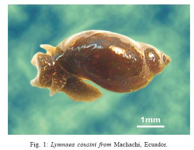
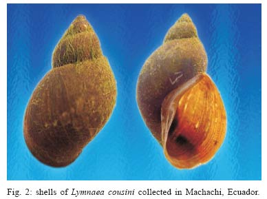
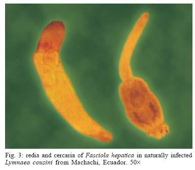
Mem Inst Oswaldo Cruz, Rio de Janeiro, Vol. 100, No. 7, November ,2005, pp. 735-737 SHORT COMMUNICATION First report of Lymnaea cousini Jousseaume, 1887 naturally infected with Fasciola hepatica (Linnaeus, 1758) (Trematoda: Digenea) in Machachi, Ecuador Ángel Villavicencio A/+, Mauricio Carvalho de Vasconcellos* Department of Veterinary Pathology, Agrary Faculty, Russian People's Friendship University, Moscow-Russia, 117-198, St. M. Maklaya 6 *Núcleo de Biologia e Controle de Endo e Ectoparasitas de Interesse Médico e Veterinário, Departamento de Biologia, Instituto Oswaldo Cruz-Fiocruz, Rio de Janeiro, RJ, Brasil Financial support: Fundacyt, Proj. 2005701 Received 22 July 2005 Code Number: oc05155 We report the first finding of Lymnaea cousini naturally infected with Fasciola hepatica in Ecuador. A sample of 70 snails was collected in April 2005 from a wetland located in a valley at approximately 3000 m a.s.l., near the locality of Machachi, Pichincha Province. The prevalence of natural infection in L. cousini was 31.43%, which is the highest value ever recorded for naturally infected lymnaeid species. Key words: Lymnaea cousini - Fasciola hepatica - Ecuador Fascioliasis, one of the main diseases of ruminants, is caused by the liver fluke Fasciola hepatica. This worldwide distributed disease occasionally affects humans and produces an important economic impact on livestock due to reduced weight gain, lowered fertility and abortion, progressive decrease in milk yield, as well as liver condemnation. The annual losses to the agricultural sector are estimated to be US$2.2 million, with over 600 million animals infected worldwide (Dalton 1999). In Ecuador the present prevalence of F. hepatica in livestock remains uncertain because most data are collected by sanitary inspectors from slaughter houses. The Andean region is the endemic area of fascioliasis, where prevalence of infection in livestock ranges from 20 to 60% and in humans from 24 to 53% (SESA 2003). This region is inhabited by 23.6 and 52.9% of the total and rural population, respectively; almost 200,000 people are infected and 1% are at risk of infection (WHO 1995). Despite fascioliasis being a serious health problem in the country, very little is known about snails of the genus Lymnaea, the intermediate hosts responsible for F. hepatica transmission. Some of the lymnaeid species occurring in Andean countries are as follows: L. truncatula Müller, 1774 from Bolivian highlands (Jabbour-Zahab et al.1997, Mas-Coma et al. 1999) and Venezuela (Pérez Mata 2003); L. columella Say, 1817 from Venezuela and Ecuador (Pérez Mata 2003); L. cubensis Peiffer, 1839 from Venezuela (Pérez Mata 2003) and L. cousini Jousseaume, 1887 (syn. L. ubaquensis Piaget, 1912; L. bogotensis Pilsbry, 1935; see Hubendick 1951 and Pointier et al. 2004). L. cousini has been found in Ecuador (Jousseaume 1887, Paraense 1995, 2004), Colombia (Piaget 1912, Pilsbry 1935), and Venezuela (Pointier at al. 2004), but no report concerning natural infection has ever been published. The aim of this study is to report the first finding of L. cousini naturally infected with F. hepatica in Ecuador. A sample of 70 snails was collected in April 2005 within a private farm where fascioliasis in cattle was previously diagnosed by faecal examination. It was located near Machachi (78°30'W, 00°30'S, 3100 m a.s.l.) in the Pichincha Province. The sampling site was a wetland placed in a relatively low valley at approximately 3000 m a.s.l. and covered an area of 4.5 m². Water in the wetland flowed slowly, had a maximum height of 10 cm and pH was 6.5. The soil is volcanic in origin, and vegetation was composed of Ray-grass (Lolium sp.) and aquatic plants. Snails collected were kept alive in plastic containers with wet cotton pads and transported to the Department of Malacology of the Instituto Oswaldo Cruz, Rio de Janeiro, Brazil. Upon arrival at the laboratory, ten specimens were relaxed, killed, and preserved according to Paraense (1984), and their taxonomic identification was performed by Dr Paraense on the basis of features of the shell and internal organs (Paraense 1995). The remaining snails shared the same external appearance as those already determined. Specimens were examined for the presence of larval stages of F. hepatica under stereoscopic microscope. All 70 collected snails belonged to L. cousini Jous-seaume, 1887 (Figs 1, 2), and 22 (31.43%) were infected with F. hepatica (Fig. 3). Shells of L. cousini specimens from Ecuador described by Jousseaume (1887) ranged 10-14 mm in height and 6-10 mm in diameter with an aperture of 7-10 mm long, while those from Venezuela varied 6.6-9.3 mm in height and 3.7-6 mm in width (Pointier et al. 2004). The surprisingly high prevalence of natural infection with F. hepatica in L. cousini may be explained by environmental conditions favouring the development of snail populations and the transmission of the parasite. Valleys in the Andean region of Ecuador are characterized by temperatures ranging from 10 to 30ºC, frequent precipitation, numerous streams, and low-permeable soil layers. In addition, anthropogenic factors involving irrigation of pasture lands by flood or ditches and inappropriate management of grazing cattle are unlikely to prevent infection in both intermediate and definite hosts. The prevalence of F. hepatica in L. cousini from Machachi, Ecuador (31.43%), is the highest value ever reported for lymnaeid snails naturally infected with this parasite. In Brazil, L. columella showed the following infection rates: 2.4% in Rio de Janeiro (Rezende et al. 1973); 5.2 and 3.9% in Minas Gerais (Coelho & Lima 2003); 1.22 and 0.14% (Ueta 1980), 5.26% (Oliveira et al. 2002) in São Paulo; 3.3% in Rio Grande do Sul (Silva Santos et al. 1987). In Corrientes, Argentina, the prevalence of L. columella was 8.8% (Prepelitchi et al. 2003). In central France, Rondelaud et al. (2001) reported a global prevalence of natural infection of 1.1% in L. truncatula and 0.3% in L. glabra. In the Russian Federation, L. truncatula populations showed infection prevalences ranging from 0.05 to 0.72% (Villavicencio 2004). ACKNOWLEDGEMENTS To Dr Wladimir Lobato Paraense, Department of Malacology, Instituto Oswaldo Cruz-Fiocruz, for his valuable help in identifying the snails. To Natalia Dondua for field assistance. To AF Barbosa for the photographs, and to all the people who lent their support and assistance during this study. REFERENCES
Copyright 2005 Instituto Oswaldo Cruz - Fiocr |
