 |
| About Bioline | All Journals | Testimonials | Membership | News |
|
||||||
|
||||||
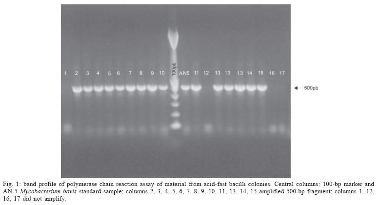
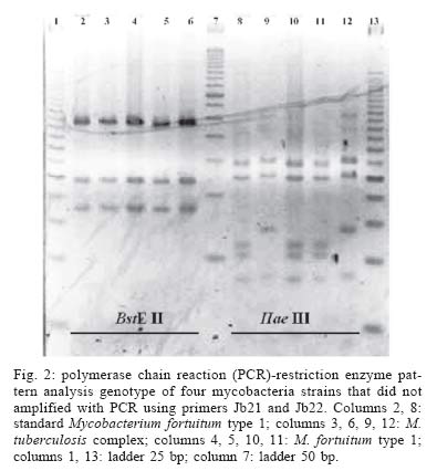
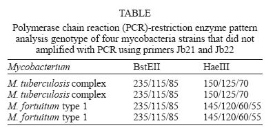
Mem Inst Oswaldo Cruz, Rio de Janeiro, Vol. 100, No. 7, November ,2005, pp. 749-752 SHORT COMMUNICATION Mycobacterium bovis identification by a molecular method from post-mortem inspected cattle obtained in abattoirs of Mato Grosso do Sul, Brazil Cristina Pires de Araújo++, Clarice Queico Fugimura Leite, Karina Andrade de Prince, Klaudia dos Santos Gonçalves Jorge*, Ana Luiza Alves Rosa Osório*/+ Faculdade de Ciências Farmacêuticas, Universidade Estadual Paulista Júlio de Mesquita Filho, Araraquara, SP, Brasil *Universidade Federal de Mato Grosso do Sul, Cidade Universitária, 79070-900 Campo Grande, MS, Brasil Financial support: Fundect Received 15 December 2004 Code Number: oc05158 The presence of Mycobacterium bovis in bovine carcasses with lesions suggestive of tuberculosis was evaluated. Seventy-two carcass samples were selected during slaughter inspection procedures in abattoirs in the state of Mato Grosso do Sul, Brazil. Seventeen (23.6%) of samples showed colonies suggestive of mycobacteria that were confirmed to be acid-fast bacilli by Ziehl-Neelsen staining. Polymerase chain reaction (PCR) using primers specific for M. bovis identified M. bovis in 13 (76.5%) isolates. The PCR-restriction enzyme pattern analysis using gene encoding for the 65-kDa protein and two restriction enzymes identified the remaining four isolates that were represented by two M. tuberculosis complex and two nontuberculous mycobacteria. The results are indicative of infection of slaughter cattle by M. bovis and other mycobacteria in the state of Mato Grosso do Sul. Key words: Mycobacterium bovis - nontuberculous mycobacteria - polymerase chain reaction - PCR-restriction enzyme pattern analysis - cattle - post-mortem inspection - abattoirs Tuberculosis plays a central role in public health and animal health because of its severity in humans, in addition to the economic losses related to affected herds (Rodriguez et al. 1999). According to WHO (1993), infection with Mycobacterium bovis is responsible for about 5% of human tuberculosis (Tb) cases in Brazil, suggesting the importance of better control of transmission from cattle to man (Parreiras et al. 2004). In Brazil, the prevalence of the disease in bovines was estimated at 1.3% from 1989 to 1998 (Brasil 2003). In the southwestern state of Mato Grosso do Sul, from 1974 to 1979, Schenk and Schenk (1982) observed prevalence of lesions suggestive of bovine Tb in 0.2% of samples collected from slaughterhouses. Post-mortem examination, carried out by sanitary inspection services, provides only a presumptive diagnosis, since the examination constitutes a simple macroscopic analysis of the lesions encountered (Brasil 2001). In this sense, the microbiological methods as diagnostic procedures should complement post-mortem inspection (Andrade et al. 1991, Corner 1994, Liébana et al. 1995, Pinto et al. 2002). Due to dysgonic and very slow growth, the identification of M. bovis by conventional biochemical methods is cumbersome and time-consuming. Direct use of polymerase chain reaction (PCR) on biological samples enables diagnosis to be reached within 48 h, but the presence of inhibitors in tissue samples could interfere with its performance (De Wit et al. 1990, Clarridge et al. 1993, Folgueira et al. 1993, Nolte et al. 1993, Liébana et al. 1995, Kirshner et al. 1996, Mangiapan et al. 1996). In this sense PCR from culture is more sensible (Sakamoto 1997). In 1995, Rodri-guez et al. described a pair of primers for the amplification of a 500-bp DNA fragment specific for M. bovis, that was applied to study M. bovis infection in cattle (Sakamoto 1997, Rodriguez et al. 1999, Sechi et al. 2000). The genotypic detection of the gene encoding for the 65-kDa protein using two restriction enzymes by PCR-restriction enzyme pattern analysis (PRA) (Telenti et al. 1993) is a reliable assay to identify species that belong to the Mycobacterium genus, although this technique fails to differentiate M. bovis from M. tuberculosis. Due to a few number of M. bovis research in Brazil, mainly in Mato Grosso do Sul, the aim of this study was to carry out the molecular identification of acid-fast bacilli isolated from tissue samples taken from carcasses of bovines during post-mortem inspection. A total of 72 samples of tuberculosis-suspected lesions were collected from five slaughterhouses in Mato Grosso do Sul from May to November 2003. Lymph nodes lesions and lung fragments were extracted from the carcasses during the slaughter inspection carried out by veterinarians of Federal Inspection Service. All samples were kept in ice on their way to the laboratory. The pathological samples were initially decontaminated with the Petroff method (Brasil 1994) and cultured on Stonebrink medium for 3 months at 37°C. Colonies positive for acid-fast bacilli (AFB) by Ziehl-Neelsen technique were submitted to molecular identification by PCR (Rodriguez et al. 1999), and PRA (Telenti et al. 1993). Briefly, for PCR identification, the mycobacterial DNA was extracted by three time boiling/freezing proceeding and amplified using the primers Jb21 (5'-TCGTCCGCTGATGCAAGTGC-3') and Jb22 (5'-CGTCCGCTGACCTCAAGAAG-3') described by Rodriguez et al. (1995). For PRA, primers for hsp65 gene Tb11 (5'-ACCAACGAT GGTGTGTCCAT-3') and Tb12 (5'-CTTGTCGAACCGCATACCCT-3') were used for amplification of four samples negative for PCR. The amplicons were fragmented by the restriction enzymes BstE II and Hae III. The length of restriction fragments were estimated with the computer software ImageMaster VDS, version 3.0 for Windows 95,NT (Pharmacia Biotech), and the patterns obtained were evaluated using the Prasite (http://app.chuv.ch/prasite/index.html). Mycobacteria were isolated from 17 (23.6%) of 72 lesion samples. All the isolates were confirmed as being AFB. Among the isolates, by PCR, 500-bp fragments suggestive of M. bovis were present in 13 (76.4%) samples (Fig. 1). From four isolates (23.6%) that lacked the 500-bp, PRA identified two as M. tuberculosis complex (MTC), and two as nontuberculous mycobacteria (M. fortuitum type 1) (Fig. 2, Table). These results demonstrate that two isolates identified as MTC by PRA may also be M. bovis that did not amplify by species specific primers (JB21/JB22). In 121 culture samples from Argentina, México, and Colombia, Rodriguez et al. (1999) obtained 100% of data concordance between the microbiological method and PCR with primers JB21/JB22. However, Sechi et al. (2000) found that in 13.3% (4/30) of samples M. bovis failed to be identified when PCR was conducted with the same primers. By using the primers internalized in the insertion sequence IS6110, Sechi et al. (2000) confirmed that the isolates that remained unidentified by JB21/JB22 belonged to the MTC. These data can justify our negative results in 11.8% (2/17) of the isolates. Possibly, these isolates lacked a target for primers JB21/JB22. The use of a single molecular technique can produce false negative results, hence the need for applying more than one type of technique. Leite et al. (2003), using culture and identification by PCR and RFLP-PCR, found 68.2% of positivity for M. bovis in pathological bovine and bubaline samples. The efficiency of the culture used as a first criterion for M. bovis identification was low (23.6%). One possible explanation is that some tissues may contain only a few live bacteria and even a short delay in getting tissues to the laboratory diagnosis might reduce chances for a successful bacterial isolation. Another reason is the sensitivity of the mycobacteria to the sodium hydroxide used in the method of Petroff (CPZ 1988, Miller et al. 2002). The results show that the PCR method used in the present work is quick and reproductive, reliable for the study of slow-growing mycobacteria, particularly in cultures where the small number of bacilli hinders identification by classical methods. Although the presence of M. bovis was confirmed in only 13 out of 72 samples, the results indicate that the slaughter cattle from Mato Grosso do Sul is a source of infection by M. bovis that can be accounted for the disease in other animal species as well as for the zoonoses occurring in this Brazilian state. The additional Mycobacterium specie identified from pathologic specimens, M. fortuitum is considered pathogenic and cause a variety of disease in humans (Wollinsky 1992). In a national surveillance of mycobacterioses, from 500 cultures of nontuberculous mycobacteria, M. fortuitum was identified in 10.8% of the cases and represented one of the most frequently identified species (Barretos & Campos 2002). In conclusion, the presence of M. bovis and other potential pathogenic mycobacteria in livestock tissue, suggests that humans may be exposed to these organisms as result of contact and ingestion. REFERENCES
Copyright 2005 Instituto Oswaldo Cruz - Fiocr |
