 |
| About Bioline | All Journals | Testimonials | Membership | News |
|
||||||
|
||||||
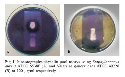
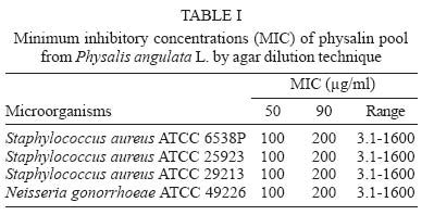
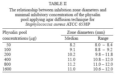
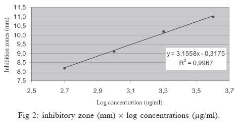
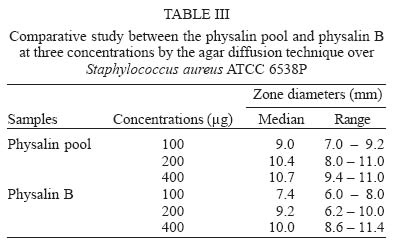
Mem Inst Oswaldo Cruz, Rio de Janeiro, Vol. 100, No. 7, November ,2005, pp. 779-782 Studies on antimicrobial activity, in vitro, of Physalis angulata L. (Solanaceae) fraction and physalin B bringing out the importance of assay determination Melissa TG Silva/*/+, Sonia M Simas**, Terezinha GFM Batista**, Paola Cardarelli***, Therezinha CB Tomassini* Programa de Pós Graduação em Vigilância Sanitária *Laboratório de Química de Produtos Naturais, Far-Manguinhos **Laboratório de Controle Microbiológico de Antibióticos ***Laboratório de Biologia Molecular-INCQS-Fiocruz, Av. Brasil 4365, 21040-900 Rio de Janeiro, RJ, Brasil Financial support: Far-Manguinhos-Fiocruz Received 16 June 2005 Code Number: oc05163 Complex physalin metabolites present in the capsules of the fruit of Physalis angulata L. have been isolated and submitted to a series of assays of antimicrobial activity against Pseudomonas aeruginosa ATCC 27853, Staphylococcus aureus ATCC 29213, S. aureus ATCC 25923, S. aureus ATCC 6538P, Neisseria gonorrhoeae ATCC 49226, Escherichia coli ATCC 8739; E. coli ATCC 25922, Candida albicans ATCC 10231 applying different methodologies such as: bioautography, dilution broth, dilution agar, and agar diffusion techniques. A mixture of physalins (pool) containing physalins B, D, F, G inhibit S. aureus ATCC 29213, S. aureus ATCC 25923, S. aureus ATCC 6538P, and N. gonorrhoeae ATCC 49226 at a concentration of 200 mg/µl, using agar dilution assays. The mixture was inactive against P. aeruginosa ATCC27853, E. coli ATCC 8739; E. coli ATCC 25922, C. albicans ATCC 10231 when applying bioautography assays. Physalin B (200 µg/ml) by the agar diffusion assay inhibited S. aureus ATCC 6538P by ± 85%; and may be considered responsible for the antimicrobial activity. Key words: Physalis angulata L. - antimicrobial methods - physalins Physalis is an important genus of the Solanaceae family. Most of the species are herbaceous annuals or perennials, native to tropical North and South America. Some species have edible fruits and the tea of their roots is considered in popular medicine. The medical uses of Physalis are numerous: a wide variety of species are used for asthma, urinary problems, rheumatism, and tumors. Their anti-inflammatory and anti-spasmodic properties are also known. Ray and Gupta (1994) and Tomassini et al. (2000) report some data on therapeutic applications and describe the pharmacological activity of the Physalis species as anti-parasitic, anti-viral, and anti-neoplasic. Trypanocidal activity was described by Freiburhaus et al. (1996) for P. angulata L. dichloromethane and ether extracts. Acetone, ethyl acetate, ethanolic extracts from leaves, stems, and roots of the specimen were assayed against Biomphalaria tenagophila yielding positive results for molluscicidal activity (Dos Santos 2003). Antimicrobial activity was described by Ongulana and Ramstad (1975) for methanol/water extracts of P. angulata against Bacillus subtilis. Cáceres et al. (1995) reported the results of the antimicrobial tests with the ethanolic extract leaves of Neisseria gonorrhoeae in agreement with the reported popular use in Guatemala. Aqueous and ethanolic extracts of P. angulata inhibited the growth of Staphylococcus aureus and Escherichia coli (Sanches et al. 1997, Silva et al. 1999). Pietro et al. (2000) described the tuberculostatic activity of P. angulata chloroform extracts from epigeal parts against Mycobacterium tuberculosis, M. avium, M. kansaii, M. malmoense, and M. intracellulare. Januário et al. (2003) reported activity against M. tuberculosis for physalins B and D. Physalin F isolated from P. angulata ethanolic extract (Chiang et al. 1993) shows a potent antineoplasic activity. Soares et al. (2003) described the immunosuppressive activity of physalins B, F and G extracted from the stems of P. angulata L. The continuous development of antibiotic resistance of pathogenic microorganisms and particularly of Streptococcus pneumoniae to penicillin (PRSP), Staphylococcus aureus to methicillin (MRSA), and Enterococcus to vancomycin (VRE) is a major health concern worldwide with economic, social and political implications. The screening of plant materials and their isolated substances for new antimicrobial compounds represent an important potential source for new effective medicines. Several techniques are routinely available to test antimicrobial properties, among which, the most popular are the following: Agar diffusion technique - Known as the Kirby-Bauer method, this assay was standardized by Bauer et al. in 1966. It is the test, which is most widely used in clinical practice and is recommended by Clinical and Laboratory Standards Institute (CLSI 2001). The method measures microbial growth inhibition at the surface of an inoculate medium around a paper disk impregnated with the antimicrobial substance at a standard concentration. The result may show, or not, the presence of an inhibition zone around the paper disk, the diameter of this zone being a good indicator of the antibiotic activity. Bioautography - This assay is a variation of the agar diffusion methodology. The sample to be analyzed is transferred from the chromatographic adsorbent to the inoculated agar. The spots containing the substances are visualized using microbial indicators (tetrazolium salts) as a growth detector of dehydrogenase activity (Kline & Golab 1965, Rahalison et al. 1994, Nostro 2000). Dilution tests - These can be applied in solid (agar dilution) or liquid (micro and macrodilution broth method) media. The results obtained allow a quantitative estimate of antimicrobial activity. Several dilutions of the antimicrobial substance are incorporated to the liquid or solid media to determine the minimal inhibitory concentration (MIC). MIC is the smallest concentration of the antimicrobial agent, which is capable of inhibiting the growth of the microorganism in vitro. These data are important to determine the optimal dosage as well as the correct administration route of the antimicrobial substance in therapy. The present study deals with the antimicrobial activity of Physalin B and enriched physalin fractions (mixture of B, D, F, and G physalins) against pathogenic gram positive and gram negative microorganisms in a search for a new antibiotic, as well as to find the best conditions for antimicrobial determination. MATERIALS AND METHODS Plant collection - P. angulata L. was collected in Belém, Pará, North of Brazil and a voucher (RFA# 23907/8) was deposited at the Botanical Department of the Universidade Federal do Rio de Janeiro. Plant extraction, fractioning, and identification - Dried and powdered capsules (1000 g) from the fruits of P. angulata L. were extracted exhaustively with hot ethanol (Soxhlet) to yield, after evaporation under reduced pressure, 101 g of crude material. This ethanolic extract was treated with lead acetate under the experimental conditions described by Tomassini et al. (1999) to yield 9.7 g of a pool of physalins from which pure physalin B (58 mg) was purified by chromatography on silica gel with elution by hexane ethyl acetate 30:70. Physalin B was identified by spectroscopic analyses and by comparison with literature data. The pool of physalins and physalin B were obtained from the P. angulata ethanolic capsule extract in 9.6 and 1.2% yield respectively. Microorganisms - The microorganisms used in the antimicrobial assay were C. albicans ATCC 10231, E. coli ATCC 8739, E. coli ATCC 25922, N. gonorrhoeae ATCC 49226, P. aeruginosa ATCC 27853, S. aureus ATCC 29213, S. aureus ATCC 25923, S. aureus ATCC 6538P. These microorganisms were obtained from the Instituto Nacional de Controle da Qualidade e Saúde (INCQS) Fiocruz, Rio de Janeiro, Brazil. Antimicrobial tests were performed by bioautography, broth dilution, agar dilution, and agar diffusion procedures. Bioautography - Tests were performed with 100 µg/ml of physalin pool methanolic solutions at: 1000 µg/ml, 2500 µg/ml, 5000 µg/ml respectively. Each aliquot (100 µl) was applied over a chromatosheet of Silica gel 60 F254 followed by elution with a system of ethyl acetate/hexane (7:3) and air-drying. The chromatogram was placed on a Petri dish and inoculated agar was distributed over the chromatogram inside the Petri dish with 40 ml of an appropriate culture medium such as Iso-sensitest agar inoculated with 0.5% from a standardized suspension for S. aureus and P. aeruginosa, 0.7% for E. coli, and Sabouraud agar 1% for a standardized suspension of C. albicans. The standardized suspensions of microorganisms were prepared by using a spectrophotometer at 25% T 580 nm (FDA 1991). For N. gonorrhoeae the chromatogram was deposited on 40 ml of solid GC agar enriched with 1% Isovitalex sowed with a culture of N. gonorrhoeae inoculum adjusted to the 0.5 McFarland turbidity standards (Bauer 1966, CLSI 1993). All plates were incubated at 35°C (29°C for C.albicans) for 18 h, with 5-8% CO2 for N. gonorrhoeae and in an ambient air incubator for the others. The inhibition zones were visualized with 2.5% methylthiazolyltetrazolium (MTT) solution to detect the dehydrogenase activity. This test was performed in duplicate. Dilution methods - Both dilutions methods, i.e, microdilution broth and agar dilution, were carried out by following the procedure outlined by CLSI (1993, 2001) using physalin pool concentrations of 1600 µg/ml, 800 µg/ml, 400 µg/ml, 200 µg/ml, 100 µg/ml, 50 µg/ml, 25 µg/ml, 12.5 µg/ml, 6.3 µg/ml, and 3.1 µg/ml. Microdilution trays were incubated at 35°C for 18 h in an ambient air incubator. Controls were prepared: one without physalins and the other without the microorganism used in the assay. This method was applied only for the aerobic microorganisms. The agar dilution technique was used for aerobic microorganisms and fastidious microorganisms. Controls with methanol/water 100, 50, 25, and 12% were used for all microorganisms. The plates were incubated at 35°C for 18 h in an ambient air incubator for aerobic pathogens at 35°C for 18 h, with 5-8% CO2 (for fastidious microorganism N. gonorrhoeae). All tests were performed six times. Agar diffusion - Tests were carried out according to the Farmacopéia Brasileira (1988) and USP XXIII Pharmacopoeia (1995) procedures using disks impregnated with the following amounts applied in methanol of the physalin pool: 1600 µg, 800 µg, 400 µg, 200 µg, 100 µg, and 50 µg. Based on the linearity range determination and values of MIC obtained from the physalins pool, the physalin B concentrations assayed were 400 µg, 200 µg, and 100 µg. The plates were incubated at 35°C for 18 h in an ambient air incubator. The zone diameters were measured with the Fisher Lilly apparatus. The tests were performed nine times. RESULTS AND DISCUSSION The bioautography screening method was the first method applied to detect the microorganism sensitivity to physalins. The bioautography involved a solvent that completely dissolved the physalin pool and pure physalin B. Bioautography as described, with fully dissolved physalins was reproducible at all concentrations used. After detection with MTT, C. albicans ATCC 10231, E. coli ATCC 8739, E. coli ATCC 25922, and P. aeruginosa ATCC 27853 did not show sensitivity to the physalin pool while N. gonorrhoeae ATCC 49226, S. aureus ATCC 29213, S. aureus ATCC 25923, S. aureus ATCC 6538P strains showed positive results (Fig. 1). The MIC for the physalin pool was based on the lowest concentration at which a positive effect was detected in bioautography assays. Due to the insolubility of physalins in water or in aqueous 1.5% dimethylsulfoxide, it was not possible to use the microdilution broth technique. Table I shows the MIC values for N. gonorrhoeae ATCC 49226, S. aureus ATCC 29213, S. aureus ATCC 25923, S. aureus ATCC 6538P for the physalin pool when applying the agar dilution assay. This technique is more versatile than microdilution broth due to the good stability of physalins in agar. The agar dilution assay suffers less from contamination and allows the determination of MIC for several microorganisms at the same time. In agreement with the USP XXIII Pharmacopoeia (1995) and the FDA Code (1991), protocols recommended for the study of antibiotic potency against the strain of S. aureus ATCC 6538P were used for the potency determination of physalin applying the agar diffusion technique. Table II shows the average values for inhibitory zones obtained after nine replications. Based on these results, it was possible to establish the linearity range presented in Fig 2. Table III shows the results of a comparative study between physalin pool and physalin B at three different concentrations using the agar diffusion technique. This result shows that, at the concentration of 200 µg/ml, pure physalin B exhibited ± 85% of the inhibitory zone observed with the total pool of physalins, at the same concentration. In the bioautography assays, the physalin pool and physalin B did not inhibit C. albicans, P. aeruginosa or E. coli strains but did reveal efficacy against N. gonorrhoeae and S. aureus. The physalin pool at 200 µg/ml yielded 100% inhibition for S. aureus ATCC 29213, S. aureus ATCC 25923, S. aureus ATCC 6538P, and N. gonorrhoeae ATCC 49226, in agar dilution tests. The above results point to the following conclusions: (a) the bioautography assay is a practical, reproducible test and easy to perform; (b) the agar dilution technique is more versatile than the broth dilution assay and does not present the problems encountered with this latter assay with the sample solution, contamination and MIC determination; (c) the agar diffusion technique can be used only for pure substances because when it is applied to mixtures containing constituents, which exhibit different diffusion factors, and, thus the results may be unreliable; (d) the methods related here-in permit the choice of the best tool for determination of the antimicrobial constituents from plant material. ACKNOWLEDGEMENTS To the National Institute of Health and Control for technical support. REFERENCES
Copyright 2005 Instituto Oswaldo Cruz - Fiocr |
