 |
| About Bioline | All Journals | Testimonials | Membership | News |
|
||||||
|
||||||
Memórias do Instituto Oswaldo Cruz, Vol. 101, No. 3, May 2006, pp. 277-279 Automated systems in the identification and determination of methicillin resistance among coagulase negative staphylococci Juliana Caierão, Silvana Superti*, Cícero AG Dias, Pedro Alves d'Azevedo/+ Departmento
de Microbiologia e Parasitologia, Fundação Faculdade
Federal de Ciências Médicas de Porto Alegre, Financial
support: CNPq, Capes Received
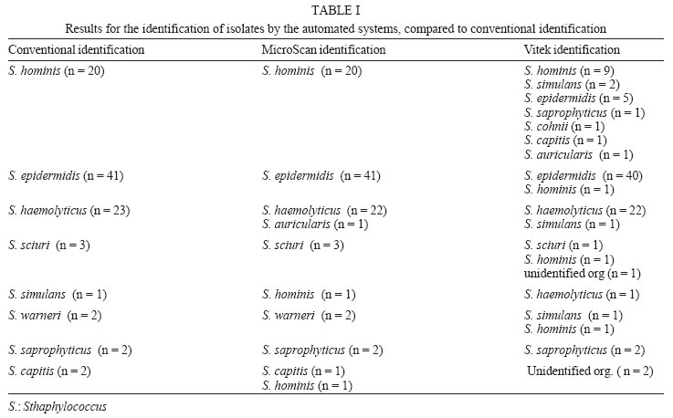
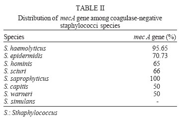
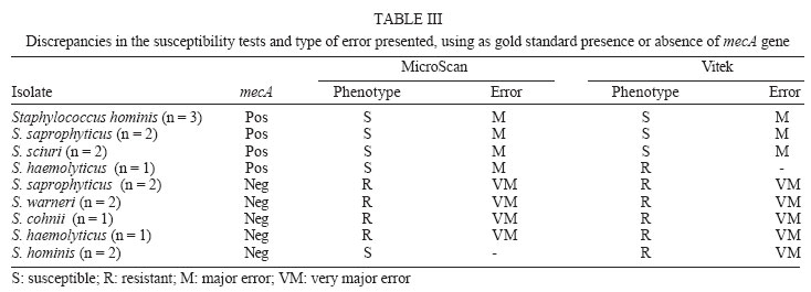
25 November 2005 Code Number: oc06049 Coagulase-negative staphylococci (CoNS) are an important cause of nosocomial bacteremia, specially in patients with indwelling devices or those submitted to invasive medical procedures. The identification of species and the accurate and rapid detection of methicillin resistance are directly dependent on the quality of the identification and susceptibility tests used, either manual or automated. The objective of this study was to evaluate the accuracy of two automated systems MicroScan and Vitek - in the identification of CoNS species and determination of susceptibility to methicillin, considering as gold standard the biochemical tests and the characterization of the mecA gene by polymerase chain reaction, respectively. MicroScan presented better results in the identification of CoNS species (accuracy of 96.8 vs 78.8%, respectively); isolates from the following species had no precise identification: Staphylococcus haemolyticus, S. simulans, and S. capitis. Both systems were similar in the characterization of methicillin resistance. The higher discrepancies for gene mec detection were observed among species other than S. epidermidis (S. hominis, S. saprophyticus, S. sciuri, S. haemolyticus, S. warneri, S. cohnii), and those with borderline MICs. Key words: coagulase-negative staphylococci - methicillin - automated systems Coagulase-negative staphylococci (CoNS) become important nosocomial pathogens, being, in many institutions, among the main etiological agents of nosocomial bacteremias (Cockerill et al. 1997, Hussain et al. 1998, Petinaki et al. 2001). Many clinical laboratories do not identify CoNS in species level when these microrganisms are detected in blood or in cerebrospinal fluid. However, as the significance of CoNS as pathogens has been increased, it has become more important to know the epidemiology and the pathogenic potential of species, individually. This might be particularly important considering blood culture isolates, since it is often difficult to determine the clinical significance of an individual isolate. There are numerous commercial systems and kits available nowadays for the identification of CoNS species (Kloos & Ban-nerman 1999). Resistance to methicillin among these microrganisms is a matter of concern because of the high and increasing levels detected. In Brazil, a multicenter study showed that methicillin resistance was observed in 87.7% of CoNS isolated from bloodstream infections (Sader et al. 1999). The accurate detection of methicillin resistance among CoNS isolates in the clinical laboratory is important to guide therapy and to promote the correct use of glycopeptides (Hussain et al. 1998, Yamazumi et al. 2001). This work was performed to evaluate the accuracy of two automated systems in the identification of species and determination of methicillin resistance, considering as gold standard the conventional biochemical tests and the characterization of mecA gene by polymerase chain reaction (PCR), respectively. MATERIALS AND METHODS Bacterial samples - Ninety-four consecutive CoNS isolates were included in the study, coming from blood cultures of patients hospitalized in the Complexo Hos-pitalar Irmandade Santa Casa de Misericórdia in Porto Alegre. The isolates were kept at -20oC in skim milk (Difco), plus 20% glycerol. The quality control of the tests was done using the Staphylococcus hominis ATCC 27844, S. epidermidis ATCC 12228, S. saprophyticus CCM 883, S. haemolyticus CCM 2737, and S. aureus ATCC 33591. Identification of bacterial isolates by conventional biochemical tests - The isolates were identified by conventional biochemical tests based on Manual for Clinical Microbiology (Bannerman 2003). The following test were used: catalase test, coagulase test, clumping factor, urease activity, ornitine decarboxilation, PYRase activity, presence of hemolysis, phosphatase activity, and fermentation of carbohydrates. Determination of susceptibility to methicillin - Susceptibility to methicillin was determined by the characterization of mecA gene by PCR. The bacterial DNA was extracted by thermal lysis according to Nunes et al. (1999), and the PCR was performed according to Santos et al. (1999), with modifications. Briefly, primers mecA1 (5'-TGGCTATCGTGTCACAATCG-3') and mecA2 (5'-CTGGAACTTGTTGAGCAGAG-3') were used for amplification of a fragment of 310 pb of the mecA gene. The solution for PCR, with a final volume of 50 µl, was composed of 10 µl of extracted DNA, 50 pmolar of each primer, 250 µmolar of each dNTP, 2 µl of magnesium chloride, 5 µl of the buffer of the enzyme, and 2.5 U of Taq DNA polymerase. The total cycle of the amplification was composed of an initial denaturation stage at 94ºC/1 min, followed by 30 cycles, with denaturation at 94ºC/15 s, annealing at 55ºC/15 s, and extension at 72ºC/5 s. The amplification products were analyzed by electrophoresis in agarose gel at 1.5% in TBE 0.5X, containing ethidium bromide (0.5 µg/ml) at 130 V and photographed under ultraviolet light. As standard of electrophoretic running, the standard molecular weight of 100 pb was used (Gibco, BRL). The strains S. aureus ATCC 33591 and S. aureus ATCC 25923 were used as positive and negative control, respectively. Identification of species and methicillin susceptibility using automated systems - The identification of species and the methicillin susceptibility were determined using automated systems MicroScan (Dade Behring), panel PC-13, and Vitek, GPS-105 (bioMérieux). The manufacturer's instructions were followed for the preparation of the inoculum and period of incubation of isolates. From the combination of the results of conventional biochemical tests, the 94 consecutive isolates of CoNS were identified as follows: 41 S. epidermidis, 23 S. haemolyticus, 20 S. hominis, 3 S. sciuri, 2 S. warneri, 2 S. saprophyticus, 2 S. capitis, and 1 S. simulans. The automated system MicroScan correctly identified 91 of the 94 isolates (accuracy of 96,8%). One isolate of S. simulans was identified as S. hominis, 1 S. haemolyticus as S. auricularis, and 1 isolate of S. capitis was mistakenly characterized as S. hominis (Table I ). On the other hand, the automated system Vitek correctly characterized 74 of the 94 isolates (accuracy of 78.7%), within 20 isolates mistakenly identified (Table I). The biochemical test that most frequently presented discordant results by the automated systems were the fermentation of mannose, threalose, and saccharose and the production of the urease enzyme. The characterization of methicillin resistance of the 94 samples 70 (74.4%) presented the, mecA gene. This gene is proportionally more frequent in the S. haemolyticus species but was widely distributed across the other species (Table II). The discrepancies in the susceptibility tests are presented in Table III, showing the types of errors ("major" or "very major") presented by the automated systems. All isolates with false-positive results presented MICs of 0.5 or 1 µg/ml, considered borderline (Table III). The automated system MicroScan characterized as resistant 62 (88.57%) of the 70 samples that carried the mecA gene. Of the 8 false-positive results, 3 were S. hominis, 1 S. haemolyticus, 2 S. saprophyticus, and 2 belonged to the S. sciuri species. The 6 false-positive results were distributed as follows: 2 S. saprophyticus, 2 S. warneri, 1 S. haemolyticus, and 1 S. cohnii. In the automated system Vitek, 63 (90%) samples were correctly characterized as resistant. The same 3 isolates of S. hominis, 2 S. saprophyticus, and 2 S. sciuri falsely characterized as susceptibile by the MicroScan system also showed false-positive results by the Vitek. The false-positive isolates by the MicroScan system also showed false-positivel results by the Vitek. Moreover, 2 isolates of S. hominis were also phenotypically resistant even without the presence of the mecA gene in their genome. DISCUSSION CoNS are the microorganisms most commonly isolated from blood cultures, representing a serious health problem in many developing countries and also in developed ones (Renneberg et al. 1995). So, there are, in the literature, many works discussing this issue. Cunha et al. (2004) present two methods modified in their laboratory to manually identify CoNS and they conclude that methods were found to be highly efficient for routine use due to their high sensitivity and specificity compared to the reference method (biochemical tests proposed by Bannerman in 2003), requiring fewer tests and thus being more economical and faster than the standard method. Here, we discuss the accuracy of automated systems, once they are faster than manual tests, being appropriate for routine laboratory. In general, the automated system MicroScan proved to be more accurate in the identification of CoNS species (accuracy of 96.8 vs 78.7%, respectively). However, the discrepant results in the identification are related to species which are less frequently isolated, considering that for the 2 most frequent isolated and more clinically relevant species S. epidermidis and S. haemolyticus the systems had similar performances. As a result of the low correlation between the presence of the mecA gene in S. epidermidis and the results of the disk diffusion test and the determination of MIC (York et al. 1996, Tenover et al. 1997), the NCCLS (1999) changed the methicillin breakpoints for CoNS isolates. Thus, automated systems had to modify their software so that they fit the new breakpoints and can effectively detect methicillin resistance among CoNS isolates. The systems presented good performance in the determination of methicillin resistance, especially for S. epidermidis. On the other hand, the interpretative criteria of the NCCLS (2004), strains related to serious infections with MICs varying from 0.5 to 2 µl/ml must be tested for the presence of the mecA gene or for the protein expressed by the gene, considering that they may present doubtful phenotypes. Considering that nowadays less frequent species of CoNS have been related to serious infections in hospital institutions, and that these have increasingly been isolated, it is very important to use automated systems which are accurate for the identification of these and for the determination of susceptibility to methicillin, leading to more rational therapy. In the present study, the MicroScan system was more accurate in the identification of CoNS strains and both presented limitations in the characterization of methicillin resistance in less frequent species. ACKNOWLEDGEMENTS To Dade Behring, to the staff of Central Laboratory of the Complexo Hospitalar Santa Casa of Porto Alegre. REFERENCES
Copyright 2006 Instituto Oswaldo Cruz - Fiocruz |
