 |
| About Bioline | All Journals | Testimonials | Membership | News |
|
||||||
|
||||||
Memórias do Instituto Oswaldo Cruz, Vol. 101, No. 3, May 2006, pp. 287-290 Antibacterial and cytotoxic activity of Brazilian plant extracts - Clusiaceae Ivana B Suffredini+, Mateus LB Paciencia, Daniela C Nepomuceno, Riad N Younes, Antonio D Varella Laboratório de Extração, Universidade Paulista, Av. Paulista 900, 1o andar, 01310-100 São Paulo, SP, Brasil Financial
support: Fapesp, Unip Received
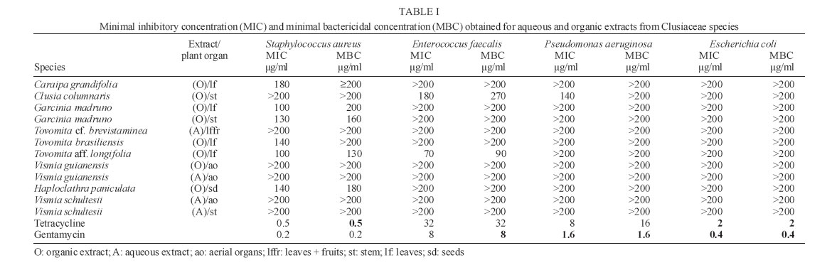
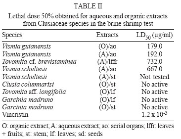
12 December 2005 Code Number: oc06051 Twelve extracts obtained from nine plants belonging to six different genera of Clusiaceae were analyzed against Gram-negative (Escherichia coli and Pseudomonas aeruginosa) and Gram-positive (Staphylococcus aureus and Enterococcus faecalis) bacteria using the microdilution broth assay. Tovomita aff. longifolia, T. brasiliensis, Clusia columnaris, Garcinia madruno, Haploclathra paniculata, and Caraipa grandifolia extracts showed significant results against the bacteria. The organic extract obtained from the leaves of T. aff. longifolia showed minimal inhibitory concentration (MIC) = 70 µg/ml and minimal bactericidal concentration (MBC) = 90 µg/ml against E. faecalis and the organic extract made with the stem of C. columnaris showed MIC = 180 µg/ml and MBC = 270 µg/ml against P. aeruginosa. None of the antibacterial extracts showed lethal activity against brine shrimp nauplii. On the other hand, both aqueous and organic extracts obtained from the aerial organs of Vismia guianensis that were cytotoxic to brine shrimp nauplii did not show a significant antibacterial activity in the assay. Key words: antibacterial - cytotoxicity - Clusiaceae - Amazon rain forest - Brazil Natural products are considered an important source of new antibacterial agents. Drugs derived from unmodified natural products or drugs semi-synthetically obtained from natural sources corresponded to 78% of the new drugs approved by the FDA between 1983 and 1994 (Cragg et al. 1997). This evidence contributes to support and quantify the importance of screening natural products. Previous screening works were done with more than 700 Amazon rain forest plant extracts, and results can be seen elsewhere (Frana & Suffredini 2002, Suffredini et al. 2002a, b 2004, Nepomuceno et al. 2003). Less than 20% of the Angiospermae have been screened for biological activity, and taking the Brazilian rain forests in concern, not much has been done so far. Brazil is home to more than 20% of the world's biodiversity, and the Amazon rain forest concentrates 17% of the biodiversity found within the country (Wilson & Peter 1988). For this reason, our research team has been systematically studying the antibacterial activity of organic and aqueous plant extracts obtained from plants native to the rain forest against Gram-positive Staphylococcus aureus ATCC 29213 (Sau) and Enterococcus faecalis ATCC 29212 (Efae) and Gram-negative Pseudomonas aeruginosa ATCC 27853 (Psa) and Escherichia coli ATCC 25922 (Ecol) with the aim of discovering new natural product compounds that can be used as antibiotics. Twelve aqueous and organic extracts obtained from plants belonging to six different genera of Clusiaceae (Vismia, Garcinia, Haploclathra, Tovomita, Caraipa, and Clusia) were submitted to the determination of the minimal inhibitory concentration (MIC) and minimal bactericidal concentration (MBC) using the microdilution broth assay (Suffredini et al. 2004). The extracts were also tested against brine shrimp nauplii in order to evaluate their potential as cytotoxic agents and to determine if there is any relationship between brine shrimp toxicity and antibacterial activity for those twelve extracts. MATERIALS AND METHODS Plant material - Plants were identified and are deposited in Herbarium Unip. Collect numbers are the following: Vismia guianensis (Aubl.) Choisy (PS 98); Tovomita cf. brevistaminea Engl. in Mart. (PS 134); Vismia schultesii N. Robson (AAO 3291); Clusia columnaris Engl. (AAO 3357); Tovomita aff. longifolia (Rich.) Hochr. (AAO3409); Garcinia madruno (Kunth) Hammel (AAO 3422), Haploclathra paniculata (Mart.) Benth. (AAO 3275); Tovomita brasiliensis (Mart.) Walp. (AAO3532) and Caraipa grandifolia Mart. (IBS 40). Plants were collected in water-flooded forests and in terra firme forests (Ferreira 1997) in the Amazon rain forest. Extraction - Plant parts were separated, dried, and ground before being submitted to a 24-h maceration with methanol:dichloromethane (1:1) followed by a 24-h maceration with Milli-Q water before being freeze dried, so as to produce two extracts from each specimen (Younes et al. 2000). Extracts were evaporated, lyophilized, and kept in a freezer at -20°C. The aqueous extract obtained from the aerial organs of V. guianensis (233.3 g) yielded 3.5% and the organic extract obtained from the aerial organs of this species (233.3 g) yielded 7.2%. The aqueous extract obtained from the leaves and fruits of T. cf. brevistaminea (165.42 g) yielded 9%. The aqueous extracts obtained from the aerial organs (1116.2 g) and stems (1338.5 g) of V. schultesii yielded 2 and 0.6%, respectively. The organic extract obtained from the seeds of H. paniculata (187.1 g) yielded 13.5%. The organic extract obtained from the stems of C. columnaris (657.8 g) yielded 5.3%. The organic extract obtained from the leaves of T. longifolia yielded 12.6%. The organic extracts obtained from the leaves (486.2 g) and stems (861.4 g) yielded 9.8 and 6%, respectively. The organic extract obtained from the leaves of T. brasiliensis (369.86 g) yielded 9.5%, and the organic extract obtained from the leaves of C. grandifolia (422 g) yielded 6.3%. The organic extract obtained from the leaves of T. longifolia (377.94 g) yielded 12.62%. Antibacterial microdilution broth assay - Microdi-lution broth assay was used to screen the extracts for their antimicrobial activity against the four bacteria. The bacterial inoculum of each ATCC strain was obtained from fresh colonies in Müeller-Hinton agar plates. Inoculums were initially prepared to a concentration of 1.5 x 10⁸ CFU/ml (0.5 MacFarland), and then diluted to 3 x 10² CFU/ml (Suffredini et al. 2004). From these diluted bacteria suspensions, 190 ml were transferred to each well of the microplates. Ten ml of the extract solutions were added to the wells and the microplates were incubated at 35ºC for 18 to 20 h. Extracts were prepared to 20 times the desired test concentration (2 mg/ml) in water or 50% DMSO solution. Results were visually analyzed and classified according to the following patterns: X = culture flocks in the bottom of the well, C = turbidity without flocculation, ++ = many culture flocks, + = few culture flocks, and L = total growth inhibition. Extracts that showed inhibitory activity in the preliminary broth assay were submitted to a subculture in Müeller-Hinton Agar, in order to evaluate bacterial growth. MIC was determined as the lowest concentration visually analyzed as "L" that implied in bacterial growth when subcultured; and MBC was determined as the lowest concentration that showed no bacterial growth in the subcultures (Suffredini et al. 2004). Gentamycin and tetracycline were used as standard drugs. Brine shrimp lethality test - Brine shrimp test was performed (Meyer et al. 1982) with modifications in the doses. Extracts diluted to concentrations of 0.5; 0.1; 0.02; 0.004; 0.008; and 0.0002 mg/ml were tested in triplicates. Living nauplii were counted after 24 h of exposure. LD50 was calculated using probit analysis (Finney 1971). Assay was validated with vincristin (Oncovin®) as the standard substance. Thin layer chromatography (TLC) analysis - A phytochemical screening by classes of constituents was done with the extracts using TLC analyses. The solvent system was ethyl acetate:formic acid:water (40:8:12), development was realized in aluminum plates coated with cellulose or silica gel GF254 (Merck). The following spray reagents were used: Kedde, Dragendorff, 10% ethanol KOH, anisaldehyde-sulphuric acid (AS) reagent and 1% methanolic diphenylboric acid-b-ethylamino ester (NP reagent). UV light at 254 nm and 366 nm was also used in order to develop the spots (Wagner & Bladt 1996). RESULTS AND DISCUSSION Results can be seen in Tables I and II. It was established that results would be considered significant if MIC or MBC ≤ 200 mg/ml. The organic extract obtained from the leaves of T. aff. longifolia showed a significant activity against E. faecalis and S. aureus. The organic extracts obtained from the leaves of T. brasiliensis and C. grandifolia showed activity against S. aureus. The organic extracts obtained from the leaves and from the stem of G. madruno were very efficient against S. aureus. Our experience of screening 1200 extracts against the four bacteria species shows that Gram-negative bacteria are hardly ever susceptible to plant extracts in doses as low as 200 mg/ml, although the organic extract obtained from the stem of G. columnaris has shown significant activity for both E. faecalis and P. aeruginosa. The organic extract obtained from the seeds of H. paniculata showed activity in the microdilution broth assay against S. aureus. Brine shrimp test (BST) is known as a low-cost test indicative of antibacterial, cytotoxic, pesticidal, and insecticidal activity and could be used as a simple method for screening antibacterial products. We could not find any correlation between brine shrimp lethal activity and the bactericidal activity in this group of the nine extracts. The four extracts whose antibacterial activity was more significant were not lethal to the nauplii. Nonetheless, the aqueous and organic extracts obtained from the aerial organs of V. guianensis showed a significant lethal activity in the BST but no significant antibacterial activity. Thus, once BST could be used to screen extracts for antibacterial activity, cytotoxicity would be extremely relevant, and paradoxally disqualify the extract to further use as antibiotics, unless chemical and structure modifications were done. Only one out of twelve extracts showed significant results against Gram-negative P. aeruginosa (the organic extract obtained from the stem of C. columnaris) and none showed activity against Gram-negative E. coli. As S. aureus and E. faecalis are important bacteria species related to human infectious diseases, we are encouraged to study the extracts in terms of their chemical constituents and to evaluate their performance in other antibacterial assays. The species investigated in the present assay were insufficiently investigated either chemical or biologically. However, correlations between isolated compounds and biological activity were reported to species belonging to the same genera of the plants recently screened. Xanthones were isolated from Tovomita species (Seo et al. 1999, Marques et al. 2000, Zhang et al. 2002), polyisoprenylated benzophenones were isolated from Clusia sp. (Lokvam et al. 2000, Porto et al. 2000), anthraquinones with antiprotozoal activity were found in V. orientalis (Mbwambo et al. 2004), and cytotoxic compounds were isolated from three species of Vismia (Hussein et al. 2003). A lupane derivative was isolated from C. densifolia (Gunasekera et al. 1983). Xanthones (Mahabusakaram et al. 2005) and benzophenones (Baggett et al. 2005) were isolated from Garcinia species, and some of them showed antibacterial activity (Rukachaisirikul et al. 2003). According to the TLC analyses performed with the extracts, nor cardiac glycosides or alkaloids were detected, except for the aqueous extract obtained from T. cf. brevistaminea, which showed a brown spot in the presence of Dragendorff reagent. Anthrones and phenolic compounds appear to be present in most of the extracts. Ten percent alcoholic KOH is considered to be reactive to anthrones (yellow), anthraquinones (red) or coumarins (blue), depending on the color of the spot observed under UV 365 nm (Wagner & Bladt 1996). Both organic extracts from the aerial organs of V. guianensis and from the leaves of T. aff. longifolia did not seem to have anthrones, anthraquinones or coumarins. On the other hand, spots with no fluorescence under UV 254 or 365 nm could be seen in these extracts and that may indicate the presence of saponins or terpenes which characteristic orange or yellow latex were found in most of the Clusiaceae (Gentry 1996). NP reagent indicates the presence of flavonoids if spots become yellow, orange or green when analyzed under UV 365 nm. Only the organic extract from the aerial organs of V. guianensis and the aqueous extracts from V. guianensis and V. schultesii did not seem to have flavonoids. The present findings support further studies related to the antibiotic activity of the organic extracts obtained from different organs of H. paniculata, C. columnaris, T. longifolia, G. madruno, T. brasiliensis, and C. grandifolia, species of Clusiaceae native to the Brazilian Amazon rain forest. The further identification of the active compounds are going to be made by bioguide fractionation, and are going to be focused in the possible presence of saponins, terpenes, coumarins, anthrones, anthraquinones, and flavonoids in the active extracts. ACKNOWLEDGEMENTS To the technicians involved in the process, Dr Nidia Cuello and Dr Volker Bittrich for reviewing the botanical material and Mrs Flávia Romano, for the English review of the manuscript. REFERENCES
Copyright 2006 Instituto Oswaldo Cruz - Fiocruz |
