 |
| About Bioline | All Journals | Testimonials | Membership | News |
|
||||||
|
||||||
Memórias do Instituto Oswaldo Cruz, Vol. 101, No. 5, August, 2006, pp. 499-501 Application of biochemical and polymerase chain reaction assays for identification of Campylobacter isolates from non-human primates Mônica de Castro Britto Vilardo, Jacqueline Darc da Silva Thomé, Wagner Thadeu Cardoso Esteves, Ana Luzia Lauria Filgueiras/+, Selma Soares de Oliveira* Laboratório
de Zoonoses Bacterianas, Departamento de Bacteriologia, Instituto Oswaldo
Cruz-Fiocruz, Av. Brasil 4365, Received 4 November
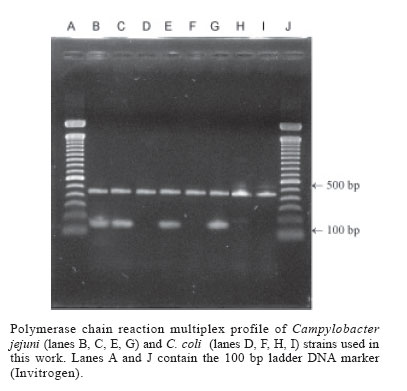
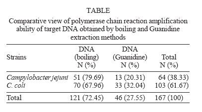
2005 Code Number: oc06084 A multiplex polymerase chain reaction (PCR) assay was performed on 167 thermophilic campylobacters isolated from non-human primates. Samples were first identified by phenotypic methods resulting in 64 Campylobacter jejuni and 103 C. coli strains. Four strains identified biochemically as C. coli, were then determined to be C. jejuni by PCR. Comparison of methodologies showed that the main discrepancies were attributed to the hippurate hydrolysis test and sensitivity to cephalothin and nalidixic acid. Analysis of data showed that the application of phenotypic methods should be supplemented by a molecular method to offer a more reliable Campylobacter identification. Key words: Campylobacter - identification - non-human primates The Campylobacter genus comprises a group of strictly related Gram-negative bacteria that are usually found colonizing the gastrointestinal tract of a variety of warm-blooded animals. For more than 40 decades Campylobacter spp. were related to veterinary diseases. However, since the development of an appropriate selective medium in the 1970s, Campylobacter isolations have increased and the bacteria have become the most commonly recognised cause of human gastroenteritis in developed countries (Butzler 2004). In spite of the relevance of Campylobacter infections, epidemiological tracing of enteritis is poorly understood in developing countries, probably due to a combination of characteristics such as the lack of seasonal variation, milder illness, the high rates of assymptomatic carriage and the high rates of infections with multiple pathogens (Taylor 1992). Furthermore, many clinical laboratories commonly do not identify Campylobacter strains to species level, since they are fastidious organisms and only a reduced number of biochemical tests are available. C. jejuni and C. coli are the thermophilic species most frequently isolated from stool samples. Although clinical manifestations of Campylobacter enteritis are very similar with acute diarrhoea, fever and mainly abdominal cramps, C. jejuni is responsible for the majority (80-90%) of infections and some serotypes are involved in GüillainBarré syndrome (Allos e Taylor 1998). Endemic campylobacteriosis is frequent with laboratory animals that are housed in groups where fecal contamination occurs. Several non-human primates mantained in captive colonies become routinely infected with multiple Penner serotypes of C. jejuni and C. coli and animal enteritis share similar clinical signs to human disease (Bryant 1983, Russel 1992). The aim of this study is to improve epidemiological studies by the identification of thermophilic Campylobacter strains isolated from non-human primates. Because discrimination among thermophilic strains is based on only a few biochemical tests which are sometimes difficult to perform and interpret, we applied a PCR assay to get a definitive sample identification. MATERIALS AND METHODS Strains - For this study 167 thermophilic Campylo-bacter isolates were selected. Strains were isolated from the faeces of Macaca mulatta, M. fascicularis, and Saimiri sp. collected during a medical management which occurred in 1999. These animals are held in a closed colony at the Primatology Department of the Center for Laboratory Animal Breeding at the Oswaldo Cruz Foundation-Fiocruz. Treatment of specimens - Bacteria were grown on Columbia agar supplemented with defibrinated sheep blood (5%) or activated coal (0.4 g%), and also included an FBP solution. Plates were incubated (42oC for 48 h) under a microaerophilic atmosphere. Campylobacter strains were identified by a classic biochemical methodology according to Filgueiras and Hofer (1989) and stored in tubes containing Peptonated water, pH 7.0-7.2, with 20% glycerol at -70oC. C. jejuni ATCC 33560 and C. coli ATCC 33559 strains were included in all tests as positive controls. Identification of Campylobacter spp. by multiplex PCR - Genotypic identification of Campylobacter strains was performed by PCR multiplex as described by Harmon et al. (1997). The primers (Invitrogen Brasil Ltda.) used were pg3 (5'- GAACTTGAACCGATTTG - 3') and pg50 (5'- ATGGGATTTCGTATTAAC - 3') for gene flaA present in C. jejuni and C. coli and primers C1 (5'- CAAATAAA GTTAGAGGTAGAATGT-3') and C4 (5'-GGATAAGCAC TAGCTAGCTGAT - 3') for a non-determined gene of C. jejuni. DNA samples - Some colonies from Columbia agar plate were resuspended in 100 µl of sterile TE buffer (10 Mm Tris, 1 Mm EDTA, pH 8.0) in order to obtain 2 x 10⁹ cells (Englen and Kelley 2000). DNA extractions for PCR assay were done by two different protocols: the boiling method of Van Eys et al. (1989), and the method of Pitcher et al. (1989), which uses guanidine thiocyanate as a chaotropic agent. The DNA concentration was estimated in the GeneQuant pro/RNA/DNA calculator - Amersham Pharmacia Biotech. DNA amplification - The amplification reaction was performed in a volume of 50 µl containg: 20 ng of sample DNA, 10 mM of Tris-HCL; 50 mM of KCl; 200 mM of (each) dATP, dCTP, dGTP, dTTP; 5.5mM of MgCl2; 20 pmoles each of C1 and C4; 40 pmoles each of pg3 and pg50 and 1.25U of Taq DNA polymerase. All components of the mix were synthesized by Invitrogen and each amplification reactions contained positive controls of C. jejuni ATCC 33560 and C. coli ATCC 33559. Reaction mixtures were amplified in a thermocycler device PTC 150 - MJ Research, Incorporation. The samples were subjected to an initial denaturation step at 94oC for 4 min followed by 25 amplification cycles, each one consisted of 1 min at 94oC, 1 min at 45oC, and 1 min at 72oC, ending with a primer extension step of 7 min at 72oC. Analysis of amplified fragments - PCR reaction products were separated through electrophoresis on a 1.5% agarose gel (TBE 0.5x), including the 100 pb ladder as molecular weight marker (Invitrogen). Agarose gels were stained in an ethidium bromide solution and visualized under UV light, in the transiluminator (Chemical Company T102 SIGMA). RESULTS All 167 strains of Campylobacter spp. were identified using both biochemical and multiplex PCR techniques. The use of multiplex PCR resulted in the identification of 64 strains as C. jejuni and 103 as C. coli. C. jejuni strains showed both 460 bp to 160 bp fragments whereas C. coli generated only the 460 bp fragment (Figure). The molecular technique and the conventional phenotypic tests were in agreement for 163 of the 167 isolates. Four strains (2.84%), firstly identified as C. coli, based on their inability to hydrolyze sodium hippurate, were later identified as C. jejuni by PCR. Multiplex PCR and sodium hippurate hydrolysis tests were repeated for three times and these strains were then recognized as hippurate-negative C. jejuni. Six strains of C. coli and two of C. jejuni that showed nalidixic acid resistance were also identified by multiplex PCR. As results were in agreement with biochemical identification these strains were then characterized as nalidixic acid resistant C. coli and C. jejuni. Initially, DNA was extracted by the boiling water method. However, for 46 (out of 167) strains, the DNA obtained by this technique was not amplified. This group of strains was then submitted to a DNA extraction by the guanidine thiocyanate method and all of them were subsequently amplified (Table). Thirty-three (71.74%), out of these 46 strains, were DNAse positive in biochemical tests. DISCUSSION Epidemiological studies have demonstrated a distinct pattern of Campylobacter enteritis between industrialized and developing countries, including clinical manifestations and relevancy of infections. Few published studies are realized in the developing world since the majority of public health laboratories do not have success in isolating campylobacters, which require an adequate selective media, temperature and microaerophilic conditions for incubation. These steps are not included in laboratory routines for common enteric pathogens. In attempt to contribute to a better understanding of campylobacteriosis in Brazil, the Zoonosis Laboratory has been undertaking studies with strains isolated from several sources. A major number of isolates was obtained from non-human primates living in closed groups, which are frequently colonized by different strains of Cam-pylobacter (Russel et al. 1988). The low biochemical activity of Campylobacter spp. and the occurence of ambiguous results makes phenotypic identification difficult. Uncommon phenotypes like C. jejuni and C. coli strains which are resistant to quinolones as well as the hippurate-negative C. jejuni strains are frequently pointed out (Rautelin et al. 1999, Nachankin et al. 2000). Molecular techniques, including the PCR, have recently been successfully used for the identification of Campylobacter species (Steinhauserova et al. 2001). There is a large use of Campylobacter flagellum genes as sequences for PCR primers because they are highly conserved within different strains (Wegmüeller et al. 1993). The use of primers pg3 and pg50 specific for C. jejuni and C. coli are described in other publications (Oyofo et al. 1992, Aquino et al. 2001) and show a high sensitivity of detection. In the present study, as well as phenotypic identification, multiplex PCR proposed by Harmon et al. (1997) was used to distinguish C. jejuni from C. coli strains. However, 46 (27.55%) strains did not give amplification when the boiling method was used for DNA extraction. As Mohran et al. (1998) related the existence of a differing ability of Campylobacter to display its genome by the boiling method, a second method using guanidine thiocyanate was chosen. The 46 strains showing a phenotype resistant to boiling were then amplified by the last method and it was observed that in this group, there was a high prevalence of DNAse producing strains (71.74%). Lai-King et al. (1997) recommended attention when processing DNAse positive Campylobacter spp. because their DNA is more difficult to detect using nucleid acid-based methods when compared with other genera. We considered that DNA extraction using guanidine thiocyanate was the best method for which it becomes possible to obtain target DNA from DNAse positive strains. The differences in Campylobacter identification by phenotypic and PCR assays have shown that an application of a molecular analysis is essential to complement classic biochemical methodology. Our results, together with the literature, suggest that multiplex PCR can be helpful in resolving ambiguous results, for characterizing uncommon strains, and for epidemiological investigations. ACKNOWLEDGMENTS To Ângela Lopes Norte, chief of International Cooperation Division from Cefet-RJ, Brazil, and Dr Lawrence Price from Health Canada, National Microbiology Laboratory for reviewing the manuscript REFERENCES
Copyright 2006 Instituto Oswaldo Cruz - Fiocruz The following images related to this document are available:Photo images[oc06084t1.jpg] [oc06084f1.jpg] |
| |||||||||
