 |
| About Bioline | All Journals | Testimonials | Membership | News |
|
||||||
|
||||||
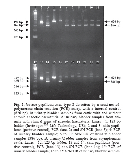
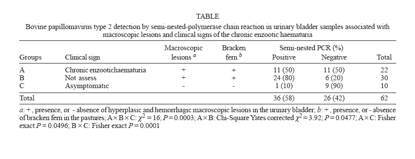
Mem Inst Oswaldo Cruz, Rio de Janeiro, Vol. 101, No. 6, September ,2006, pp. 635-638 Bovine papillomavirus type 2 detection in the urinary bladder of cattle with chronic enzootic haematuria J Sheila R Wosiacki, Marlise P Claus, Alice F Alfieri, Amauri A Alfieri+ Laboratório de Virologia Animal, Departamento
de Medicina Veterinária Preventiva, Universidade Estadual
de Londrina, Received 7 March 2006 Code Number: oc06107 The bovine papillomavirus type 2 (BPV-2) involvement in the aetiology of chronic enzootic haematuria associated to bracken fern ingestion has been suggested for a long time. However, a few reports have shown the presence of the BPV-2 in urinary bladder tumors of cattle. The aim of this study was to investigate the presence of the BPV-2 infection in the urinary bladder of cattle with chronic enzootic haematuria in Brazilian cattle herds. Sixty-two urinary bladders were collected from adult cattle in beef herds from the north region of the state of Paraná, Brazil. According to clinical and pathological finds the specimens were distributed in three groups: the group A was constituted by 22 urinary bladders with macroscopic lesions collected at necropsy of cattle with clinical signs of chronic enzootic haematuria; the group B by 30 urinary bladders with macroscopic lesions collected in a slaughterhouse of cows coming from bracken fern-endemic geographical region; and the group C (control) by 10 urinary bladders without macroscopic lesions collected from asymptomatic cattle in a bracken fern-free geographical region. By a semi-nested polymerase chain reaction (PCR) assay, with an internal control, a fragment of the BPV-2 L1 gene with 386 bp length was amplified in 36 (58%) urinary bladder. The rate of BPV-2 positive urinary bladders was 50% (11/22) for group A, 80% (24/30) for group B, and 10% (1/10) for group C (control). The rate of the positive results found in groups A and B that included urinary bladder samples with macroscopic lesions was 67% (35/52) and the detection of the BPV-2 in both groups was significantly higher (P < 0.05) than in the control group. RFLP with Rsa I and Hae III enzymes evaluated the specificity of the BPV-2 amplicons. The PCR internal control that amplified a 626 bp fragment of the ND5 gene of the bovine mitochondrial genome was amplified in all analyzed samples and excluded false-negatives or invalid results in the semi-nested PCR. These results suggest the BPV-2 involvement in the chronic enzootic haematuria aetiology and open the perspective of the development of new strategies for the control of this disease that is the major cause of economical losses in beef herds from many Brazilian geographical regions. Key words: cattle-viruses - urinary bladder - chronic enzootic haematuria - bovine papillomavirus type 2 - semi-nested-polymerase chain reaction . The chronic enzootic haematuria causes high economical losses in cattle herds around the world. The disease is clinically characterized by intermittent haematuria caused by malignant lesions in the urinary bladder, followed by anemia, progressive emaciation, and death (Hopkins 1986). The bovine papillomavirus type 2 (BPV-2) infection and the chronic intoxication by bracken fern (Pteridium aquilinum sub. caudatum var. arachnoideum) ingestion were associated with urinary bladder lesions and the clinical signs of chronic enzootic haematuria in adult cattle. Evidences indicate that the malignant progression of urinary bladder lesions is dependent on an inter-relationship between the BPV-2 infection and carcinogenic, mutagenic, and immunosuppressive compounds of the bracken fern (Reddy & Fialkow 1983, Campo 1995, 1997). However, few studies were accomplished on the aetio-logical aspects of chronic enzootic haematuria, and the aetiology and pathogeny of this disease are not yet completely known. The BPV-2 is a small oncogenic DNA virus member of the genus Deltapapillomavirus in the Papillomaviridae family (Fauquet et al. 2004). It is a fibropapillomavirus that can cause common skin papillomas, fibropapillomas in the alimentary tract and in the urinary bladder of cattle (Campo 1997). The diagnosis of the BPV-2 infection has been made through the identification of the viral DNA in tissue fragments using techniques such as the Southern blot and the polymerase chain reaction (PCR) (Campo et al. 1992, Bloch et al. 1997, Borzacchiello et al. 2003, Wosiacki et al. 2005). Due to the importance of chronic enzootic haematuria in cattle herds, mainly in beef cows, from different regions around the world studies to contribute with the definition of the urinary bladder neoplastic lesions aetiology are necessary. This study was carried out with the objective to detect BPV-2 in urinary bladder of cattle with macroscopic lesions of chronic enzootic haematuria collected in necropsies and in slaughterhouses from bracken fern-endemic geographical region. MATERIALS AND METHODS Positive control - As BPV-2 positive control a skin papilloma sample from the cervico-dorsal area of a seven-month-old calf was used. The macroscopic aspects and the histopathological findings were similar to the skin fibropapillomas caused by the BPV-2. The BPV-2 L1 gene was identified in this skin papilloma sample by a semi-nested PCR (SN-PCR) assay, and characterized by using of restriction fragment length polymorphism (RFLP) and sequence analysis (Wosiacki et al. 2005). Specimens collection - Sixty-two urinary bladders were collected from adult cows in beef cattle herds from the north region of the state of Paraná, Brazil. According to clinical and pathological finds the specimens were distributed in three groups: (i) the group A containing 22 urinary bladders with macroscopic hyperplasic and hemorrhagic lesions collected at necropsy of cows with clinical signs of chronic enzootic haematuria; (ii) the group B consisted of 30 urinary bladders with macroscopic lesions collected in slaughterhouses of cows coming from bracken fern-endemic geographical region; (iii) the group C (control) was constituted of 10 urinary bladders without macroscopic lesions collected in a slaughterhouse from asymptomatic cattle in a bracken fern-free geogra-phical region. BPV-2 detection - Fragments of urinary bladder samples were triturated in phosphate-buffered saline (PBS, pH 7.2) and the suspensions (10-20% w/v) were centrifuged for 15 min at 1500 g at 4oC. Aliquots of 250 µl of the supernatant were treated with lysis buffer (10 mM Tris; 1 mM EDTA; 0.5% Nonidet P40; 1% SDS; and 0.001 mg proteinase K). After homogenization, the samples were incubated at 56oC for 30 min. For DNA extraction a combination of phenol/chloroform/isoamyl alcohol and silica/guanidine isothiocyanate methods was performed according to Alfieri et al. (2004). First, the samples were treated with an equal volume of phenol/chloroform/isoamyl alcohol (25:24:1), homogenized, and heated at 56oC for 15 min (Sambrook & Russell 2001). Next, the samples were centrifuged at 10,000 g for 10 min and the aqueous phase processed in silica/guanidine isothiocyanate (Boom et al. 1990). The DNA was eluted in 125 µl of ultrapure (Milli-QÒ) sterile water and stored at _20oC until use. Aliquots of ultrapure sterile water were included as negative controls in all the DNA extraction procedures. The BPV-2 L1 gene detection was carried out by a SN-PCR performed according to Wosiacki et al. (2005). In the first round (PCR), the BPVP1 (5-TGT TCC CAA AGT GTC TG-3, nt. 5771-5787) and BPVP2 (5-CAT TTT GAG GTA GTC TGG-3, nt. 6299-6282) primers were used to amplify a fragment of 529 bp. In the second round (SN-PCR) the BPVP1 primer and the BPVP3 (5-ATT CTA AAG GAG GAC ACG-3, nt. 6156-6139) primer amplify a 386 bp size fragment. In the first round all the reactions were carried out with internal control primers BOV1 (5-ATA CGC CTT CAT TAC CAG-3, nt. 12.231-12.248), and BOV 2 (5-TTG AAT GGA GTA GTG CTG-3, nt. 12.856-12.839) that amplified a 626 bp fragment of the ND5 gene of the bovine mitochondrial genome that was used in a multiplex reaction. In the PCR assay 2.5 µl of extracted DNA, 0.4 pmol of each BPVP1, BPVP2, BOV1, and BOV2 primers, 1 x PCR buffer (20 mM Tris-HCl pH 8.4 and 50 mM KCl), 2.5 mM MgCl2, 300 µM each dNTP (InvitrogenTM Life Technologies, US), 1.25 units of Platinun Taq DNA Polymerase (InvitrogenTM) and ultrapure sterile water to a final volume of 25 µl were used. The amplification was performed in a thermocycler (PTC 200 - MJ Research Co. Water Town, MA, US) and consisted of the following time and temperature conditions: one step of 2 min/94oC followed by 40 cycles at 1 min/94oC, 1 min/55oC and 1 min/72oC. For the SN-PCR 1 µl of PCR product, 0.4 pmol of BPVP1 and BPVP3 primers, 1 x PCR buffer, 2 mM MgCl2, 100 µM each dNTP, 0.75 units of Platinum Taq DNA Polymerase, and ultrapure sterile water to a final volume of 25 µl were used. The conditions of amplification consisted in one step of 2 min/94oC followed by 30 cycles at 30 s/94oC, 30 s/55oC, and 1 min/72oC. The RFLP was assessed by the analysis of the BPV-2 SN-PCR amplicons with the Rsa I and Hae III enzymes (InvitrogenTM). The digestion was made during 1 h at 37oC according to manufacturer's instructions. Amplicons of SN-PCR and RFLP were analyzed by electrophoresis in a 2.5% agarose gel in TBE buffer pH 8.4 (89 mM Tris; 89 mM boric acid; 2 mM EDTA) stained with 0.5 µg/ml ethydium bromide and visualized under UV light. Statistical analysis - The association between the BPV-2 detection in urinary bladder samples and the three groups included in this study was analyzed by the Chi-Square (χ2) or the Fisher exact test. The statistical analysis was done using the software EpiInfo 3.3.2 significance level of 5%. RESULTS A 386 bp specific amplicon of the BPV-2 L1 gene was detected in 58% (36/62) (IC 95% = 44.84-77.48) of the urinary bladder samples (Fig.1). The Table shows the distribution of SN-PCR positive results by group (A, B, and C) of cattle included in this study. The rate of BPV-2 positive urinary bladder with macroscopic lesions (n = 52, groups A and B) was 67%. In the control group C in which only urinary bladder samples without macroscopic lesions were included, the rate of BPV-2 positive samples was 10%. The association of BPV-2 diagnosis in urinary bladders and macroscopic lesions was significant (P = 0.0003). The specificity of the L1 gene amplicons was confirmed by RFLP. As in silico analysis the amplicon of 386 bp yielded fragments of 85 and 301 bp length with Rsa I, and of 147 and 239 bp with Hae III enzymes (Fig.2). A 626 bp fragment of internal control was amplified in the first amplification round in all clinical samples included in this study. DISCUSSION The involvement of papillomaviruses in the aetiology of chronic enzootic haematuria associated to bracken fern ingestion has been suggested for a long time (Jarrett et al. 1978, Pachauri et al. 1981, Moura et al. 1988). However, the difficulty of the BPV diagnosis by conventional virological methods, such as virus isolation in cell culture, stimulated the development and assessment of molecular diagnostic techniques and some reports have shown the involvement of the BPV-2 in the aetiology of urinary bladder neoplastic lesions in cattle (Campo et al. 1992, Stocco dos Santos et al. 1998, Borzacchiello et al. 2003, Wosiacki et al. 2005). In this study we used a SN-PCR assay to assess the presence of the BPV-2 in the urinary bladder samples with hyperplasic and hemorrhagic macroscopic lesions collected from adult cattle in geographical regions where the chronic enzootic haematuria is endemic in cattle herds. All these samples were obtained from cows raised in highly infested pastures with bracken fern and in a geographical region where the chronic enzootic haematuria clinical disease is frequent and represent the major cause of mortality, mainly in cows from beef herds. Campo et al. (1992) detected the BPV-2 DNA by a Southern blot assay in 46% (7 of 15) of the natural tumors cases and in 69% (9 of 13) of the experimentally induced lesions in immunosuppressed animals, suggesting a close association between BPV-2 and bovine bladder tumor. Twenty percent of positive samples were found in the control group, suggesting the viral persistence that can be activated when the animal is exposed to the bracken co-carcinogens and immunosuppressants compounds. In Italy, Borzacchiello et al. (2003) detected the BPV-2 by a PCR assay in 76.7% (46/60) and 50% (17/34) of cattle urinary bladder samples with and without neoplastic lesions, respectively. Their control group consisted of urinary bladder samples without macroscopic lesions. However, these samples were obtained in the same endemic geographical area as the urinary bladders with macroscopic lesions, which explain the high rate of positive samples in the control group The detection rate of papillomaviruses DNA in urinary bladder samples with lesions varies substantially in different studies. The major problem may be discrepancies in the number of samples assessed, in the diagnostic techniques, and in the interpretation of the result. However, in this study SN-PCR false-negatives results were not generated by nucleic acid extraction failure, variable (low or high) amounts of DNA or by the presence of inhibitors of the PCR reaction. The internal control of the PCR reaction was amplified in all the urinary bladder samples. The papillomaviruses DNA persists in tumor cells in episomal and/or integrative forms (Klimov et al. 2002). Although viral DNA remains exclusively episomal in benign papillomas, integration of a partially deleted viral genome is present in most human cervical cancers. Integration of human papillomavirus (HPV) DNA into the host genome occurs early in cancer development and is probably an important event in malignant transformation of cervical cancer (Cullen et al. 1991, Yoshinouchi et al. 1999, Lukaszuk et al. 2003). For BPV-2, no studies of the phenomenon of viral integration in samples of urinary bladder tumors were found. However the genetic analyses of BPV-2 and BPV-1 have demonstrated the highest homology and similarity of these viruses. Genetic studies have mapped the BPV-1 and three independent transforming proteins were found and encoded as E5, E6, and E7 genes. The E5 gene is the major transforming gene of BPV-1. This gene is highly conserved among the group of papillomaviruses and can affect the activity and metabolism of growth factor receptors (Campo 2002). Studies in animal model have clearly demonstrated the contribution of host genetics, chemical carcinogens, and immunosuppression to the conversion of papillomaviruses induced benign lesions into malignant tumors. Most of the papillomaviruses basic knowledge is resulting from the BPV study. In some geographical regions around the world the chronic enzootic haematuria is a major cause of death in adult cattle, and no treatment is known for this disease at the moment. However, few epidemiological researches about this disease have been accomplished. The evidence of this study contributed with the demonstration of the viral aetiology in chronic enzootic haematuria of cattle herds in north region of the state of Paraná, Brazil. The studies for the definition of viral aetiology of chronic enzootic haematuria can contribute for proposals of vaccine development for the BPV-2 infection control in herds from endemic regions and also to reduce the severity of chronic enzootic haematuria clinical signs in cattle around the world. REFERENCES
Copyright 2006 Instituto Oswaldo Cruz - Fiocruz The following images related to this document are available:Photo images[oc06107t1.jpg] [oc06107f2.jpg] [oc06107f1.jpg] |
| |||||||||
